Review of Critical Care Medicine

  • Blog Stats

    • 741,078 hits
  • Disclaimer

    The author does not take over any guarantee for the topicality, the correctness, completeness or quality of the information, made available. Liability claims against the author, concerning damage of idealistic or of material kind, which was caused by the use or not use of the presented information and/or by the use of incorrect and incomplete information, are in principle impossible, so far as not a deliberate or roughly negligent fault can be proved on the part of the author. The documents and graphics on this Web site can be affected by technical inaccuracies or misprints, for which we don't assume any liability. Furthermore,quotation from a book or the incidental capturing of copyrighted material in a segment of a MCQ should be considered as "FAIR USE".It is only for nonprofit educational purposes.The display is a regular part of the systematic instructional activities of this non-profit educational website and is meant entirely as help to solve the ever-challenging MCQs.Due respect has been given to great medical authors and their names displayed prominently alongside quotations/pictures or paragraphs.
  • Member of The Internet Defense League

  • Follow Review of Critical Care Medicine on WordPress.com
  • April 2024
    M T W T F S S
    1234567
    891011121314
    15161718192021
    22232425262728
    2930  
  • Recommended Books

  • Webchat

Posts Tagged ‘Medical MCQs’

Trauma MCQs & Notes

Posted by Dr KAMAL DEEP on June 3, 2011

BLUNT TRAUMA ABDOMEN

image

Blunt abdominal trauma initially is evaluated by FAST examination in most major trauma centers, and this has largely supplanted DPL .FAST is not 100% sensitive, however, so diagnostic peritoneal aspiration is still advocated in hemodynamically unstable patients without a defined source of blood loss to rule out abdominal hemorrhage FAST is used to identify free intraperitoneal fluid in Morison’s pouch, the left upper quadrant, and the pelvis. Although this method is exquisitely sensitive for detecting intraperitoneal fluid of >250 mL, it does not reliably determine the source of hemorrhage nor grade solid organ injuries.28,29 Patients with fluid on FAST examination, considered a “positive FAST,” who do not have immediate indications for laparotomy and are hemodynamically stable undergo CT scanning to quantify their injuries. Injury grading using the American Association for the Surgery of Trauma grading scale is a key component of nonoperative management of solid organ injuries. Additional findings that should be noted on CT scan in patients with solid organ injury include contrast extravasation (i.e., a “blush”), the amount of intra-abdominal hemorrhage, and presence of pseudoaneurysms . CT also is indicated for hemodynamically stable patients for whom the physical examination is unreliable. Despite the increasing diagnostic accuracy of multislice CT scanners, CT still has limited sensitivity for identification of intestinal injuries. Bowel injury is suggested by findings of thickened bowel wall, “streaking” in the mesentery, free fluid without associated solid organ injury, or free intraperitoneal air.30 Patients with free intra-abdominal fluid without solid organ injury are closely monitored for evolving signs of peritonitis; if patients have a significant closed head injury or cannot be serially examined, DPL should be performed to exclude bowel injury

Blunt injuries to the abdomen – (JIPMER 82,80)
a)May cause peritonitis
b)Rarely need urgent laparotomy
c)May cause intestinal obstruction
d)May cause gastroduodenal ulceration

Blunt trauma secondary to motor vehicle accidents, motorcycle accidents, falls, assaults, and striking of pedestrians remains the most frequent mechanism of abdominal injury. Penetrating abdominal wounds are usually caused by either gunshot or stab wounds and by a significantly smaller number of shotgun wounds.

Based on the high frequency of intra-abdominal organ injury after gunshot wounds, mandatory abdominal exploration, with the rare exception of tangential and superficial wound trajectories restricted to the right upper quadrant, remains the standard form of management. Stab wounds to the abdomen, however, carry a significantly lower risk of intra-abdominal organ injury than do gunshot wounds, and several studies have recently favored a more selective approach, as opposed to mandatory exploratory laparotomy.

The impetus for nonoperative management of solid organ injury in stable blunt trauma patients has expanded to penetrating trauma as well. With improved imaging, more stable patients sustaining a single solid organ injury after stab and gunshot wounds to the abdomen will be treated conservatively.

The diagnostic approach differs for penetrating trauma and blunt abdominal trauma. As a rule, minimal evaluation is required before laparotomy for gunshot or shotgun wounds that penetrate the peritoneal cavity, because over 90% of patients have significant internal injuries.

In contrast to gunshot wounds, stab wounds that penetrate the peritoneal cavity are less likely to injure intra-abdominal organs.

Blunt abdominal trauma initially is evaluated by FAST examination in most major trauma centers, and this has largely supplanted DPL . FAST is not 100% sensitive, however, so diagnostic peritoneal aspiration is still advocated in hemodynamically unstable patients without a defined source of blood loss to rule out abdominal hemorrhage

Hemodynamically stable patients sustaining blunt trauma are adequately evaluated by abdominal ultrasound or CT, unless other severe injuries take priority and the patient needs to go to the operating room before the objective abdominal evaluation. In such instances, DPL or focused abdominal sonography for trauma (FAST) is usually performed in the operating room to rule out intra-abdominal bleeding requiring immediate surgical exploration.

Hemodynamically stable blunt trauma patients are evaluated by ultrasound in the resuscitation room, if available, or by DPL to rule out intra-abdominal injuries as the source of blood loss and hypotension.

Hypotensive patients with isolated penetrating abdominal trauma who are hypotensive or in shock or have peritoneal signs should go to the operating room despite the mechanism of injury. Stab wound victims without peritoneal signs, evisceration, or hypotension benefit from wound exploration and DPL. Gunshot wound victims should generally undergo exploration

MC abdominal organ injured in blunt trauma abdomen is – (PGI 99)
a) Spleen b) Liver
c) Pancreas d) Stomach

The spleen is the intra-abdominal organ most frequently injured in blunt trauma. Suspicion of a splenic injury should be raised in any patient with blunt abdominal trauma. History of a blow, fall, or sports-related injury to the left side of the chest, flank, or left upper part of the abdomen is usually associated with splenic injury. The diagnosis is confirmed by abdominal CT in a hemodynamically stable patient or during exploratory laparotomy in an unstable patient with positive DPL findings.

The small bowel is the most frequently injured organ after penetrating injuries.The colon is the second most frequently injured organ after gunshot wounds and the third after stab wounds to the abdomen.

Suspicion of a splenic injury should be raised in any patient with blunt abdominal trauma.

More than 70% of all stable patients are currently being treated by means of a nonoperative approach. The classic criteria for nonoperative treatment include hemodynamic stability, negative abdominal examination, absence of contrast extravasation on CT, absence of other clear indications for exploratory laparotomy or associated injuries requiring surgical intervention, absence of associated health conditions that carry an increased risk for bleeding (coagulopathy, hepatic failure, use of anticoagulants, specific coagulation factor deficiency), and injury grade I to III.

Recent series have also indicated that nonoperative management should be performed in patients older than 55 years, those with a large hemoperitoneum, and patients with injury grades IV and V, which in the past have been relative contraindications.

Splenectomy is indicated for hilar injuries, pulverized splenic parenchyma, or any injury of grade II or higher in a patient with coagulopathy or multiple injuries. The authors use autotransplantation of splenic implants to achieve partial immunocompetence in younger patients.Drains are not used. Partial splenectomy can be employed in patients in whom only the superior or inferior pole has been injured. Hemorrhage from the raw splenic edge is controlled with horizontal mattress sutures, with gentle compression of the parenchyma.As in repair of hepatic injuries, in splenorrhaphy hemostasis is achieved by topical methods (electrocautery; argon beam coagulation; application of thrombin-soaked gelatin foam sponges, fibrin glue, or BioGlue), envelopment of the injured spleen in absorbable mesh, and pledgeted suture repair.

Postsplenectomy sepsis is caused by encapsulated bacteria, Streptococcus pneumoniae, Haemophilus influenzae, and Neisseria meningitidis, which are resistant to antimicrobial treatment. In patients undergoing splenectomy, prophylaxis against these bacteria is provided via vaccines administered optimally at 14 days.

Table 20-11 Spleen Injury Scale (1994 Revision)
GRADE[*] TYPE OF INJURY DESCRIPTION OF INJURY
I Hematoma Subcapsular, <10% surface area
Laceration Capsular tear, <1 cm in parenchymal depth
II Hematoma Subcapsular, 10%-50% surface area; intraparenchymal, <5 cm in diameter
Laceration Capsular tear, 1-3 cm in parenchymal depth and not involving a trabecular vessel
III Hematoma Subcapsular, >50% surface area or expanding, ruptured subcapsular or parenchymal hematoma; intraparenchymal hematoma, ≥5 cm or expanding
Laceration >3 cm in parenchymal depth or involving the trabecular vessels
IV Laceration Laceration involving the segmental or hilar vessels and producing major devascularization (>25% of spleen)
V Laceration Completely shattered spleen
Vascular Hilar vascular injury that devascularizes the spleen
From Moore EE, Cogbill TH, Jurkovich GJ, et al: Organ injury scaling: Spleen and liver (1994 revision). J Trauma 38:323-324, 1995
* Advance one grade for multiple injuries up to grade III.

Investigation of choice for blunt trauma abdomen in unstable patient – (PGI 2000)
a) X-ray abdomen b) USG
c) Diagnostic Peritoneal lavage d) MRI e) CT scan

A 30 year old female comes with hypovolemic shock after blunt trauma of the abdomen. An emergency USG of abdomen shows splenic tear. Which of the following is to be done – (PGI 01)
a)CECT of the abdomen
b)Diagnostic lavage of peritoneal cavity before proceeding
c)Monitor patient to assess for progression
d)Immediate surgery
e)Chest X-ray

Commonly injured in blunt abdominal injury is – (PGI 01)
a) Midileum b) Prox. jejunum
c) Midjejnum d) Distal ileum e) Ileocecal junction

The postulated mechanisms involved in blunt intestinal injury include the following:

1.Crushing injury of the bowel between the vertebral bodies and the blunt object, such as a steering wheel or handlebars

2Deceleration shearing of the small bowel at fixed points, such as the ligament of Treitz and the ileocecal valve and around the mesenteric artery

3.Closed-loop rupture caused by a sudden increase in intra-abdominal pressure

Preferred incision for abdominal exploration in Blunt injury abdomen is – (AI 07)
a)Always Midline incision
b)Depending upon the organ
c)Transverse incision
d)Paramedian

Emergent Abdominal Exploration

Abdominal exploration in adults is performed using a generous midline incision because of its versatility. For children under the age of 6, a transverse incision may be advantageous. Making the incision is faster with a scalpel than with an electrosurgical unit; incisional abdominal wall bleeding should be ignored until intra-abdominal sources of hemorrhage are controlled. Liquid and clotted blood is evacuated with multiple laparotomy pads and suction to identify the major source(s) of active bleeding. After blunt trauma the spleen and liver should be palpated and packed if fractured, and the infracolic mesentery inspected to exclude injury. In contrast, after a penetrating wound the search for bleeding should pursue the trajectory of the penetrating device. If the patient has an SBP of <70 mmHg when the abdomen is opened, digital pressure or a clamp should be placed on the aorta at the diaphragmatic hiatus. After the source of hemorrhage is localized, direct digital occlusion (vascular injury) or laparotomy pad packing (solid organ injury) is used to control bleeding .If the liver is the source in a hemodynamically unstable patient, additional control of bleeding is obtained by clamping the hepatic pedicle (Pringle maneuver) .Similarly, clamping the splenic hilum may more effectively control bleeding than packing alone. When the spleen is mobilized, it should be gently rotated medially to expose the lateral peritoneum; this peritoneum and endoabdominal fascia are incised, which allows blunt dissection of the spleen and pancreas as a composite from the retroperitoneum .

image

Pringle

The four points of probe placement in Focused Abdominal Sonogram for trauma (FAST) in blunt thoraco-abdominal trauma are – (Karnataka 04)
a) Epigastrium (R) hypochondrium, (L) Lower chest, hypogastrium
b) Epigastrium, (R) and (L) Hypochondria, (R) Iliac fossa
c) Epigastrium, (R) and (L) Lumbar regions, hypogastrium
d) Hypogastrium (R) and (L) Lumbar regions, (R) lower chest

True in Pancreas trauma – (PGI June 06)
a)Solitary involvement common
b)Blunt injury usual cause
c)Always surgery needed
d)Amylase increase
in 90% cases
e)HRCT is investigation of choice

Pancreatic injury is rare and accounts for approximately 10% to 12% of all abdominal injuries. The great majority of such injuries are caused by penetrating mechanisms and are often associated with significant injuries involv-ing other intra-abdominal organs. Blunt trauma to the abdomen caused by a direct blow or seat belt injury may compress the pancreas over the vertebral column and result in pancreatic disruption. Major abdominal vascular injuries are present in more than 75% of cases of penetrating pancreatic trauma, and injuries to solid organs and hollow viscera are common after blunt trauma.

Increased levels of serum and urinary amylase after a blunt injury are not diagnostic, but a persistent elevation suggests pancreatic injury. Contrast-enhanced duodenography may reveal widening of the C-loop. DPL is not sensitive enough for the diagnosis of retroperitoneal injuries, but this test may be positive because of the high frequency of associated injures and should prompt abdominal exploration. Abdominal CT is of potential value, but its role is still unclear. The diagnosis of a pancreatic injury with the use of newer-generation CT scanners has improved significantly; however, some injuries may be identified only during follow-up scans obtained because of changes in clinical status

Isolated pancreatic injuries are rare.

The most frequent complications after pancreatic trauma are pancreatic fistula and peripancreatic abscess. These complications occur in approximately 35% to 40% of patients sustaining pancreatic injuries. Pancreatic fistulas, if well drained, will close spontaneously in the majority of patient

In Renal injury following blunt injury to abdomen which is not done – (AIIMS 92)
a)Prophylactic nephrectomy
b)Diagnostic peritoneal lavage
c)IVP
d)Exploratory laparotomy

Forty eight hours after sustaining a blunt abdominal injury, a 15 year old boy presents with hematuria and pain in the left side of abdomen. On examination, he has a pulse rate of 96/minute with a BP of 110/70 mmHg. His Hb is 10-8 gm% with a PCV of 31%. The blood urea is 32 mg%. Abdominal examination revealed tenderness in left lumbar region but no palpable mass. The most appropriate investigation to diagnose and find the extent of renal injury would be –
a)Sonographic evaluation of abdomen (UPSC 05)
b)Intravenous pyelography
c)Contrast enhanced computed tomography
d)MR urography

Cullen’s sign in seen in – (Kerala 94)
a)Acute cholecystitis
b)Acute pancreatitis
c)acute haemorrhagic pancreatitis
d)Blunt injury abdomen

Best diagnostic aid in blunt trauma abdomen is –
a)CT scan (AIIMS 87)
b)4 quadrant aspiration
c)Pertioneeal lavage
d)Ultrasound

Commonest cause of hemobilia is – (JIMPER 92)
a) Gall stones b) Trauma
c) Cholangitis d) Hepatoma

Hemobilia is defined as bleeding into the biliary tree from an abnormal communication between a blood vessel and bile duct. It is a rare condition that is often difficult to distinguish from common causes of gastrointestinal bleeding. The most common causes of hemobilia in modern times are iatrogenic trauma, accidental trauma, gall-stones, tumors, inflammatory disorders, and vascular disorders.

Portal venous bleeding into the biliary tree is rare, minor, and self-limited unless the portal pressure is elevated. Arterial hemobilia, the most common source, can be dramatic, however. Clinical sequelae of hemobilia are related to blood loss and the formation of potentially occlusive blood clots in the biliary tree. The classic triad of symptoms and signs of hemobilia are upper abdominal pain, upper gastrointestinal hemorrhage, and jaundice.

When hemobilia is suspected, the first evaluation is upper gastrointestinal endoscopy, which rules out other sources of hemorrhage and may visualize bleeding from the ampulla of Vater. Upper endoscopy is only diagnostic of hemobilia in about 10% of cases, however. If upper endoscopy is diagnostic and conservative management is planned, no further studies are necessary. Ultrasound or CT may be helpful in demonstrating intrahepatic tumor or hematoma. Evidence of active bleeding into the biliary tree may be seen on contrast-enhanced CT in the form of pooling contrast, intraluminal clots, or biliary dilation. CT may also show risk factors associated with hemobilia, such as cavitating central lesions and aneurysms. Arterial angiography is now recognized as the test of choice when significant hemobilia is suspected and will reveal the source of bleeding in about 90% of cases. Cholangiography demonstrates blood clots in the biliary tree, which may appear as stringy defects or smaller spherical defects. The latter may be difficult to distinguish from stones.


In India, Splenectomy is most commonly performed
for – (AIIMS 92)
a) Hydatid cyst b) Carcinoma thyroid
c) Trauma d) Portal hypertension

Which of the following statements related to gastric injury is not true? AI2007
A.Mostly related to penetrating trauma
B.Treatment is simple debridement and suturing
C.Blood in stomach is always related to gastric injury
D.Heals well and fast

Sabiston:- Gastric injuries often result from penetrating trauma. Less than 1% of such wounds are due to blunt trauma secondary to motor vehicle accidents, falls, cardiopulmonary resuscitation, or interpersonal violence.

Most penetrating wounds are treated by débridement of the wound edges and primary closure in layers. Injuries with major tissue loss may best be treated by gastric resection.

During initial evaluation a nasogastric tube should be inserted, and if the aspirate is positive for blood, injury to the stomach should be suspected.

Ten days after a splenectomy for blunt abdominal trauma, a 23-year-old man complains of upper abdominal and lower chest pain exacerbated by deep breathing. He is anorectic but ambulatory and otherwise making satisfactory progress. On physical examination , his temperature is 38.2°C(108°C)rectally, and he has decreased breath sounds at the left lung base. His abdominal wound appears to be healing well, bowel sound are active and there are no perito-neal signs. Rectal examination is negative. The W.B.C. count is 12,500 mm3with a shift to left. Chest X-rays show platelike atelectasis of the left lung field. Abdominal X-rays show a nonspecific gas pattern in the bowel and an air-fluid level in the left upper quadrant. Serum amylase is 150 Somogyi units dl (normal 60 to 80). The most likely diagnosis is AI 2002
A.Subphrenic abscess
B.Pancreatitis
C.Pulmonary embolism
D.Subfascial wound infection

It has been noted that splenic absence provides a relative dead space in the left upper quadrant, which often becomes occupied with blood clot or serum, creating a potential for subphrenic abscess.

A case of blunt trauma is brought to the emergency, in a state of shock; he is not responding to IV crystal-loids; next step in his management would be: AI 2001
A.Immediate laparotomy
B.Blood transfusion
C.Albumin transfusion
D.Abdominal compression

Which of the following is true about renal trauma AI1995
A.Urgent IVP is indicated
B.Exploration of the kidney to be done in all cases
C.Lumbar approach to kidney is preferred
D.Renal artery aneurysm is common

The workup of patients with suspected urinary tract injuries depends on hemodynamic status. Patients sustaining penetrating abdominal injuries requiring immediate exploratory laparotomy may undergo one-shot IVP. Victims of blunt trauma with blood at the urethral meatus should undergo urethrocystography to rule out the presence of a urethral injury before bladder catheterization.Once urethral injury has been ruled out, cystography is performed by injecting 250 to 300 mL of contrast medium through the Foley catheter to maximally distend the bladder. Films should be obtained after full distention and after emptying the bladder. This postvoid film is important to identify posterior extravasation of contrast that is not seen on AP films obtained when the bladder is maximally distended Patients with pelvic fractures involving the anterior arch are particularly likely to have an associated bladder injury

A patient sustained Traumatic injury to major abdominal vessels. It has been planned to explore the
Suprarenal Aorta, the Caeliac Axis, the Superior Mesentric Artery, and the Left Renal Artery. What maneuvre for exposure is recommended: AI2007
A.Cranial visceral Rotation
B.Caudal visceral Rotation
C.Left Medial Visceral Rotation
D.Right Medical Visceral Rotation

image

A left medial visceral rotation is used to expose the abdominal aorta.

image

A right medial visceral rotation is used to expose the infrahepatic vena cava.


image

FAST Examination

Focused assessment of the sonographic examination of the trauma patient (FAST) is a rapid diagnostic examination to assess patients with potential thoracoabdominal injuries. The test sequentially surveys for the presence or absence of blood in the pericardial sac and dependent abdominal regions, including the right upper quadrant (RUQ), left upper quadrant (LUQ), and pelvis. Surgeons perform FAST during the American College of Surgeons’ advanced trauma life support (ATLS) secondary survey. Although minimal patient preparation is needed, a full urinary bladder is ideal to provide an acoustic window for visualization of blood in the pelvis.

FAST is designed to assess fluid accumulation (presumed to be blood) in dependent areas of the pericardial sac and abdomen while the patient is in the supine position. It is important to note that FAST needs to be performed in a specific sequence. The pericardial area is visualized first so that blood within the heart can be used as a standard to set the gain. Most modern US machines have presets so that gain does not need to be reset each time that the machine is turned on. Periodically, however, especially if multiple types of examinations are performed with different transducers, gain needs to be checked to make sure that intracardiac blood appears anechoic. This maneuver ensures that hemoperitoneum will also appear anechoic and will therefore be readily detected on the US image. The abdominal part of FAST begins with a survey of the RUQ, which is the location within the peritoneal cavity where blood most often accumulates and is therefore readily detected with FAST. Investigators from four level I trauma centers examined true-positive US images of 275 patients who sustained either blunt (220 patients) or penetrating (55 patients) injuries. They found that regardless of the injured organ (with the exception of patients who had an isolated perforated viscus), blood was most often identified on the RUQ image of FAST.This can be a time-saving measure because when hemoperitoneum is identified on a FAST examination in a hemodynamically unstable patient, that image alone, in combination with the patient’s clinical picture, is sufficient to justify an immediate abdominal operation

The technique of performing FAST is well documented. US transmission gel is applied to four areas of the thoracoabdomen, and the examination is conducted in the following sequence: pericardial area, RUQ, LUQ, and pelvis ( Fig. 13-3 ). A 3.5-MHz convex transducer is oriented for sagittal views and positioned in the subxiphoid region to identify the heart and examine for blood in the pericardial sac. Normal and abnormal views of the pericardial area are shown in Figure 13-4 . The subcostal image is not usually difficult to obtain, but a severe chest wall injury, a very narrow subcostal area, subcutaneous emphysema, or morbid obesity can prevent a satisfactory examination. Both of the latter conditions are associated with poor imaging because air and fat reflect the wave too strongly and prevent penetration into the target organ. If the subcostal pericardial image cannot be obtained or is suboptimal, a parasternal US view of the heart is performed.

30 year old person met with a roadside accident.On admision his pulse rate was 120/minute, BP was 100/60 mmHg.Ultrasonagraphy examination revealed laceration of the lower pole of spleen and haemoperitoneum. He was resuscitated with blood and fluid. Two hours later, his pulse was 84/minute and BP was 120/70 mm Hg. The most appropriate course of management in this case would be-(ICS 98)
a)Exploring the patient followed by splenectomy
b)Exploring the patient followed by excusion of the lower pole of spleen
c)Splenorrhaphy
d)Continuation of conservatve treatment under close monitoring system and subsquent surgery if further inficated

Ultrasound.

It can be done expeditiously and is as accurate as DPL in detecting hemoperitoneum. It can also evaluate the liver and the spleen once free fluid is identified; however, this is not its main purpose. Portable machines can be used in the resuscitation area or in the emergency department in a hemodynamically unstable patient without delaying the resuscitation. Another advantage of ultrasound over DPL is its noninvasiveness. No further workup is necessary after a negative ultrasound in a stable patient. CT of the abdomen usually follows positive ultrasound findings in a stable patient. The advantages and disadvantages of abdominal ultrasound are listed in Box 20-3 . Its sensitivity ranges from 85% to 99% and its specificity from 97% to 100%.

Advantages and Disadvantages of Ultrasound
Advantages

Noninvasive
Does not require radiation
Useful in the resuscitation room or emergency department
Can be repeated
Used during initial evaluation
Low cost

Disadvantages

Examiner dependent
Obesity
Gas interposition
Lower sensitivity for free fluid <500 mL
False-negatives: Retroperitoneal and hollow viscus injuries

Abdominal Computed Tomography

CT is the most frequently used method to evaluate a stable blunt abdominal trauma patient. The retroperitoneum is best evaluated by CT. The indications and contraindications for abdominal CT are listed in Box 20-4 . The drawback of CT is the need to transport the patient to the radiology department.CT also evaluates solid organ injury, and in a stable patient with positive ultrasound findings, it is indicated to grade organ injury and to evaluate contrast extravasation. If contrast extravasation is seen, even with minor hepatic or splenic injuries, exploratory laparotomy or, more recently, angiography and embolization are indicated. Another indication for CT is in the evaluation of patients with solid organ injuries initially treated nonoperatively who have a falling hematocrit. The most important disadvantage of CT is its inability to reliably diagnose hollow viscus injury ( Box 20-5 ). Usually, the presence of free abdominal fluid on CT without solid organ injury should raise suspicion for mesenteric, intestinal, or bladder injury, and exploratory laparotomy is often warranted.

Indications and Contraindications for Abdominal Computed Tomography
Indications

Blunt trauma
Hemodynamic stability
Normal or unreliable physical examination
Mechanism: Duodenal and pancreatic trauma

Contraindications

Clear indication for exploratory laparotomy
Hemodynamic instability
Agitation
Allergy to contrast media


Advantages and Disadvantages of Abdominal Computed Tomography
Advantages

Adequate assessment of the retroperitoneum
Nonoperative management of solid organ injuries
Assessment of renal perfusion
Noninvasive
High specificity

Disadvantages

Specialized personnel
Hardware
Duration: Helical versus conventional
Hollow viscus injuries
Cost


One of the most intriguing problems regarding the objective evaluation of blunt abdominal trauma by CT is what to do when free fluid without signs of solid organ or mesenteric injury is found

Diagnostic Peritoneal Lavage

DPL is a rapid and accurate test used to identify intra-abdominal injuries after blunt trauma in a hypotensive or unresponsive patient without obvious indication for abdominal exploration. Standard criteria for positive DPL findings in blunt trauma include aspiration of at least 10 mL of gross blood, a bloody lavage effluent, a red blood cell count greater than 100,000/mm3, a white blood cell count greater than 500/mm3, amylase level greater than 175 IU/dL, or detection of bile, bacteria, or food fibers. The indications and contraindications for DPL are listed in Box 20-2 . DPL is highly sensitive to the presence of intraperitoneal blood; however, its specificity is low, and because positive DPL findings prompt surgical exploration, a significant number of explorations will be nontherapeutic.

Box 20-2

Indications and Contraindications for Diagnostic Peritoneal Lavage
Indications

Equivocal physical examination
Unexplained shock or hypotension
Altered sensorium (closed head injury, drugs, etc.)
General anesthesia for extra-abdominal procedures
Cord injury

Contraindications

Clear indication for exploratory laparotomy
Relative contraindications:

Previous exploratory laparotomy
Pregnancy
Obesity

Blunt hepatic injuries in hemodynamically stable patients without other indications for exploration are best served by a conservative, nonoperative approach. [49] [50] [51] These stable patients without peritoneal signs are better evaluated by ultrasound, and if abnormalities are found, a CT scan with contrast should be obtained  In the absence of contrast extravasation during the arterial phase of the CT scan, most injuries can potentially be treated nonoperatively. The classic criteria for nonoperative treatment of liver injuries include hemodynamic stability, normal mental status, absence of a clear indication for laparotomy such as peritoneal signs, low-grade liver injuries (grade I-III), and transfusion requirements of less than 2 units of blood. Recently, these criteria have been challenged and a broader indication for nonoperative management has been used. It has been demonstrated that most of these patients are monitored by serial hematocrit and vital signs rather than by serial abdominal examinations, which is the reason why intact mental status is not the sine qua non for nonoperative management. Furthermore, if the hematocrit drops, most patients will undergo a repeat CT scan to evaluate and quantify the hemoperitoneum. The overall reported success of nonoperative management of blunt hepatic injuries is greater than 90% in most series. Breaking it down by injury grade, the success rate of nonoperative treatment of injury grades I to III approaches 95%, whereas for injury grades IV and V the success rate decreases to 75% to 80%. With the use of angiography and superselective embolization in patients with persistent bleeding, the success rate may in fact be higher.

DPL is a rapid and accurate test used to identify intra-abdominal injuries after blunt trauma in a hypotensive or unresponsive patient without obvious indication for abdominal exploration. Standard criteria for positive DPL findings in blunt trauma include aspiration of at least 10 mL of gross blood, a bloody lavage effluent, a red blood cell count greater than 100,000/mm3, a white blood cell count greater than 500/mm3, amylase level greater than 175 IU/dL, or detection of bile, bacteria, or food fiber

For patients undergoing DPL evaluation, laboratory value cutoffs are different for those with thoracoabdominal stab wounds and for those with standard anterior abdominal stab wounds (see Table 7-6). An RBC count of >10,000/L is considered a positive finding and an indication for laparotomy; patients with a DPL RBC count between 1000/L and 10,000/L should undergo laparoscopy or thoracoscopy. Patients with stab wounds to the right upper quadrant can undergo CT scanning to determine trajectory and confinement to the liver for potential nonoperative care.Those with stab wounds to the flank and back should undergo triple-contrast CT to detect occult retroperitoneal injuries of the colon, duodenum, and urinary tract.

Table 7-6 Criteria for “Positive” Finding on Diagnostic Peritoneal Lavage
Anterior Abdominal Stab Wounds Thoracoabdominal Stab Wounds
Red blood cell count >100,000/mL >10,000/mL
White blood cell count >500/mL >500/mL
Amylase level >19 IU/L >19 IU/L
Alkaline phosphatase level >2 IU/L >2 IU/L
Bilirubin level >0.01 mg/dL >0.01 mg/dL

Treatment of choice for stab injury caecum –
a)Caecostomy (Al 89)
b)Ileo-transverse anastomosis
c)Transverse colostomy
d)Sigmoid colostomy

Sabiston:-Penetrating colon injuries requiring resection (colostomy versus primary anastomosis) were recently evaluated in a prospective multicenter study. The type of colon management was not found by multivariate analysis to be a risk factor for abdominal complications. The authors concluded that once resection is necessary, the surgical method of colon management does not affect the incidence of abdominal complications, irrespective of associated risk factors, and that primary anastomosis should be considered in all patients.

Haemostasis in scalp wound is best achived by –
a)Direct presure over the wound (AIIMS 79,
b)Catching and crushing the PGI 85) bleeders by haemostats
c)Eversion of galea aponeurotica
d)Coagulation of bleeders

In penetrating injury of abdomen commonly affected is – (AIMS 94)
a) Liver b) Large bowel
c) Duodenum d) Small intestines

Death in blunt trauma chest is due to -(AIIMS 98)
a) Rupture oesophagus
b) Tracheobronchial injury c)Pulmonary contusions
d)Chylothorax

A 12 hour old bullet injury to the left colon is ideally treated by – (AIIMS 99)
a)Primary closure without drainage
b)Primary closure with drainage
c)Resection of affected segment with upper segment colostomy and lower segment as a mucus fistula
d)Primary repair with loop colostomy

Currently, three methods for treating colonic injuries are used: primary repair, end colostomy, and primary repair with diverting ileostomy. Primary repairs include lateral suture repair or resection of the damaged segment with reconstruction by ileocolostomy or colocolostomy. All suturing and anastomoses are performed using a running single-layer technique . The advantage of definitive treatment must be balanced against the possibility of anastomotic leakage if suture lines are created under suboptimal conditions. Alternatively, although use of an end colostomy requires a second operation, an unprotected suture line with the potential for breakdown is avoided. Numerous large retrospective and several prospective studies have now clearly demonstrated that primary repair is safe and effective in virtually all patients with penetrating wounds.Colostomy is still appropriate in a few patients, but the current dilemma is how to select which patients should undergo the procedure. Currently, the overall physiologic status of the patient, rather than local factors, directs decision making. Patients with devastating left colon injuries requiring damage control are clearly candidates for temporary colostomy. Ileostomy with colocolostomy, however, is used for most other high-risk patients

Primary repair can be selected when known associated complicating factors have been excluded. General criteria for primary repair include early diagnosis (within 4-6 hours), absence of prolonged shock or hypotension, absence of gross contamination of the peritoneal cavity, absence of associated colonic vascular injury, less than 6 units of blood transfused, and no requirement for the use of mesh to permanently close the abdominal wall.Most patients with low-risk penetrating colonic injuries can be treated by primary closure or resection and primary anastomosis by following these guidelines. High-risk colon injuries or those associated with severe injuries will benefit from resection and colostomy. Exteriorization of the colonic repair has been performed infrequently because of extremely high rates of failure, repair breakdown, and infectious complications. Some surgeons use different approaches to treat injuries on the right side than on the left side of the colon; however, no prospective randomized data are available to compare primary repair performed on right-sided colonic injuries with end-colostomy for left-sided injuries

Which one of the following veins should be avoided for intravenous infusion in the managment of abdominal trauma – (UPSC 2001)
a) Cubital b) Cephalic
c) Long saphenous d) External jugular

Ans. is ‘d’ i.e., External jugular [Ref C.S.D.T. 8th/e p. 178]
Percutoneously placed central venous cathater should not be used for initial Resusctitation, because lines are too long to permit rapid infusion and complications may occurs that can not be tolrated in emergency situation

Schwartz:- A rule of thumb to consider is placement of femoral access for thoracic trauma and jugular or subclavian access for abdominal trauma. However, placement of jugular or subclavian central venous catheters provides a more reliable measurement of central venous pressure (CVP), which is helpful in determining the volume status of the patient and excluding cardiac tamponade. In hypovolemic patients under 6 years of age, an intraosseous needle can be placed in the proximal tibia (preferred) or distal femur of an unfractured extremity

Which one of the following is not a part of the Revised Trauma score – (UPSC 2001)
a)Glasgow coma scale
b)Systolic blood pressure
c)Pulse rate
d)Respiratory rate

Following trauma, which hormone is not released—
a) Thyroxine b) Glucagon (AI 92)
c) ADH d) GH

Posted in Medical | Tagged: , , , , , , , , | 14 Comments »

Surgery MCQs (Arterial,Venous Disorders & Lymphatic Disorders)

Posted by Dr KAMAL DEEP on May 31, 2011

Commonest cause of A-V fistulae is – (AI 88)
a) Congenital b) Traumatic
c) Surgical creation d) Tumour erosion

Congenital A-V fistulas in the thigh will be associated with all except – (PGI 89)
a)Increased cardiac output
b)Increased skin tempreture
c)Gigantism of limb
d)Superficial venous engorgement

Continuous murmur is not found in- (AIIMS 89)
a)PDA
b)Systemic A-V fistula
c)Rupture of sinus of valsalva
d)Double outlet right ventricle

AV fistula leads to all except – (AIIMS 98)
a)Sinus tachycardia
b)Increased preload
c)Cardiac arrythm ias
d)Increased cardiac output

AV fistula causes – (PGI 98)
a) dec Diastolic b) inc Venous return
c) dec Venous congestion d) inc Systolic fillin

Nicoladoni branham sign is – (PG198)
a)Compression cause bradycardia
b)Compression cause tachycardia
c)Hypotension
d)Systolic filling

True regarding AV fistula is – (PGI 02)
a)Leads to cardiac failure
b)Causes local gigantism
c)Can cause ulcers
d)Cause excess bleeding on injury

e)Closes spontaneously

Complications arising out of A – V fistula done for renal failure include the following EXCEPT –
a)Infection (Jipmer 03)
b)Thrombosis
c)High output cardiac failure
d)Necrosis of the distal part

Ans. is ‘d’ i.e., Necrosis of the distal part [Ref Sabiston 161"/e p.1456; Love & Bailey 246/e p.950 ; 23"//e p.23]

Pulsating varicose vein in a young adult is due to-
a)Arteriovenous fistula (AIIMS 92)
b)Sapheno femoral incompetence
c)Deep vein thrombosis
d)Abdominal tumour

In Osler Weber Rendu syndrome A.V. fistulas occur.

The clinical features depend on the location and size of the fistula. Frequently, a pulsatile mass is palpable, and a thrill and bruit lasting throughout systole and diastole are present over the fistula. With longstanding fistulas, clinical manifestations of chronic venous insufficiency, including peripheral edema; large, tortuous varicose veins; and stasis pigmentation become apparent because of the high venous pressure. Evidence of ischemia may occur in the distal portion of the extremity. Skin temperature is higher over the arteriovenous fistula. Large arteriovenous fistulas may result in an increased cardiac output with consequent cardiomegaly and high-output heart failure.

The diagnosis is often evident from the physical examination. Compression of a large arteriovenous fistula may cause reflex slowing of the heart rate ( Nicoladoni -Branham sign).

Physiological effect
The combination of an uncontrolled leak from the high-pressure arterial system and an enhanced venous return and venous pressure results in an increase in pulse rate and cardiac output. The pulse pressure is high if there is a large and persistent shunt. Left ventricular enlargement and, later, cardiac failure occur. A congenital fistula in the young may cause overgrowth of a limb. In the leg, indolent ulcers may result from relative ischaemia below the short circuit.
Clinical signs
Clinically, a pulsatile swelling may be present if the lesion is relatively superficial. On palpation, a thrill is detected and auscultation reveals a buzzing continuous bruit. Dilated veins may be seen, in which there is a rapid blood flow. Pressure on the artery proximal to the fistula causes the   swelling to diminish in size, the thrill and bruit to cease, the pulse rate to fall [known variously as Nicoladoni’s (1875) or Branham’s (1890) sign] and the pulse pressure to return to normal.

220px-Radiocephalic_fistula_svg

AV (arteriovenous) fistulas are recognized as the preferred access method. To create a fistula, a vascular surgeon joins an artery and a vein together through anastomosis. Since this bypasses the capillaries, blood flows rapidly through the fistula. One can feel this by placing one’s finger over a mature fistula. This is called feeling for "thrill" and produces a distinct ‘buzzing’ feeling over the fistula. One can also listen through a stethoscope for the sound of the blood "whooshing" through the fistula, a sound called bruit.

Fistulas are usually created in the nondominant arm and may be situated on the hand (the ‘snuffbox‘ fistula’), the forearm (usually a radiocephalic fistula, or so-called Brescia-Cimino fistula, in which the radial artery is anastomosed to the cephalic vein), or the elbow (usually a brachiocephalic fistula, where the brachial artery is anastomosed to the cephalic vein). A fistula will take a number of weeks to mature, on average perhaps 4–6 weeks. During treatment, two needles are inserted into the fistula, one to draw blood and one to return it.

The advantages of the AV fistula use are lower infection rates, because no foreign material is involved in their formation, higher blood flow rates (which translates to more effective dialysis), and a lower incidence of thrombosis. The complications are few, but if a fistula has a very high blood flow and the vasculature that supplies the rest of the limb is poor, a steal syndrome can occur, where blood entering the limb is drawn into the fistula and returned to the general circulation without entering the limb’s capillaries.

Maximum tourniquet time for the upper limb is –
a) 1/2 hour b) 1 hr (JIPMER 87)
c) 1-1/2 hrs.
d) 2 hrs
e) 2-1/2 hrs

Sabiston:-  Tourniquet Application:-       The tourniquet is used to provide a bloodless field so that clear visualization of all structures in the operative field is obtained. Penrose drains, rolled rubber glove fingers, or commercially available tourniquets can be used on digits. Great care must be taken in using any constrictive device on digits because narrow bands cause direct injury to underlying nerves and digital vessels. With the use of an arm tourniquet, the skin beneath the cuff must be protected with several wraps of cast padding. During skin preparation, this area must be kept dry to prevent blistering of the skin under an inflated cuff over moist padding. The cuff selected needs to be as wide as the diameter of the arm. Standard pressures used are 100 to 150 mm Hg greater than systolic blood pressure. The cuff is deflated every 2 hours for 15 to 20 minutes (5 minutes of reperfusion for every 30 minutes of tourniquet time) to revascularize distal tissues and to relieve pressure on nerves locally before reinflating the cuff for more extensive procedures.  Exsanguination of the extremities is performed by wrapping the extremity with a Martin’s bandage in all cases, except those involving infection or tumors. In these latter cases, because of the possibility of embolization by mechanical pressure, exsanguination by bandage wrapping needs to be avoided. Simple elevation of the extremity for a few minutes before tourniquet inflation suffices.

Bailey:-  Tourniquet:-    A bloodless field is essential for accurate surgery. A well-padded tourniquet above the elbow, inflated to 75 mmHg pressure over the systolic blood pressure, is usually satisfac­tory. The time should not exceed 2 hours. An Esmarch bandage or a rubber-tube exsanguinator are effective, but should be avoided for tumour or infection cases lest the pathology is spread systemically. In the finger, a tourniquet can be made by placing a sterile glove on the patient, snipping off the tip and then rolling the glove down to the base of the finger.


Thromboembolism after pelvic surgery is usually from the veins – (A189)
a) iliac b) Calf
c) Femoral d) Pelvic

An obese patient develops acute oedematous lower limb following a Pelvic surgery. Deep vein thrombosis is suspected . The most useful investigation in this case would be – (UPSC 2002)
a) Doppler imaging b) Fibrinogen uptake
c) Venography d) Plethysmography

Which of these is not a risk factor for thromboembolism – (TN 2001)
a)Myocardial infarction
b)Hypertension
c)Estrogen therapy
d)Superficial thrmbophlebitis

Most common cause of death in patients with
Burger’s disease is – (AIIMS 87)
a) Gangrena b) Pulmonary embolism
c) Myocardial infarction d) Carcinoma lung

Which of the following best responds to sympathectomy – (JIPMER 86)
a) Burger’s disease b) Hyperhydrosis
c) Raynaud’s disease d) Acrocyanosis

Intermittent claudication at the level of the hip indicates – (PGI 87)
a)Popliteal artery occlusion
b)Bilateral iliac artery occlusion
c)Common femoral occlusion
d)superficial femoral artery occlusion

The artery commonly involved in cirsoid aneurysm is – (PGI 88)
a) Occipital b) Superficial temporal
c) Internal carotid d) External carotid

Plusating tumours include all except – (PGI 88)
a)Bone sarcoma
b)Osteoclastoma
c)Secondaries from hyper nephromas
d)Secondary from prostate

Preferred material for femoro popliteal bypass –
a) Dacron b) PTFE (PGI 89)
c) Saphenous vein d) Gortex

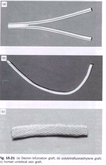
Prosthetic materials For bypass of the aortoiliac segment the favoured material is Dacron (Fig. 15.21a). Prostheses come in two types: woven and knitted. Woven grafts tend to leak less when first exposed to blood flow during surgery, but newer knitted prostheses may be sealed with gelatin or collagen by the manufacturer and may leak even less than their woven counterparts. In the final analysis, there is probably little to choose between any of the styles of Dacron graft; all achieve satisfactory results. For bypass in the femoropopliteal region, if autogenous long saphenous vein (or other veins such as the short saphenous or arm vein) is not available, PTFE (Fig. 15.21b) or glutaraldehyde-tanned, Dacron-supported, human umbilical vein (Fig. 15.21c) may be employed. In general, any vein used requires a diameter of at least 3.5mm. For profundaplasty, a small piece of vein may be used or, alternatively, PTFE or Dacron. Suture materials for vascular surgery are usually monofilament in nature; polypropylene has been particularly popular. In the aorta it is usual to use 2/0 or 3/0 polypropylene. In the femoral artery at the groin it is usual to use 4/0 or 5/0 polypropylene. Finer sutures, up to 7/0, may be needed further down the limb. PTFE may (alternatively) be stitched using a suture of the same material. PTFE sutures tend to cause less bleeding through stitch holes in the graft substance.

image

Most common cause of aneurysm of abdominal aorta is – (A196)
a) Trauma b) Atherosclerosis
c) Syphilis d) Cystic medial necrosis

The most common complication of an aortic aneurysm size 8 cm is – (Delhi, PG 96)
a) Rupture b) Intramural thrombosis
c) Embolism d) Calcification

Management of a cause of iliac artery embolism requires – (JIPMER 81, UPSC 86)
a)Embolectomy
b)Injection of vasodilators
c)Hypotensive therapy
d)Sympathectomy

Intermittent claudicatin is caused by – (TN 89)
a)Venous occlusion
b)Arteria insufficiency
c)Nerual compression
d)Muscular dystrophy

Burger’s disease is seen in – (PGI 88)
a) Only male b) Age less than 40
c) Age more than 40 d) Smoker

In the abdomen, aneurysms of the …. commonly occur next only to the aorta …. – (PGI 88)
a)Internal iliac artery
b)External iliac artery
c)Splenic artery
d)Inferior mesentric artery

In extraperitoneal approach, to left sympathectomy the following may be injured –
a) Ureter b) Gonadal vessels
c) A+B d) IVC

Lumbar sympathectomy is indicated in – (TN 90)
a)Intermittent claudication
b)TAO with skin changes
c)Burger’s disease
d)Raynaud’s disease

Commonest site of throboangitis obliterans is –
a) Femoral artery b) Popiteal artery (A190)
c) iliac artery d) Pelvic vessels


Treatment of acute femoral embolus is- (AIIMS 91)
a)Warfarin
b)Heparin
c)Immediate embolectomy
d)Embolectomy after 5 days bed rest

Ganglion which is spared in Lumbar sympathetomy is – (JIPMER 92)
a) Ll b) L2
c) L3 d) L4

Surgical lumbar sympathectomy may be indicated in arterial disease. Surgical or chemical (phenol injection) sympathectomy may be used to treat rest pain or other troublesome sensory symptoms in arterial disease or in causalgia. The segment of the chain including the second and third lumbar ganglia is removed: preservation of the first lumbar ganglion is said to lessen the risk of ejaculatory problems 
image

Vessels most commonly involved in thrombo angitis obliterans – (AIIMS 92)
a)ilio-femoral
b)Aorto-iliac
c)Femora popliteal
d)Arterior and Posterior tibial

Following are used in treatment of Buergers disease except – (Al 93)
a) Trental b) Anticoagulation
c) Sympathectomy d) Antiplatelets

Buerger’s disease affects all except – (PGI 01)
a) Small arteries b) Small veins
c) Medium -size arteries d) Multiparity
e) First pregnancy after 30 years

Buerger’s disease is associated with – (rGI 02)
a) smoking b) Poor nutrition
c) Alcohol d) Prolonged standing
e) Superficial thrombophlebitis

The most common cause of peripheral limb ischaemia in India is – (AIIMS NOV 05)
a) Trauma b) Altherosclerosis
c) Buerger’s disease d) Takayastu’s disease

A 45-year-old male having a long history of cigarette smoking presented with gangrene of left foot. An amputation of the left foot was done. Representative sections from the specimen revealed presence of arterial thrombus with neutrophilic infiltrate in the arterial wall. The inflammation also extended into the neighbouring veins and nerves. The most probably diagnosis is – (AIIMS 06)
a)Takayasu arteritis
b)Giant cell arteritis
c)Hypersensitivity angiitis
d)Thromboangiitis obliterans

Ans. is ‘d’ i.e., Thromboangiitis obliterans [Ref: Hariison 166/e p. 1487; Schwartz 86/e p. 792]

THROMBOANGIITIS OBLITERANS :- Thromboangiitis obliterans (Buerger’s disease) is an inflammatory occlusive vascular disorder involving small and medium-sized arteries and veins in the distal upper and lower extremities. Cerebral, visceral, and coronary vessels may be affected rarely. This disorder develops most frequently in men <40 years. The prevalence is higher in Asians and individuals of eastern European descent. While the cause of thromboangiitis obliterans is not known, there is a definite relationship to cigarette smoking in patients with this disorder. In the initial stages of thromboangiitis obliterans, polymorphonuclear leukocytes infiltrate the walls of the small and medium-sized arteries and veins. The internal elastic lamina is preserved, and a cellular, inflammatory, thrombus develops in the vascular lumen. As the disease progresses, mononuclear cells, fibroblasts, and giant cells replace the neutrophils. Later stages are characterized by perivascular fibrosis, organized thrombus, and recanalization. The clinical features of thromboangiitis obliterans often include a triad of claudication of the affected extremity, Raynaud’s phenomenon, and migratory superficial vein thrombophlebitis. Claudication is usually confined to the calves and feet or the forearms and hands because this disorder primarily affects distal vessels. In the presence of severe digital ischemia, trophic nail changes, painful ulcerations, and gangrene may develop at the tips of the fingers or toes. The physical examination shows normal brachial and popliteal pulses but reduced or absent radial, ulnar, and/or tibial pulses. Arteriography is helpful in making the diagnosis. Smooth, tapering segmental lesions in the distal vessels are characteristic, as are collateral vessels at sites of vascular occlusion. Proximal atherosclerotic disease is usually absent. The diagnosis can be confirmed by excisional biopsy and pathologic examination of an involved vessel. There is no specific treatment except abstention from tobacco. The prognosis is worse in individuals who continue to smoke, but results are discouraging even in those who do stop smoking. Arterial bypass of the larger vessels may be used in selected instances, as well as local debridement, depending on the symptoms and severity of ischemia. Antibiotics may be useful; anticoagulants and glucocorticoids are not helpful. If these measures fail, amputation may be required.

Lumbar sympathectomy:- Operative method. Using a transverse loin incision, an extraperitoneal approach is used in which the colon and peritoneum, to which the ureter clings, are stripped medially so as to expose the inner border of the psoas muscle .The sympathetic trunk lies on the sides of the bodies of the lumbar vertebrae; on the right side it is overlapped by the vena cava. Lumbar veins are apt to cross the trunk superficially. The sympathetic trunk is divided on the side of the body of the fourth lumbar vertebra. It is then traced upwards to be divided above the large second lumbar ganglion, which is easily recognised by the number of white rami which join it. Care should be taken not to mistake small lymph nodes, lymphatics, the genitofemoral nerve or the occasional tendinous strip of the psoas minor for the sympathetic chain. It is possible to perform the operation via an endoscope after the creation of a suitably expanded retroperitoneal tissue plane. Along with a decline in the recognised indications for sympathectomy there has been a move away from the operative approach in favour of the less hazardous chemical (phenol) sympathectomy.
Chemical method. This is contraindicated in patients taking anticoagulants. Under radiographic fluoroscopic control, with the   patient in the lateral position, local anaesthetic is injected. A long spinal needle is then inserted  to seek the side of the vertebral body and to pass alongside it to reach the lumbar sympathetic chain. After confirming the needle position by injection of contrast agent, approximately 5 ml of phenol in water is injected. This is usually done at two sites: beside the bodies of the second and fourth lumbar vertebrae. Great care is needed to avoid penetrating the aorta, cava or ureter; the plunger of the syringe must always be drawn back before injection to exclude the presence of blood.

In a lumbar sympathectomy the sympathetic chain in its usual position is likely to confused with the – (pal 81, AIIMS 80, 82)

a) Ureter b) Psoas minor
c) Genitofemoral nerve
d) Ilioinguinal nerve e) Lymphatics

The commonest cause of aneurysm formation
is – (JIPMER 80, Delhi 89)
a) Gun shot injury b) Syphilis
c) Congenital factors
d) Atherosclerosis

Bullet wounds near major blood vessels should
be explored only if- (PGI 81, AMC 85)
a)The extremity is cold
b)The fingers or toes are paralysed
c)The pulse is weakened
d)There in no pulse
e)In all cases regardiess of physical findings

A knitted Dacron artery graft (PGI 99, AIIMS 84)
a)Is not porous
b)Is eventually dissolived by tissue reaction
c)Never gets infected
d)Can be easily incised and the opening resutured

The sequence of symptoms in pulmonary embolism is – (JIPMER 89, DNB 90)
a)Fever, pain, dyspnoea
b)Fever, dyspnoea
c)Dysponea, pain, haemoptysis
d)Dysponea, cough, purulent sputum

A useful through temporary improvement in a patient’s ischaemic foot can be attained by giving intravenously – (PGI 79, Delhi 84)
a) 10% Mannitol b) 10% Dextrose
c) Dextran 40 d) Dextran 100

Diabetic gangrene is due to – (Kerala 94)
a)’schema
b)Increased blood glucose
c)Altered defence by host and neuropathy
d)All of the above

All are true about Embolic Arterial occlusion except-
a)No previous history (JIPMER 95)
b)Muscles are unaffected
c)Pulse is absent
d)Anaesthesia is present

All are true about Raynauds phenomena except-
a)Exposure to cold aggravate (Kerala 95)
b)Spasm of vessels
c)More common is females
d)Atherosclerosis of vessels

The commonest site of iodgement of a pulmonary embolus is in the territory at – (UPSC 95)
a) Rt. lower lobe b) Rt. upper lobe
c) Lt. lower lobe
d) Lt. upper lobe

Kaposi sarcoma is commonly seen in – (AMU 95)
a) Upper limbs
b) Lower limbs
c) Head and Neck d) Trunk

Which of the following causes meximum bleeding-
a)Partial arterial severing (PGI 95)
b)Complete arterial severing
c)Artery caught between fractured ends of bones
d)Intimal tear

In a 40 years old male thrombus in the common femoral artery is because of – (AIIMS 97)
a)Atheroma
b)Thrombangits obliterans
c)Reynauds disease
d)Abdominal mass

One of the following is not indicated for arterial
leg ulcer – (PGI 96)
a) Debridement b)
Elevation of limb
c) Head end of bed is raised d) Low dose aspirin

Not used as graft material in peripheral vascular disease – (PGI 97)
a) Dacron graft b) Vein
c) PTFE d) PVC

The commonest cause of arterio-venous fistula is-
a)Penetrating injury (MP 97)
b)Congenital
c)Neoplasmic invasion of an artery and adjacent vein
d)Aneurysm of the artery eroding a vein

AV fistula leads to all except – (AIIMS 98)
a)Sinus tachycardia
b)Increased preload
c)Cardiac arrythm ias
d)Increased cardiac output

Commonest peripheral aneurysm is – (SCTIMS 98) a) Popliteal b) Femoral
c) Carotid d) iliac

Popliteal aneurysm-All are true except-
a)Presents as a swelling (SCTIMS 98) behind the knee
b)Presents with symptoms due to complication
c)Surgery is indicated in case of complication
d)Uncommon among peripheral aneurysm

Graft used in infra inguinal by pass is 4Jipmer 2K)
a) PTFE b) Dacron
c) Autologous vein d) Autologous artery

True about Erythrocyanosis except — (A.P 96)
a)Affects young girls
b)Cold peripheries
c)Palpable pulses
d)Ulceration & gangrene of fingers

Diabetic gangrene is due to AfE (TN 86)
a)Vasospasm
b)Atherosclerosis
c)Peripheral neuritis
d)Increased sugar in blood

Pseudo aneurysms are most commonly due- (Jipmer a) Atherosclerosis b) Trauma 93)
c) Congenital deficiency d) Infections

Bilateral pulseless disease in upper limbs in caused
by- (PGI 97)
a) Aortoarteritis b) Coarctation of aorta
c) Fibromuscular dysplsia d) Buerger’s disease

Abdominal Aneurysm is characterized by all except – (PGI 2000)
a)Elective surgery complication should be < 5%
b)Emergency surgery complication < 10%
c)Rarely asymptomatic before rupture

d)Bigger the size it is more prone to rupture

Dissection of which artery is seen in pregnancy –
a) Carotid artery b) Aorta (PGI 2000)
c) Coronary A d) Femoral artery

Peripheral arterial occlusion (Sudden onset) is characterized all except – (PGI 2000)
a) Paresthesia b) Rubor
c) Pallor d) Pain

In which one of the following conditions Dactylitis CANNOT be seen- (UPSC 02)
a) Sickle – cell anemia b) Beta thalassemia
c) Congenital syphilis d) Tuberculosis
e) Sarcoidosis

Not seen in sudden onset peripheral arterial occlusion – (Kerala 04)
a) Pain b) Rubor
c) Pallor d) Anesthesia

Drug used for Burger’s disease – (MAHE 05)
a) Xanthinol micotinate b) Propranolol
c) GTN d) All the above

Pseudoarterial aneurysm in drug abuser’s seen in -a) Radial b) Brachial (PGI June 05)
c) Femoral d) Carotid
e) Pedal

Syndrome of internal iliac artery occlusion manifested by – (PGI June 05)
a)Pain in calf
b)Absent pulse at the dorsalis pedis artery
c)Intermittent claudication

d)Gangrene

The most common cause of peripheral limb ischaemia in India is – (AIIMS NOV 05)
a) Trauma b) Altherosclerosis
c) Buerger’s disease d) Takayastu’s disease

Lumbar sympathectomy is of value in the management of- (AI 05)
a)Intermittent claudication
b)Distal ischaemia affecting the skin of the toes
c)Arteriovenous Fistula
d)Back pain

The most common cause of acquired arteriovenous fistuala is – (Al 06)
a) Bacterial infection b) Fungal infection
c) Blunt trauma d) Penetrating trauma

Etiopathogenesis of diabetic foot include the following except- (UPSC 07)
a)Myelopathy
b)Osteoarthropathy
c)Microangiopathy
d)Infection

Fogarty’s catheter is used for – (UPSC 07)
a)Drainage of urinary bladder
b)Parenteral hyperalimentation
c)Removal of embolus from blood vessels
d)Ureteric catheterisation

Pseudoaneurysms in IV drug abusesrs seen commonly in – (PGI June 07)
a)Brachial artery
b)Radial artery
c)Femoral artery

Treatment of femoral artery naeurysm –
a)Ultrasound guided compression of the neck of aneurysm (PGI June 07)
b)Thrombin injection
c)Bypass graft repair


Venous Disorders:-

Bailey & Love,Gray, Sabiston

imageimageimageimage 

Perforators are not present at – (AIIMS Nov 07)
a) Ankle b) Medial calf
c) Distal to calf d) Below inguinal ligament


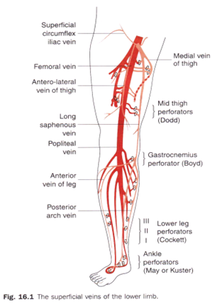
The superficial veins are in the subcutaneous connective tissue and are interconnected with and ultimately drain into the deep veins. The superficial veins form two major channels-the great saphenous vein and the small saphenous vein. Both veins originate from a dorsal venous arch in the foot:

  • the great saphenous vein originates from the medial side of the dorsal venous arch, and then ascends up the medial side of the leg, knee, and thigh to connect with the femoral vein just inferior to the inguinal ligament;
  • the small saphenous vein originates from the lateral side of the dorsal venous arch, ascends up the posterior surface of the leg, and then penetrates deep fascia to join the popliteal vein posterior to the knee; proximal to the knee, the popliteal vein becomes the femoral vein.

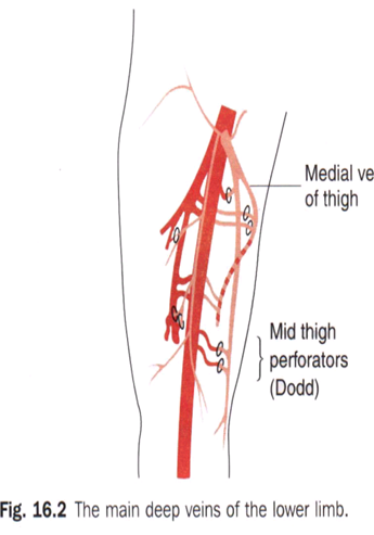
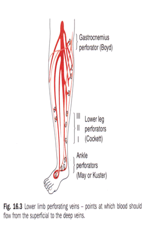
The superficial and deep veins join at a number of points. The short saphenous vein terminates at the saphenopopliteal junction (SPJ) and the long saphenous vein at the saphenofemoral junction (SFJ) in the groin. Here the flow in the superficial veins joins that in the deep veins. There is, in addition, a number of places in the calf and thigh where flow in the superficial veins may also join that in the deep veins. These is the ankle, calf and thigh communicating or perforating veins (Fig. 16.3). The names of these veins come from their course from the superficial to the deep venous system in which they perforate the deep fascia of the leg. Near the ankle are the Cockett perforating veins, near the knee the Boyd perforators and in the thigh the Hunterian perforating vein. All veins in the upper and lower limbs contain valves every few centimetres which ensure that blood flows towards the heart.

Multiple perforator veins traverse the deep fascia to connect the superficial and deep venous systems. Clinically important perforator veins are the Cockett and Boyd perforators. The Cockett perforator veins drain the medial lower leg and are relatively constant. They connect the posterior arch vein (a tributary to the GSV) and the posterior tibial vein. They may become varicose or incompetent in venous insufficiency states. The Boyd perforator veins connect the GSV to the deep veins approximately 10 cm below the knee and 1 to 2 cm medial to the tibia.

Earliest sign of deep vein thrombosis is(AIIMS 87) a) Calf tenderness b) Rise in temperature c) Swelling of calf muscle d) Homan’s sign

 Injection sclerotherapy for varicose veins is by using – (PGI 88)
a) Phenol b) Absolute alcohol
c) 70% alcohol d) Ethanolamine oleate

Sclerotherapy acts by destroying the venous endothelium. Sclerosing agents include hypertonic saline, sodium tetradecyl sulfate, and polidocanol. Concentrations of 11.7 to 23.4% hypertonic saline, 0.125 to 0.250% sodium tetradecyl sulfate, and 0.5% polidocanol are used for telangiectasias. Larger varicose veins require higher concentrations: 23.4% hypertonic saline, 0.50 to 1% sodium tetradecyl sulfate, and 0.75 to 1.0% polidocanol.83 Elastic bandages are wrapped around the leg after injection and worn continuously for 3 to 5 days to produce apposition of the inflamed vein walls and prevent thrombus formation. After the bandages are removed, elastic compression stockings should be worn for a minimum of 2 weeks. Complications from sclerotherapy include allergic reaction, pigmentation, thrombophlebitis, DVT, and possible skin necrosis.

White leg is due to – (TN 90)
a)Femoral vein thrombosis and lymphatic obstruction
b)Deep femoral vein thrombosis
c)Lymphatic obstruction only
d)None of the above

All of the following are seen in deep vein thrombosis except – (Al 90)
a) Pain b) Discolouration
c) Swelling d) Claudication

The following is the commonest site for venous ulcer- (A IIMS 91)
a)Instep of foot
b)Lower 1/3 leg and ankle
c)Lower 2/3 of leg
d)middle 1/3 of leg

The most important perforator of the Lower limb is between – (ALL INDIA 92 )
a)Long saphenous and posterior tibial vein
b)Short saphenous and posterior tibial vein
c)Short saphenous and popliteal vein
d)Long saphenous and femoral vein

Best method for diagnosis of Deep vein thromvosis is – (JIPMER 92)
a)Doppler examination
b)Plethysmography
c)Contrast phlebography
d)1131 Fibrinogen studies

Commonest complication varicose vein stripping is-
a) Thrombo embolism b) Hemorrhage
c) Ecchymosis d) Infection

Investigation of choice for diagnosis of deep vein thrombosis – (AIIMS 92)
a) Venogram
b) Doppler
c) Isotope scan d) Homans sign

Pulsating varicose vein in ayoung adult is due to-
a)Arteriovenous fistula (AIIMS 92)
b)Sapheno femoral incompetence
c)Deep vein thrombosis
d)Abdominal tumour

Which is not used in treatment of Superficial venous thrombosis – (AIIMS 92)
a)Immediate anticoagulation
b)Rest and elevation
c)Analgesics
d)Treat assosiated malignancy

Most common complication of varicose vein stripping is – (JIPMER 78, AIMS 79,92)
a) Infection b) Haemorrhage
c) Ecchymosis d) Thrombo embolism

An operated case of varicose veins has a recurrence rate of – (AIIMS 80, AP 89)
a) About 10% b) About 25%
c) About 50% d) Over 60%

What is acceptable in the management of femoral vein thrombosis – (AIIMS81, PGI 86)
a)Bed rest and spiral elastic bandages
b)A venogram
c)Thrombectomy
d)Embolectomy
e)A mobin udin umbrella inserted into the vein

Operative Venous Thrombectomy

In patients with acute iliofemoral DVT, surgical therapy is generally reserved for patients who worsen with anticoagulation therapy and those with phlegmasia cerulea dolens and impending venous gangrene. If the patient has phlegmasia cerulea dolens, a fasciotomy of the calf compartments is first performed. In iliofemoral DVT, a longitudinal venotomy is made in the common femoral vein and a venous balloon embolectomy catheter is passed through the thrombus into the IVC and pulled back several times until no further thrombus can be extracted. The distal thrombus in the leg is removed by manual pressure beginning in the foot


Operations for varicose veins are best accomplished by – (PGI 81, AIIMS 84, 86)
a)Stripping
b)Multiple subcutaneos ligatures
c)Subfascial ligatures
d)Division and ligation at the superficial venous system

A 60-years old male has been operated for carcinoma of caecum and right hemicolectomyhas been done. On the fourth post – oprative day, the patient develops fever and pain in the legs. The most important clinical entity one should lookfor is – (UPSC 96)
a)Urinary tract infection
b)Intravenous line infection
c)Chest infection
d)Deep vein thrombosis

All of following may be predisposing factors for deep vein thrombosis except – (AIIMS 95)
a) Oral contrceptives b) Nephrotic syndrome
c) Sickle cell anemia d) Thrombocytosis

The duration of heparin therapy in deep vein thrombosis is – (CUPGEE 96)
a) 7 – 10 days b) 15-20 days
c) 3-4 days d) 1 month

Cocket & Dodd’s operation is for (AP 96)
a)Saphenofemoral flush ligation
b)Subfascial ligation
c)Deep vein thrombosis
d)Diabetic foot

In obstruction of inferior vena cava there is -(A197)
a)Prominent thoraco epigastric vein
b)Caput medusa
c)Hemorrhoids
d)Esophageal varices

Most accurate & non invasive method for diagnosing deep vein thrombosis – (JIPMER 98)
a)Doppler duplex
b)Plethesmography
c)Radioactive labelled fibrinogen
d)Angiography

Most common site for venous thrombosis -(JIPMER a) Popliteal vein b) Soleal vein 98)
c) Femoral vein d) Internal iliac vein

Deep vein thrombosis is caused by all except –
a)Lower limb trauma (AIIMS 98)
b)Hip and pelvic surgery
c)Subungual melanoma
d)Cushing’s syndrome

Which of the following test is used to detect perforator incompetence in varicose- (JIPMER 2K)
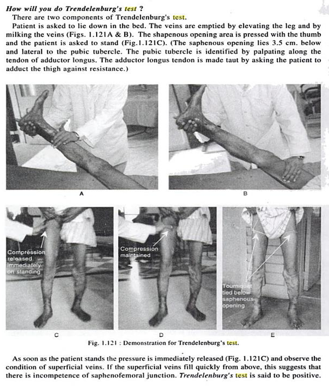
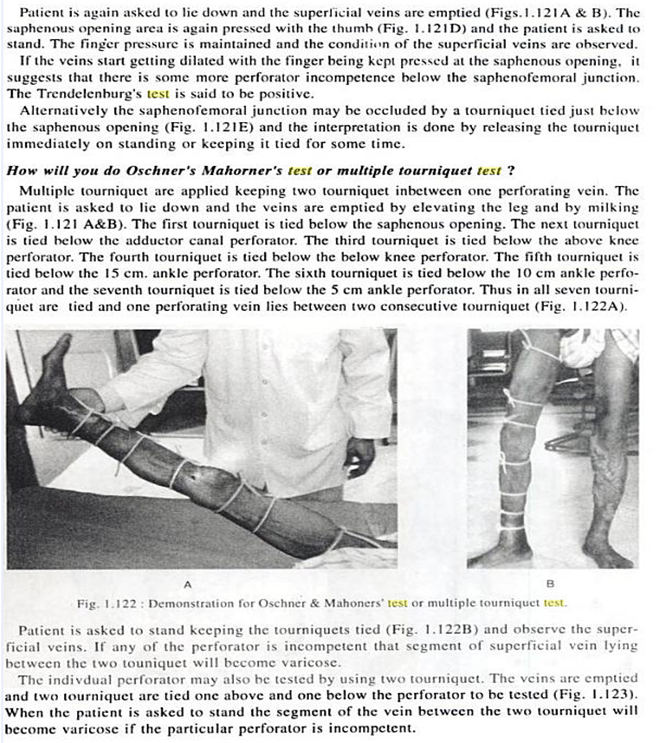
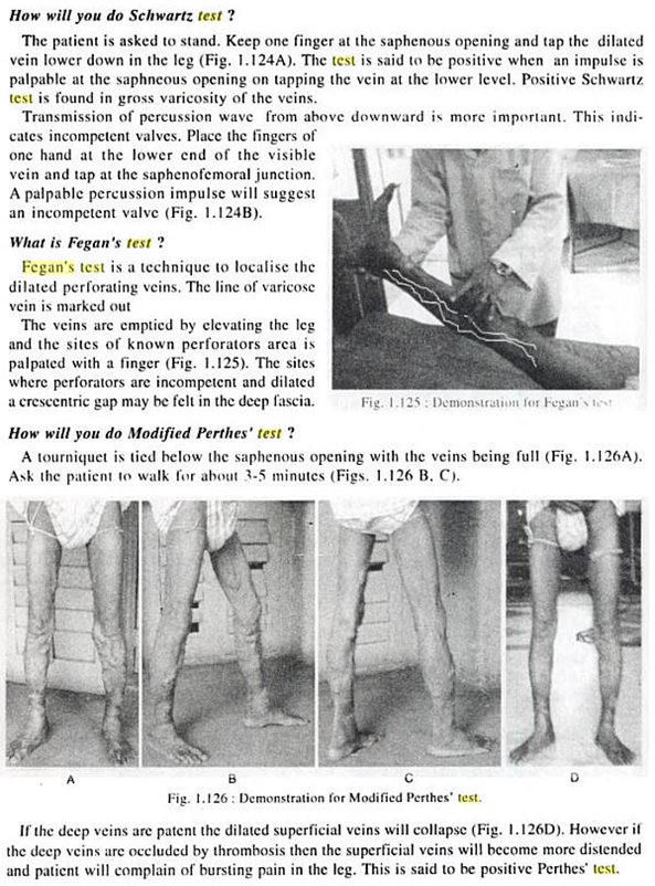
a) Trendelenberg test b) Fegan’s test- (localise)
c) Morissey’s test d) Homan’s test

image

image

image

image

image


The deficiency of all of the following factors increases the incidence of thrombus formation except – (UPSC 2K)
a) Lipoprotein A b) Protein – C
c) Anti – thrombin III d) Protein – S

The most common vein to get thrombosed is the – (AIIMS 99)
a) Long saphenous b) Short saphenous
c) Both d) Posterior tibial

Brodie -Trendlenburg test demonstrates-
a)Mid – thigh perforation (ORRISA 98)
b)Deep vein thrombosis
c)Sapheno — femoral incompetence
d)Calf perforators

An intern was doing saphenous cannulation for a burns patient. Then the patient developed sudden onset of pain along the medial border of the correponding foot. Which nerve must have been accidentally ligated – (AIIMS 2K)
a) Sural nerve b) Deep peroneal nerve
c) Saphenous nerve d) Genicular nerve

In DVT all are seen except (CMC 2001)
a)High fever
b)Increased temperature at site
c)Pain
d)Tenderness

Low Grade Pyrexia is seen not High.

An obese patient develops acute oedematous lower limb following a Pelvic surgery. Deep vein thrombosis is suspected . The most useful investigation in this case would be – (UPSC 2002)
a) Doppler imaging b) Fibrinogen uptake
c) Venography d) Plethysmography

In a patient on anticoagulant therapy, the INR is maintained at – (UPSC 2002)
a)1.5 to 2.5 times the normal
b)2.5 to 3.5 times the normal
c)3.5 to 4.5 times the normal
d)4.5 to 5.5 times the normal

DVT, investigation of choice is – (PGI 97)
a) Doppler b) Plethysmography
c) Venography d) X-ray

In diabetic ulcer, following site is involved-(PGI 97)
a) Heel b) Head of metatarsal
c) Webs d) Tips of toes

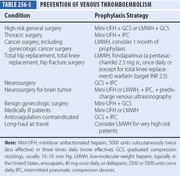
For prophylaxis of deep vein thrombosis used is –
a)Warfarin (PGI 97)
b)Heparin
c)Pneumatic shock garment
d)Graded stocking

Effective methods of VTE prophylaxis involve the use of one or more pharmacologic or mechanical modalities. Currently available pharmacologic agents include low-dose UFH, LMWH, synthetic pentasaccharides, and vitamin K antagonists. Mechanical methods include intermittent pneumatic compression (IPC) and graduated compression stockings. Aspirin therapy alone is notadequate for DVT prophylaxis. These prophylaxis methods vary with regard to their efficacy, and the 2008 ACCP Clinical Practice Guidelines stratify their uses according to the patient’s level of risk.

Harrison:-

image

Schwartz:-

Thromboembolism Risk and Recommended Thromboprophylaxis in Surgical Patients


Level of Risk Approximate DVT Risk without Thromboprophylaxis (%) Suggested Thromboprophylaxis Options
Low risk:- <10 No specific thromboprophylaxis
Minor surgery in mobile patients   Early and "aggressive" ambulation
Moderate risk:- 10–40 LMWH (at recommended doses), LDUH bid or tid, fondaparinux
Most general, open gynecologic, or urologic surgery   Mechanical thromboprophylaxis
Moderate VTE risk plus high bleeding risk    
High risk:- 40–80 LMWH (at recommended doses), fondaparinux, oral vitamin K antagonist (INR 2–3)
Hip or knee arthroplasty, hip fracture surgery    
Major trauma, spinal cord injury   Mechanical thromboprophylaxis
High VTE risk plus high bleeding risk    

DVT = deep vein thrombosis; INR = International Normalized Ratio; LDUH = low-dose unfractionated heparin; LMWH = low molecular weight heparin; VTE = venous thromboembolism.


Deep vein thrombosis is best diagnosed by-(PGI 97)
a) Plethysmography b) Duplex ultrasound
c) Radionuclide scan d) CT scan

Which is true regarding Trendelenburg operation – (PGI 01)
a)Sripping of the superficial varicose vein
b)Flush ligation of the superficial varicose vein
c)Ligation of the perforators
d)Ligation of small tributaries at the distal end of superficial varicose vein
e)Ligation of short saphenous vein

Surgical treatment of varicose veins
Surgical treatment of varicose veins is widely used and is effective in removing varicose veins of the main saphenous trunks, as well as their tributaries, down to a size of about 3 mm. Veins smaller than this are best treated by sclero­therapy. Surgical removal of varices is inappropriate where these form a major part of the venous drainage of the limb, for example where a deep vein thrombosis has destroyed the main axial limb veins and the patient relies on the superficial veins. This possibility may be suggested by the patient’s medical history and can be confirmed by duplex ultrasonography or venography.
The main principles of surgical treatment are to ligate the source of the venous reflux (usually the SFJ or the SPJ) and to remove the incompetent saphenous trunks and the associated varices. Sapheno-femoral ligation alone, sometimes referred to as a ‘Trendelenburg procedure’, is associated with a high rate of recurrence of varices. Recent research has shown that it is necessary to remove the long saphenous vein to ensure that as much venous reflux as possible is eliminated. Similarly, communications between the many deep veins in the popliteal fossa and the short saphenous vein mean that some patients develop recurrences in the short saphenous vein due to the re-establishment of reflux from these veins. This problem may be eliminated by removing the short saphenous vein. Removal of the saphenous veins has the disadvantage that both veins are accompanied by a nerve that may be damaged in the vein stripping operation. To avoid nerve injury the long saphenous vein should not be removed below mid-calf level and great care should be exercised in removing the short saphenous vein.

Sensory nerve injury is seen occasionally after removal of varicose veins. The saphenous nerve and its branches accompany the long saphenous vein in the calf, the sural nerve accompanies the short saphenous vein. Damage to the main part of these nerves occurs in about 1 per cent of operations, but small areas of anaesthesia may occur more frequently (in up to 10 per cent of patients). The adoption of inverting stripping techniques and avoidance of stripping the long saphenous vein below mid-calf level have reduced the risk of damage to these nerves. All patients should be warned before surgery that they may experience small areas of numbness and tingling after the operation. These changes are usually reversible but can be quite persistent.


For which of the following venous abnormality is surgery indicated – (PGI 01)
a)Deep vein incompetence with DVT
b)Deep vein incompetence without DVT
c)Varicosity > 3 cm
b) Varicosity < 3 cm
e) Saphenofemoral incompetence

Imp:- Surgical treatment of varicose veins is widely used and is effective in removing varicose veins of the main saphenous trunks, as well as their tributaries, down to a size of about 3 mm. Veins smaller than this are best treated by sclero­therapy. Surgical removal of varices is inappropriate where these form a major part of the venous drainage of the limb, for example where a deep vein thrombosis has destroyed the main axial limb veins and the patient relies on the superficial veins. This possibility may be suggested by the patient’s medical history and can be confirmed by duplex ultrasonography or venography
Saphenous vein ligation and stripping is still the more commonly performed procedure worldwide, and it may be the preferred therapy for patients with GSVs of very large diameter (>2 cm).

Migratory thrombophlebitis is seen most commonly with- (PGI 02)
a) Pancreatic ca b) Testicular ca
c) Gastric ca d) Breast ca
e) Liver ca

Brodie-Tredenlenburg test is positive in – (PGI 02)
a)Sapheno-Femoral incompetence
b)Perforator competence above knee
c)Deep vein incompetence
d)Perforator competence below knee
e)SVT

Varicose veins are seen in – (PGI 02)
a) DVT
b) Superficial venous thrombosis

c)AV fistula
d)Prolonged standing
e)Obesity

Gold standard diagnostic test in varicose veins is
a)Photoplethysmography (Jipmer 03)
b)Duplex imaging
c)Ultrasonography
d)Radio – labeled fibrinogen study

The initial therapy of documented deep venous thrombosis in a post operative case is –
a)Subcutaneous heparin therapy (Karnataka 03)
b)Intravenous heparin therapy
c)Thropmbolytic therapy with urokinase
d)Aspirin therapy

Which one of the following reagents is not used as sclerosant in the treatment of bleeding varices –
a) Ethyl alcohol .b) Ethanolamine oleate (ICS 05)
c) Phenol d) Sodium morrhuate

Site of diabetic for ulcer – (PGI June 05)
a) Medial malleolus b) Lateral malleolus
c) Heel d) Head of metatarsal
e) Head of toes

Harrison:-Approximately 15% of individuals with DM develop a foot ulcer (great toe or MTP areas are most common), and a significant subset will ultimately undergo amputation (14–24% risk with that ulcer or subsequent ulceration). Risk factors for foot ulcers or amputation include: male sex, diabetes >10 years’ duration, peripheral neuropathy, abnormal structure of foot (bony abnormalities, callus, thickened nails), peripheral arterial disease, smoking, history of previous ulcer or amputation, and poor glycemic control. Large callouses are often precursors to or overlie ulcerations.

Which of the following statements is true regarding fat embolism – (AJIMS NOV 05)
a)Most patients with major trauma involving long bones have urinary fat globules
b)All patients with urinary fat globules develop fat embolism
c)Peak incidence of respiratory insufficiency for pulmonary fat embolism is around day 7 after injury
d)Heparin as an anticoagulant decreases mortality and morbidity in fat embolism syndrome

Fat Embolism::- A certain degree of lung dysfunction occurs in all patients after long bone fractures, but clinically significant fat embolism syndrome as such develops in only 10 to 15 percent of these patients. Signs include hypoxia, tachycardia, mental status changes, and petechiae on the conjunctiva, axilla, or upper thorax. Fat globules in the urine are nondiagnostic, but lung infiltrates seen on chest radiograph confirm the presence of lung injury.
The pathophysiology of fat embolism represents capillary endothelial breakdown causing pericapillary hemorrhagic exudates most apparent in the lungs and brain. Pulmonary edema and hypoxemia occur as a result of pulmonary exudates. Hypoxia and areas of cerebral edema may account for the variable neurologic abnormalities seen.
The more severe cases of fat embolism involve fractures of the femur and tibia. Delays in fixation of bones and extensive reaming of the medullary canals contribute to perioperative morbidity and to the severity of fat embolism syndrome. Efforts to surgically correct fractures early and minimize trauma to the bone marrow lessen the degree of fat/bone marrow embolism. Patients with coexisting lung injury are at additional risk of fat embolism. Evidence suggests that fat may pass to the systemic circulation through a patent foramen ovale  or by transpulmonary passage.The chemical composition of the fat may even contribute to this process.  For this reason, it is preferable to minimize pulmonary artery hypertension to reduce transpulmonary passage of fat and limit pulmonary endothelial transudation of fluid.
Treatment includes early recognition, oxygen administration, and judicious fluid management. Corticosteroids in large doses shortly after major trauma have been found to minimize the clinical presentation of fat embolism but are probably not necessary in most cases if oxygen therapy is administered. With appropriate fluid management, adequate ventilation, and the prevention of hypoxemia, outcome is usually excellent.


Which of the following is true about varicocele except ? (Manipal 06)
a)Incompetent valves of testicular vein are responsible for varicocele
b)90% are on the left side
c)Asymptomatic cases require surgery
d)Femoral catheterization with spermatic vien ablation is done in recurrence

Which one of the following is the investigation of choice for suspected deep vein thrombosis of the lower extremity ? (UPSC 07)
a)Radioactive labelled fibrinogen uptake
b)Ascending contrast phlebography
c)D-dimer estimation
d)Duplex ultrasonography

With reference to varicocele, which one of the following is not true of it ? (UPSC 07)
a)Varicosity of cremastric veins
b)Left side is affected usually
c)Feels like a bag of worms
d)May lead to infertility


All are done for a case of deep vein thrombosis except – (MAHE 07)
a) Thrombolytic therapy b) Bandage
c) Heparin d) Bed rest

The practice of having a patient “out of bed into a chair” is one of the most thrombogenic positions that one could order a patient into. Sitting in a chair with the legs in a dependent position causes venous pooling, which in the postoperative milieu could easily be a predisposing factor in the development of thromboembolism

Treatment regimens may include antithrombotic therapy, vena caval interruption, catheter-directed or systemic thrombolytic therapy, and operative thrombectomy

studies, as well as the current ACCP guidelines, suggest that catheter-directed thrombolysis (with adjunctive angioplasty, venous stenting, and pharmacomechanical fragmentation and extraction) may be useful in selected patients with extensive iliofemoral DVT. Patients should have a recent onset of symptoms (<14 days), good functional status, decent life expectancy, and low bleeding risk.

Table 24-2 Risk Factors for Venous Thromboembolism


Acquired Inherited
Advanced age Factor V Leiden
Hospitalization/immobilization Prothrombin 20210A
Hormone replacement therapy and oral contraceptive use Antithrombin deficiency
Protein C deficiency
Pregnancy and puerperium Protein S deficiency
Prior venous thromboembolism Factor XI elevation
Malignancy Dysfibrinogenemia
Major surgery Mixed Etiology
Obesity Homocysteinemia
Nephrotic syndrome Factor VII, VIII, IX, XI elevation
Trauma or spinal cord injury Hyperfibrinogenemia
Long-haul travel (>6 h) Activated protein C resistance without factor V Leiden
Varicose veins  
Antiphospholipid antibody syndrome  
Myeloproliferative disease  
Polycythemia

Early in the course of DVT development, venous thrombosis is thought to begin in an area of relative stasis, such as a soleal sinus vein or immediately downstream of the cusps of a venous valve in the axial calf veins. Isolated proximal DVT without tibial vein thrombosis is unusual. Early in the course of a DVT, there may be no or few clinical findings such as pain or swelling. Even extensive DVT may sometimes be present without signs or symptoms. History and physical examination are therefore unreliable in the diagnosis of DVT. In addition, symptoms and signs generally associated with DVT, such as extremity pain and/or swelling, are nonspecific. In large studies, DVT has been found by venography or DUS in ≤50% of patients in whom it was clinically suspected.Objective studies are therefore required to confirm a diagnosis of DVT or to exclude the presence of DVT.

Investigation of Choice-Duplex USG

With major advances in technology of imaging, magnetic resonance venography has come to the forefront of imaging for proximal venous disease. The cost and the issue of patient tolerance due to claustrophobia limit the widespread application, but this is changing. It is a useful test for imaging the iliac veins and the IVC, an area where duplex ultrasound is limited in its usefulness.

Gold standard:-Injection of contrast material into the venous system is obviously and understandably the most accurate method of confirming DVT and the location.Not used usually.

Clinical symptoms may worsen as DVT propagates and involves the major proximal deep veins. Massive DVT that obliterates the major deep venous channel of the extremity with relative sparing of collateral veins causes a condition called phlegmasia alba dolens or white leg .This condition is characterized by pain, pitting edema, and blanching. There is no associated cyanosis. When the thrombosis extends to the collateral veins, massive fluid sequestration and more significant edema ensues, resulting in a condition known as phlegmasia cerulea dolens.Phlegmasia cerulea dolens is preceded by phlegmasia alba dolens in 50 to 60% of patients. The affected extremity in phlegmasia cerulea dolens is extremely painful, edematous, and cyanotic, and arterial insufficiency or compartment syndrome may be present. If the condition is left untreated, venous gangrene can ensue, leading to amputation

SVT

Superficial vein thrombophlebitis (SVT) most commonly occurs in varicose veins but can occur in normal veins. When SVT recurs at variable sites in normal superficial veins, it may signify a hidden visceral malignancy or a systemic disease such as a blood dyscrasia and/or a collagen vascular disease. This condition is known as thrombophlebitis migrans. SVT also frequently occurs as a complication of indwelling catheters, with or without associated extravasation of injected material. Upper extremity vein thrombosis has been reported to occur in 38% of patients with peripherally inserted central catheters; 57% of these developed in the cephalic vein . Finally, suppurative SVT may occur in veins with indwelling catheters and may be associated with generalized sepsis.

Rx of SVT by schwartz:- In patients with SVT not within 1 cm of the saphenofemoral junction, treatment consists of compression and administration of an anti-inflammatory medication such as indomethacin. In patients with suppurative SVT, removal of any existing indwelling catheters is mandatory, and excision of the vein may be necessary. If the SVT extends proximally to within 1 cm of the saphenofemoral junction, extension into the common femoral vein is more likely to occur. In these patients, anticoagulation therapy for 6 weeks and GSV ligation appear equally effective in preventing thrombus extension into the deep venous system.

Rx of SVT by Harrisons:- Treatment is primarily supportive. Initially, patients can be placed at bed rest with leg elevation and application of warm compresses. Nonsteroidal anti-inflammatory drugs may provide analgesia but may also obscure clinical evidence of thrombus propagation. If a thrombosis of the greater saphenous vein develops in the thigh and extends toward the saphenofemoral vein junction, it is reasonable to consider anticoagulant therapy to prevent extension of the thrombus into the deep system and a possible pulmonary embolism.

____________________________________________________________________________________

image

Elastic compression stocking with zippered side to facilitate treatment of chronic venous insufficiency

Compression therapy is most commonly achieved with graduated elastic compression stockings. Graduated elastic compression stockings, initially developed by Conrad Jobst in the 1950s, were made to simulate the gradient of hydrostatic forces exerted by water in a swimming pool. Elastic compression stockings are available in various compositions, strengths, and lengths, and can be customized for a particular patient.

To improve compliance, patients should be instructed to wear their stockings initially only as long as it is easily tolerated and then gradually to increase the amount of time the stockings are worn. Alternatively, patients can be fitted with lower-strength stockings initially followed by introduction of higher-strength stockings over a period of several weeks. Many commercially available devices, such as silk inner toe liners, stockings with zippered sides,and metal fitting aids ,are available to assist patients in applying elastic stockings.

image

Metal fitting aid to assist in placement of elastic compression stockings

Lymphatic Disorders

Lymphedema is extremity swelling that results from a reduction in lymphatic transport, with resultant pooling of lymph within the interstitial space. It is caused by anatomic problems such as lymphatic hypoplasia, functional insufficiency, or absence of lymphatic valves.

The original classification system, described by Allen, is based on the cause of the lymphedema.

Primary lymphedema is further subdivided into

1. Congenital lymphedema may involve a single lower extremity, multiple limbs, the genitalia, or the face. The edema typically develops before 2 years of age and may be associated with specific hereditary syndromes (Turner syndrome, Milroy syndrome, Klippel-Trénaunay-Weber syndrome).The familial version of congenital lymphedema is known as Milroy’s disease and is inherited as a dominant trait

2.Lymphedema praecox is the most common form of primary lymphedema, accounting for 94% of cases. Lymphedema praecox is far more common in women, with the gender ratio favoring women 10:1. The onset is during childhood or the teenage years, and the swelling involves the foot and calf. The familial version of lymphedema praecox is known as Meige’s disease.

3.Lymphedema tarda is uncommon, accounting for <10% of cases of primary lymphedema. The onset of edema is after 35 years of age.

Secondary lymphedema is far more common than primary lymphedema. Secondary lymphedema develops as a result of lymphatic obstruction or disruption. Axillary node dissection leading to lymphedema of the arm is the most common cause of secondary lymphedema in the United States. Other causes of secondary lymphedema include radiation therapy, trauma, infection, and malignancy. Globally, filariasis (caused by Wuchereria bancrofti, Brugia malayi, and Brugia timori) is the most common cause of secondary lymphedema.In developed countries, the most common causes of secondary lymphedema involve resection or ablation of regional lymph nodes by surgery, radiation therapy, tumor invasion, direct trauma, or less commonly, an infectious process.

Lymphoscintigraphy has emerged as the test of choice in patients with suspected lymphedema.It cannot differentiate between primary and secondary lymphedemas; however, it has a sensitivity of 70% to 90% and a specificity of nearly 100% in differentiating lymphedema from other causes of limb swelling.

Email to Colleague Print Version

Malignant cell in Hodgkin’s lymphoma is –

a) Reed sternberg cell b) Lymphocytes 85)
c) Histiocyte d) Reticulum cells

Chronic lymphedema predisposes to all except – (PGI 89)
a) Lymphangiosarcoma b) Marjolins ulcer
c) Recurrent infections d) Thickening of skin
In the lower extremity the swelling involves the dorsum of the foot, and the toes have a squared-off appearance. In advanced cases, hyperkeratosis of the skin develops, and fluid weeps from lymph-filled vesicles

Recurrent cellulitis is a common complication of lymphedema. Repeated infection results in further lymphatic damage, worsening existing disease. The clinical presentation of cellulitis ranges from subtle erythema and worsening of edema to a rapidly progressive soft tissue infection with systemic toxicity

Commonest cause of unilareral pedal edema in india is – (A190)
a) Filariasis b) Post traumatic
c) Post irradiation d) Milroy’s disease

All are true about congenital lymphedema except-
a)It is bilateral (AI 91)
b)Involve lower limb
c)Almost always manifests before puberty
d)Acute lymphangitis may occur
All are true?

The commonest cause for lymphedema of upper limb is – (AI 91)
a) Filariasis b) Congenital
c) Neck surgery d) Post mastectomy irradiation

Commonest cause of upper limb lymphedema is –
a) Congenital b) Filariasis (AI 92)
c) Post mastectomy d) Irradiation

???

ANS given in all guides is Filariasis but it should be Post Mastectomy.

Secondary lymphedema, more common than the primary form, usually develops following disruption or obstruction of lymphatic pathways associated with a disease process, or following surgery or radiotherapy. Worldwide, filariasis is the most common cause of secondary lymphedema usually affecting lower limbs.In the United States, the most common secondary lymphedema develops in the upper extremity following axillary lymph node dissection.The incidence varies considerably, depending on the definition of edema. Results of one series of published reports reveal a 14 percent rate of secondary lymphedema in postmastectomy patients who also had undergone irradiation therapy.

Milroys disease is – (JIPMER 92)
a)Edema due to filareasis
b)Post cellulitic lymphedema
c)Congenital lymphedema
d)Lymphedema following surgery

Investigation of choice in detecting small para-aortic lymph node is – (JIPMER 92)
a) Ultra sound scan b) CT scan
c) Lymphangiography d) Arteriography

Not Sure

Contrast lymphangiography. Although few centres now perform this technique, it remains the standard by which all other lymphatic imaging is judged and provides precise information about the anatomy of the lymphatic system. It is now generally reserved for preoperative evaluation of patients with megalymphatics who arc being considered for bypass or fistula ligation.

CT scanning may be useful in identifying pathologic intra-abdominal lymph nodes and masses and in identifying the extent and localization of fluid. Lymphangiography and lymphoscintigraphy may help localize lymph leaks and obstruction; this information is particularly useful for surgical planning.

Milroys disease is lymphedema which is-(AMU 85)
a)Familial
b)Follows filariasis
c)Follows erysipelas
d)A sequele to white leg

Lymphangiography of the leg is performed by –
a)An injection of sodium diatrizoate (Hypaque) subcutaneously between the toes
b)Injecting sodium diatrizoate retrogradely under pressure into a small vein on the dorsum of the foot
c)Dissecting lymphatics through an incision on the dorsum of the foot
d)The use of an infusion pump

Direct-contrast lymphangiography provides the finest details of the lymphatic anatomy. However, it is an invasive study that involves exposure and cannulation of lymphatics at the dorsum of the forefoot, followed by slow injection of contrast medium (ethiodized oil).

Radiologic lymphology is performed by first visualizing the lymphatics by injecting colored dye into the hand or foot. The visualized lymphatic segment is exposed through a small incision and cannulated with a 27- to 30-gauge needle. An oil-based dye is then injected slowly into the lymphatics over several hours. The lymphatic channels and nodes are then visualized with traditional radiographs . Lymphangiography is reserved for patients with lymphangiectasia or lymphatic fistulas, and patients who are being considered for microvascular reconstruction

Under local anaesthesia, a small transverse incision is made in the dorsum of the foot after I ml of isosulphan blue has been injected subcutaneously to identify the lymphatics. Lymphatics are dissected out under loupe magnification and a 30G needle used to infuse lipid-soluble contrast at a rate of I ml in 8 minutes to a maximum of 7 ml (taking about 1 hour) into each limb

Finding the cause of unilateral lympoedema of the leg includes – (JIPMER 78,79, PGI 85)
a)Taking a family history
b)Looking for chronic infection in the foot
c)Looking for early malignant disease of the testis…(Not early.It should have been late metastatic )
d)Looking for filariasis
e)Performing a casoni

image

Treatment of Acute lymphangitis requires –
a)Antibiotic and rest (JIPMER 81, AMC 84)
b)Immediate lymphangiography
c)Immediate multiple incisions
d)No special treatment

Acute inflammation of the Iymphatics
Acute lymphangitis occurs when a deep or superficial infec­tion, often due to Streptococcus pyogenes or Staphylococcus aureus, spreads to the draining lymphatics and lymph nodes (lymphadenitis) where an abscess may form. Eventually this may progress to bacteraemia or septicaemia. The normal signs of infection (rubor, calor, dolor) are present and a red streak is seen in the skin along the line of the inflamed lymphatic. The part should be rested to reduce lymphatic drainage, elevated to reduce swelling and the patient treated with intravenous antibiotics based upon actual or suspected sensitivities. Failure to improve within 48 hours suggests inappropriate antibiotic therapy, the presence of undrained pus either in the lymph nodes or at the site of primary infection, or the presence of an underlying systemic disorder (malignancy, immunodeficiency). The lymphatic damage caused by acute lymphangitis may lead to recurrent attacks of infection and lymphoedema.
image


Total dose of radiation in Hodgkins dosease is – (JIPMER 95)
a) 500 -1000 rad b) 1000 – 2000 rad
c) 3000 – 5000 rad d) 5000 – 7000 rad

The most important prognostic indicator of Hodgkin’s lymphoma is – (TN 95)
a)Lymphocytic predominance histology
b)Visceral involvement
c)Hepatic involvement
d)Involvement of spleen

Diagnosis of Hodgkin’s disease is.confirmed by –
a)CT scan (PGI 9 7)
b)Bone marrow biopsy
c)Lymph node biopsy
d)Lymphangiography

Grade I lymphedema means – (JIPMER 2K)
a)Pitting edema upto the ankle
b)Pitting edema upto the knee
c)Non-pitting edema
d)Edema disapearing after overnight rest

image

Lymphovenous anastomosis is done for – (PGI 97)
a)Filarial lymphoedema
b)Lymphoid cyst
c)Cystic hygroma
d)Malignant lymphoedema

Surgery

A variety of surgical procedures have been devised for the treatment of lymphedema. Surgical treatment involves either excision of extra tissue or anastomosis of a lymphatic vessel to another lymphatic or vein.125 In excisional procedures, part or all of the edematous tissue is removed. This does not improve lymphatic drainage but debulks redundant tissue. The microsurgical procedures involve the creation of a lymphaticolymphatic or lymphaticovenous anastomosis, which theoretically improves lymphatic drainage. No long-term follow-up data are available for these interventions, and therefore operative therapy for lymphedema is not well accepted worldwide. Furthermore, operative intervention can further obliterate lymphatic channels, worsening the edema.126

Summary

Lymphedema is a chronic condition caused by ineffective lymphatic transport, which results in edema and skin damage. Lymphedema is not curable, but the symptoms can be controlled with a combination of elastic compression stockings, limb elevation, pneumatic compression, and massage. Controlling the edema protects the skin and potentially prevents cellulitis

True about lymphangioma – (PGI 03)
a)It is a malignant tumour
b)It is a congenital sequestration of lymphatic
c)Cystic hygroma is a lymphangioma

d)Laser excision is done
e)Sclerotherapy is commonly done

The most common site of lymphangiosarcoma is
a)Liver (UPSC 04)
b)Spleen
c)Post mastectomy edema of arm
d)Retroperitoenum

Commonest cause of A/C Lymphadenitis in India –
a)Barefoot walking (MAHE 05)
b)TB
c)Staphylococcal skin infection
d)Lymphoma

All of the following soft tissue sarcomas have a propensity for lymphatic spread except –
a)Neurofibrosarcoma (AiimsNov 05)
b)Synovial sarcoma
c)Rhabdomyosarcoma
d)Epitheloid sarcoma

A 40 year old man presented with a flat 1 cm x 1 cm scaly, itchy black mole on the front of thigh. Examination did reveal any inguinal lymphadenopathy. The best course of management would be- (UPSC 07)
a)FNAC of the lesion
b)Incision biopsy
c)Excisional biopsy
d)Wide excision with inguinal lymphadenectomy

In India, what is the most common cause of unilateral lymphoedema of lower limb ? (UPSC 07)
a)Lymphoedema tarda
b)Carcinoma of penis with metastatic nodes
c)Filariasis
d)Tubercular lymphadenopathy

Posted in Medical | Tagged: , , , , , , | 18 Comments »

Surgery MCQs (Burns)

Posted by Dr KAMAL DEEP on May 30, 2011

Exposure treatment is done for burns of the –
a)Upper limb (JIPMER 87)
b)Lower limbs
c)Thorax
d)Abdomen
e)Head & neck

Circumferential (encircling) burns of the trunk are generally unsuited for exposure because all of the burned surfaces cannot be adequately exposed to the air at one time.

Granulating surfaces should never be exposed.


Rule of nine to estimate surface area of a burnt patient was introduced by – (AIIMS 84)
a) Mortiz kaposi b) Alexander wallace
c) Joseph lister d) Thomas barclay

Deep skin burns is treated with – (AIIMS 91)
a)Split thickness graft
b)Full thickness graft
c)Amniotic membrane
d)Synthetic skin derivatives

In burns heat loss is by/ due to – (PGI 80,
a)Dilatation of veins DELHI 80,92)
b)Shock
c)Exposed area by evaporation
d)None of the above

The cold water treatment of burns has the disadvantage that it increase the chances of –
a) Pain b) Exudation (PGI 81, AIIMS 83)
c) Infection d) None of the above

Pus in burns form in – (PGI 79, DELHI 89)
a) 2-3 Days b) 3-5 days
c) 2-3 weeks d) 4

One of folowing is not seen in severe burns -(Al 96)
a) Hypovolemia b) Sepsis
c) Duodenal ulcer d) Hyperthemia

Undue restlessness in a patient during the immediate post burn period is often a manifestitation of-(Karp
a) Hypoxia b) Hypovolemia 95)
c) Hyperkalemia d) Anxiety


image

Sabiston:- First-degree burns are, by definition, injuries confined to the epidermis. These burns are painful and erythematous, blanch to the touch, and have an intact epidermal barrier. Examples include sunburn or a minor scald from a kitchen accident. First-degree burns do not result in scarring, and treatment is aimed at comfort with the use of topical soothing salves, with or without aloe, and oral nonsteroidal anti-inflammatory agents.

Second-degree burns are divided into two types: superficial and deep. All second-degree burns have some degree of dermal damage, and the distinction is based on the depth of injury into this structure. Superficial dermal burns are erythematous and painful, blanch to touch, and often blister. Examples include scald injuries from overheated bathtub water and flash flame burns from open carburetors. These wounds spontaneously re-epithelialize from retained epidermal structures in the rete ridges, hair follicles, and sweat glands in 7 to 14 days. After healing, these burns may result in some slight skin discoloration over the long term. Deep dermal burns into the reticular dermis appear more pale and mottled, do not blanch to touch, but remain painful to pinprick. These burns heal in 14 to 35 days by re-epithelialization from hair follicles and sweat gland keratinocytes, often with severe scarring as a result of the loss of dermis

Third-degree burns are full thickness through the epidermis and dermis and are characterized by a hard, leathery eschar that is painless and black, white, or cherry red. No epidermal or dermal appendages remain; thus, these wounds must heal by re-epithelialization from the wound edges. Deep dermal and full-thickness burns require excision with skin grafting to heal the wounds in timely fashion.

Fourth-degree burns involve other organs beneath the skin, such as muscle, bone, and brain.

Schwartz:- Burn wounds are commonly classified as superficial (first degree), partial thickness (second degree), full thickness (third degree), and fourth-degree burns, which affect underlying soft tissue. Partial-thickness burns are then classified as either superficial or deep partial thickness burns by depth of involved dermis. Clinically, first-degree burns are painful but do not blister, second-degree burns have dermal involvement and are extremely painful with weeping and blisters, and third-degree burns are hard, painless, and nonblanching

Because full-thickness grafts are impractical for most burn wounds, split-thickness sheet autografts harvested with a power dermatome make the most durable wound coverings and have a decent cosmetic appearance.

Schwartz:- Thighs make convenient anatomic donor sites, which are easily harvested and relatively hidden from an aesthetic standpoint. The thicker skin of the back is useful in older patients, who have thinner skin elsewhere and may have difficulty healing donor sites. The buttocks are an excellent donor site in infants and toddlers.

The scalp is also an excellent donor site; the skin is thick and there are many hair follicles so it heals quickly. It has the added advantage of being completely hidden once hair regrows.

Areas of cosmetic importance such as the face, neck, and hands should be grafted with nonmeshed sheet grafts to ensure optimal appearance.


All requires hospitalization except – (Al 91)
a)5% Burns in children
b)10% Scalds in children
c)Electrocution
d)15% Deep burns in adults

Indications for specialist referral in burns –
a)> 20% superficial burn in adult (PGI 04)
b)Only palms
c)Scalds on head and face
d)10% burns in infants
e)10% deep burns in adult

A burn patient is referred when – (PGI 04)
a)10% superficial burn in child
b)Scald in face

c)25% superficial burn in adult
d)25% deep burn in adult
e)Burn in palm

Improvements in burn care originated in specialized units specifically dedicated to the care of burned patients. These units consist of experienced personnel with resources to maximize outcome from these devastating injuries ( Box 22-1 ). Because of these specialized resources, burned patients are best treated in such places. Patients with the following criteria are referred to a designated burn center:

1. Partial-thickness burns greater than 10% TBSA
2. Burns involving the face, hands, feet, genitalia, perineum, or major joints
3. Any full-thickness burn
4. Electrical burns, including lightning injury
5. Chemical burns
6. Inhalation injury
7. Burns in patients with preexisting medical disorders that could complicate management, prolong recovery, or affect outcome
8. Any patient with burns and concomitant trauma (e.g., fractures) in which the burn injury poses the greater immediate risk for morbidity and mortality. In such cases, if the trauma poses the greater immediate risk, the patient may be initially stabilized in a trauma center before being transferred to a burn unit. Physician judgment is necessary in such situations, and decisions must be made in concert with the regional medical control plan and triage protocols
9. Burned children in hospitals without qualified personnel or equipment to care for children
10. Burns in patients who will require special social, emotional, or long-term rehabilitative intervention


In 3’rd degree burns, all are seen except – (PGI 99)
a)Vesicles are absent
b)Painful
c)Leathery skin
d)Reddish due to Hb Infiltration

Late deaths in burns is due to – (PGI 99)
a) Sepsis b) Hypovolemia
c) Contractures d) Neurogenic

Burns with vesiculation, destruction of the epidermis and upper dermis is – (PGI 99)
a) 1″ degree b) 2nd degree
c) 3rd degree d) 4th degree

True about burns – (PGI 2000)
a)Hyperglycemia is seen in early burns
b)Child with burns should have damp dressing
c)Chemical powder burns should be kept dry
d)3rd degree burns are painfull

Metabolic derangements in severe burns are all except- (PGI 2000)
a)cortico steroid secretion
b)Hyperglycaemia
c)secretion of HCl
d)Neutrophil dysfunction

Superficial burns; true is/are – (PGI 01)
a)Always requires skin grafting
b)Dry & inelastic
c)Blister formation
d)Painless
e)Can be healed within 7 to 10 days

Schwartz:- Clinically, first-degree burns are painful but do not blister, second-degree burns have dermal involvement and are extremely painful with weeping and blisters, and third-degree burns are hard, painless, and nonblanching

Love and Bailey:-Superficial burns These have the ability to heal themselves by epithelialisation alone. Epidermal burns look red, are painful, blisters are not present, and they heal rapidly without sequelae. Superficial dermal burns are blistered and painful; they should heal by epithelialisation within 14 days without scarring, but some­times leave long-term pigmentation changes.burns heal in 14 to 35 days

Hence superficial burns include first degree as well as second degree superficial ones with superficial dermis involvement.


True statement about burn resuscitation -(PGI 03)
a)Colloid preferred in initial 24 hrs
b)Colloid preferred if burnt area is > 15 % of total BSA
c)Half of the calculated fluid given in initial 8 hrs.
d)Diuretics should be given to all pt of electric burn.


The ideal temperature of water to cool the burnt surface is – (UPSC 02)
a) 15° b) 10°
c) 8° d) 6°

The best guide to adequate tissue perfusion in the fluid management of a patient with burns, is to ensure a minimum hourly urine output of- (Karn a) 10-30 ml b) 30-50 ml 04)
c) 50-70 ml d) 70-100 ml

Bailey:- Fluid resuscitation:- It is important at an early stage to secure large-bore intra­venous lines. Samples are taken for haemoglobin, urea and electrolytes, and blood cross-matching. Blood gases and blood analysis for carbon monoxide or cyanide poisoning are required in the unconscious patient. Having estimated the percentage burned surface area and measured the body weight, initial fluid resuscitation can be planned. The simplest formula (for adults) is: 3—4 ml/kg body weight/% burn/in the first 24 hours.Half of this volume is given in the first 8 hours and the rest in the next 16 hours. Timings begin from the time of the burn, not the start of resuscitation. Hartmann solution is preferred, but other isotonic fluids may be used. Metabolic fluid requirements are also needed. Formulae are only a guide and the adequacy of fluid resuscitation is monitored by regular clinical assessment. A urinary catheter is essential. Urine output is the best guide to adequate tissue perfusion; in an adult one should aim for 30—50 ml/hour.

Schwartz:- As in any critically ill patient, the target MAP is 60 mmHg to ensure optimal end-organ perfusion. Goals for urine output should be 30 mL/h in adults and 1 to 1.5 mL/kg per hour in pediatric patients. Because blood pressure and urine output may not correlate perfectly with true tissue perfusion, the search continues for other adjunctive parameters that may more accurately reflect adequate resuscitation. Some centers have found serum lactate to be a better predictor of mortality in severe burns; others have found that base deficit may be a better predictor of eventual organ dysfunction and mortality. Burned patients with normal blood pressures and serum lactate levels may still have compromised gastric mucosal blood flow. However, continuous measurement of gastric mucosal pH is logistically difficult and has not been widely implemented.

A third degree cirumferential burn in the arm and forearm region, which of the following is most important for monitoring – (U.P.P.GM.E.E. 04)
a)Blood gases
b)Carboxy-oxygen level
c)Macroglobiunria cryoglobinuria
d)Peripheral pulse and circulation

In 3″ degree burns, all are seen except –
a)Vesicles are absent (UPPGMEE 04)
b)Painful
c)Leathery skin
d)Reddish due to Hb infiltration

IV rules for burns – (MAHE 05)
a)% body surface area X weight in pounds X 4 = Volume in ml
b)% body surface area X weight in Kgs X 4 = Volume in Lts
c)% body surface area X weight in Kgs X 5 = Volume in ml
d)% body surface area X weight in Kgs X 4 = Volume in ml

Ans. is ‘d’ i.e., % body surface area X weight in Kgs [Ref : Bailey & Love 24th/e p. 272] X 4 = Volume in ml

A myriad of formulas exist for calculating fluid needs during burn resuscitation, suggesting that no one formula benefits all patients. The most commonly used formula, the Parkland or Baxter formula, consists of 3 to 4 mL/kg per percent burned of lactated Ringer’s, of which half is given during the first 8 hours postburn, and the remaining half over the subsequent 16 hours. The concept behind the continuous fluid needs are simple. The burn (and/or inhalation injury) drives an inflammatory response that leads to capillary leak; as the plasma leaks into the extravascular space, crystalloid administration maintains the intravascular volume. Therefore, if a patient receives a large fluid bolus in a prehospital setting or emergency department, that fluid has likely leaked into the interstitium and the patient will still require ongoing burn resuscitation, according to the estimates.

In a patient with the burn wound extending into the superficial epidermis without involving the dermis would present with all of the following EXCEPT –
a)Healing of the wound (SGPGI 05) spontaneously without scar formation
b)Anaesthesia at the site of burns
c)Blister formation
d)Painful

What is the most important aspect of management of burn injury in the first 24 hours ? (UPSC 07)
a) Fluid resuscitation b) Dressing
c) Escharotomy d) Antibiotics

The initial colonization of a burn is by which micro organisms – (JIPMER 80, UPSC 87)
a) Proteus b) Pseudomonas
c) Staphylococcus d) E. coli

Cardiac arrest, ECG changes occurs in – (UP 07)
a) Thermal burn b) Electrical burn
c) Cold burn d) Ionising radiation injury

Stress ulcers seen in burns are – (PGI 2000)
a) Curling’s ulcer b) Cushing’s ulcer
c) Meleney’s ulcer d) Rodent ulcer

‘Sterile needle test’ helps in differentiating –
a)Healing proces (JIPMER 81, AIIMS 86 )
b)Depth of burns
c)Degenerative proces
d)Infection

Which of the following is not true of Curling’s ulcer – (KA RNAT 96)
a)Seen in burned patients
b)Are solitary penetrating ulcer
c)Are shallow multiple erosions
d)Has also been described in children after head injury or craniotomy

An intern was doing saphenous cannulation for a burns pareint. Then the jpatient developed sudden onset of pain along the medial border of the correponding foot. Which nerve must have been accidentally ligated – (AIIMS 2K)
a) Sural nerve b) Deep peroneal nerve
c) Saphenous nerve d) Genicular nerve

Head & neck involvement in burns in infant is –
a) 9% b) 18% (PGI 2000)
c) 27% d) 32%

its 21%

An adult whose both lower limbs are charred along with genitalia has – – – -burns -(PGI 80, AIIMS 84)
a) 18% b) 19%
c) 36% d) 37%

Calculate the percentage of burns on the head, neck and face in a child of one year – (Al.. 88)
a) 10% b) 16%
c) 13% d) 15%…..NONE

Children have a relatively larger proportion of body surface area in their head and neck, which is compensated for by a relatively smaller surface area in the lower extremities. Infants have 21% of TBSA in the head and neck and 13% in each leg, which incrementally approaches the adult proportions with increasing age. The Berkow formula is used to accurately determine burn size in children

Berkow Diagram to Estimate Burn Size (%) Based on Area of Burn in an Isolated Body Part[*]
BODY PART 0-1 yr 1-4 yr 5-9 yr 10-14 yr 15-18 yr ADULT
Head 19 17 13 11 9 7
Neck 2 2 2 2 2 2
Anterior trunk 13 13 13 13 13 13
Posterior trunk 13 13 13 13 13 13
Right buttock 2.5 2.5 2.5 2.5 2.5 2.5
Left buttock 2.5 2.5 2.5 2.5 2.5 2.5
Genitalia 1 1 1 1 1 1
Right upper arm 4 4 4 4 4 4
Left upper arm 4 4 4 4 4 4
Right lower arm 3 3 3 3 3 3
Left lower arm 3 3 3 3 3 3
Right hand 2.5 2.5 2.5 2.5 2.5 2.5
Left hand 2.5 2.5 2.5 2.5 2.5 2.5
Right thigh 5.5 6.5 8 8.5 9 9.5
Left thigh 5.5 6.5 8 8.5 9 9.5
Right leg 5 5 5.5 6 6.5 7
Left leg 5 5 5.5 6 6.5 7
Right foot 3.5 3.5 3.5 3.5 3.5 3.5
Left foot 3.5 3.5 3.5 3.5 3.5 3.5
* Estimates are made, recorded, and then summed to gain an accurate estimate of the body surface area burned.

imageimagerule of 9

Sabiston                                                                                                       Schwartz                                                                                                               Love & Bailey

Schwartz has made the Rule of 9 depiction wrong by giving front and back of lower limb 18% each while its only 9% only.

Determination of burn size estimates the extent of injury. Burn size is generally assessed by the so-called rule of nines .In adults, each upper extremity and the head and neck are 9% of TBSA, the lower extremities and the anterior and posterior aspects of the trunk are 18% each, and the perineum and genitalia are assumed to be 1% of TBSA. Another method of estimating smaller burns is to consider the area of the open hand (including the palm and extended fingers) of the patient to be approximately 1% of TBSA and then transpose that measurement visually onto the wound for a determination of its size. This method is helpful when evaluating splash burns and other burns of mixed distribution.

Rule of Nines

Assessment of the burn area
An approximate clinical rule in wide use is the ‘rule of nines’ which acts as a rough guide to body surface area (Fig. 14.1). The examining doctor should assess the total area involved and how much of the area is partial thickness and how much full thickness. As a general rule, an adult with more than 20 per cent of the body surface involved or a child with more than 10 per cent of body surface area involved will require intravenous fluid replacement. However, an intravenous access line may be necessary for adequate analgesia for much smaller areas of burn and many children in particular will require fluid replacement because of vomiting. For smaller percentages than the above, it is necessary to maintain an adequate oral intake of fluid. The prognosis depends upon the percentage body surface area burned. A rough guide is that if the age and percentage add together to a score of 100 then the burn is likely to be fatal. A child may therefore survive a large burn, but even a small burn in an elderly patient is potentially fatal.
Intravenous access in a burnt child may be difficult. Both rectal and intraosseous infusion (into the upper third of the tibia) offer useful alternatives


Generalised diffuse peritonitis has been compared to second and third degree burns of- (AIMS 84)
a) 13% b) 30 %
c) 45% d) 60 %

Posted in Medical | Tagged: , , , , , , , , | 11 Comments »

Surgery MCQs (Grafts)

Posted by Dr KAMAL DEEP on May 27, 2011

Grafts:-

 Skin grafting dates back >3000 years to India, where forms of the technique were used to resurface nasal defects in thieves who were punished for their crimes with nose amputation.


Full thickness skin graft can be taken from the following sites except – (AIIMS 87)  ?
a) Elbow b) Back to neck
c) Supraclavicular area d) Upper eyelids

Free skin graft is rejected on – (AIMS 89)  ?
a) Muscle b) Fat
c) Deep fascia d) Dermis

Skin graft for facial wounds is taken from – (AIIMS 92)
a) Medial aspect of thigh b) Cubital fossia
c) Groin d) Post auricular region

The best skin graft for open wounds is – (A193)
a) Isograft b) Homograft
c) Allograft d) Autograft

The organism causing destruction of skin grafts is – (PGI 95)
a) Streptococcus b) Staphylococcus
c) Pseudomonas d) Clostridium

For on open wound of leg with exposure of bone, treatmen of choice – (AIIMS 96)
a) Partial skin graft b) Complete skin graft
c) Pedicle graft d) Reverdin graft

(Reverdin is credited with performing the first "fresh skin" allograft, and in 1869 while working in Paris, introduced the "pinch graft", which is a procedure for removing tiny pieces of skin from a healthy area of the body and seeding them in a location that needs to be covered. This procedure is sometimes referred to as a "Reverdin graft". His name is also associated with the "Reverdin suture needle", which is a specialized surgical needle.)

Graft is not taken up on the following-(AIIMS 96)
a) Fat b) Muscle
c) Deep fascia d) Skull bone

Man sustained an injury with loss of skin cover exposing bone of 10×10 cms. The best treatment is –
a)Full thickness graft (AIIMS 99)
b)Pedicle graft
c)Amnion
d)Split thickness skin graft

Skin graft survival in the first 48 hrs is dependent on – (AIIMS 99)
a)Random connection between
host & donor capillaries
b)Plasmatic imbibition
c)Saline in dressing
d)Development of new blood vessels

Skin grafting is not done in infection with-(MP 2K)
a)Pseudonmonas aeroginosa
b)Staph. Aureus
c)Beta hemolytic streptococci
d)E. coli

Split skin graft can be applied over – (PGI 99)
a) Muscle b) Bone
c) Cartilage d) Eyelid

Best procedure to be done after an injury to leg associated with exposure of underlying bone and skin loss – (MAHA 05)
a) Pedicle flap b) Split skin grafting
c) Full thickness grafting d) Skin flap

Dacron vascular graft is – (Al 06)
a) Nontextile synthetic b) Textile synthetic
c) Nontextile biologic d) Textile biologic

Which one of the following statements about Mesh Skin Grafts is not correct? – (UPSC 06)
a)They permit coverage of large areas -True
b)They allow egrees of fluid collections under the graft) -True
c)They contract to the same degree as a grafted sheet of skin
d)They "take" satisfactorily on a granulating bed

Split skin grafts in young children should be harvested from – (UPSC 07)
a) Buttocks b) Thigh
c) Trunk d) Upper limb

Wolfe grafts is – (UP 07)
a)Full thickness -skin grafts
b)Partial thickness skin grafts
c)Split-skin grafts
d)Pedicled flap

Deep skin burns is treated with – (AIIMS 91)
a)Split thickness graft
b)Full thickness graft
c)Amniotic membrane
d)Synthetic skin derivatives

For aortic graft the best material available is – (JIPMER 81, Delhi 79, 92)
a) Dacron b) Artery
c) Vein d) None

A knitted Dacron artery graft (PGI 99, AIIMS 84)
a)Is not porous
b)Is eventually dissolived by tissue reaction
c)Never gets infected
d)Can be easily incised and the opening resutured

Not used as graft material in peripheral vascular disease – (PGI 97)
a) Dacron graft b) Vein
c) PTFE d) PVC

Graft used in infra inguinal by pass is 4Jipmer 2K)
a) PTFE b) Dacron
c) Autologous vein d) Autologous artery

Most common artery used for coronary artery bypass graft is – (Rohtak 97)
a) Int. Mammary artery b) Intercostal artery
c) Radial artery d) Dorsalis pedisartery
e)Brachial artery


Skin Grafts and Skin Substitutes

Discussion of skin grafting requires a basic review of skin anatomy. Skin is comprised of 5% epidermis and 95% dermis. The dermis contains sebaceous glands, whereas sweat glands and hair follicles are located in the subcutaneous tissue. The dermal thickness and concentration of skin appendages vary widely from one location to another on the body. The skin vasculature is superficial to the superficial fascial system and parallels the skin surface. The cutaneous vessels branch at right angles to penetrate subcutaneous tissue and arborize in the dermis, finally forming capillary tufts between dermal papillae.4

Each technique has advantages and disadvantages. Selection of a particular technique depends on the requirements of the defect to be reconstructed, the quality of the recipient bed, and the availability of donor site tissue.

Type Description Thickness (in)
Split thickness Thin (Thiersch-Ollier) 0.006–0.012
  Intermediate (Blair-Brown) 0.012–0.018
  Thick (Padgett) 0.018–0.024
Full thickness Entire dermis (Wolfe-Krause) Variable
Composite tissue Full-thickness skin with additional tissue (subcutaneous fat, cartilage, muscle) Variable

Split-Thickness Grafts

Split-thickness skin grafting represents the simplest method of superficial reconstruction in plastic surgery. Many of the characteristics of a split-thickness graft are determined by the amount of dermis present. Less dermis translates into less primary contraction (the degree to which a graft shrinks in dimensions after harvesting and before grafting), more secondary contraction (the degree to which a graft contracts during healing), and better chance of graft survival. Thin-split grafts have low primary contraction, high secondary contraction, and high reliability of graft take, often even in imperfect recipient beds. Thin grafts, however, tend to heal with abnormal pigmentation and poor durability compared with thick-split grafts and full-thickness grafts. Thick-split grafts have more primary contraction, show less secondary contraction, and may take less hardily. Split grafts may be meshed to expand the surface area that can be covered. This technique is particularly useful when a large area must be resurfaced, as in major burns. Meshed grafts usually also have enhanced reliability of engraftment, because the fenestrations allow for egress of wound fluid and excellent contour matching of the wound bed by the graft. The fenestrations in meshed grafts re-epithelialize by secondary intention from the surrounding graft skin. The major drawbacks of meshed grafts are poor cosmetic appearance and high secondary contraction. Meshing ratios used usually range from 1:1.5 to 1:6, with higher ratios associated with magnified drawbacks.

Full-Thickness Grafts

By definition full-thickness skin grafts include the epidermis and the complete layer of dermis from the donor skin. The subcutaneous tissue is carefully removed from the deep surface of the dermis to maximize the potential for engraftment. Full-thickness grafts are associated with the least secondary contraction upon healing, the best cosmetic appearance, and the highest durability. Because of this, they are frequently used in reconstructing superficial wounds of the face and the hands. These grafts require pristine, well-vascularized recipient beds without bacterial colonization, previous irradiation, or atrophic wound tissue.

Graft Take:- Differences in Schwartz and Sabiston

Graft Take

Schwartz:- Skin graft take occurs in three phases, imbibition, inosculation, and revascularization. Plasmatic imbibition refers to the first 24 to 48 hours after skin grafting, during which time a thin film of fibrin and plasma separates the graft from the underlying wound bed. It remains controversial whether this film provides nutrients and oxygen to the graft or merely a moist environment to maintain the ischemic cells temporarily until a vascular supply is re-established. After 48 hours a fine vascular network begins to form within the fibrin layer. These new capillary buds interface with the deep surface of the dermis and allow for transfer of some nutrients and oxygen. This phase, called inosculation, transitions into revascularization, the process by which new blood vessels either directly invade the graft or anastomose to open dermal vascular channels and restore the pink hue of skin. These phases are generally complete by 4 to 5 days after graft placement. During these initial few days the graft is most susceptible to deleterious factors such as infection, mechanical shear forces, and hematoma or seroma

Special considerations in choosing a skin graft donor site include skin quality and color from the donor region that will best match the recipient site. For example, skin harvested from the blush zone above the clavicles is best suited for facial grafting. Skin grafts harvested from areas caudal to the waist will result in tallow discoloration and possible unwanted hair growth. Because split-thickness donor sites will permanently scar, it is wise to choose a donor site that can be concealed. When a large amount of graft is needed, the thighs and buttocks are areas that can be hidden with everyday clothes. The inner arm and groin crease are each fine sources for full-thickness grafts because both areas offer relatively glabrous skin sources, the donor sites of which can be easily hidden with clothes. One often overlooked split-thickness donor site is the scalp, taking extreme care to avoid taking the graft below the level of the hair follicle; this donor site heals quickly, painlessly, and with imperceptible scar consequences.


Sabiston :- The skin graft must be applied to a well-vascularized recipient wound bed. It will not adhere to exposed bone, cartilage, or tendon devoid of periosteum, perichondrium, or peritenon, respectively, or devoid of its vascularized perimembranous envelope.There are three steps in the “take” of a skin graft: imbibition, inosculation, and revascularization. Imbibition occurs up to 48 hours after graft placement and involves the free absorption of nutrients into the graft. Inosculation designates the time period when donor and recipient capillaries become aligned. There remains a debate as to whether new channels are formed or if preexisting channels reconnect. Finally, after about 5 days, revascularization occurs, and the graft demonstrates both arterial inflow and venous outflow.

Time period and Graft take not well explained in bailey .

Sabiston:- By examining the skin graft before the fourth postoperative day, a hematoma or seroma can be evacuated, and the mechanical obstruction to revascularization of the graft is thus removed. Some surgeons make stab incisions in the graft preemptively to create small outlets for fluid to drain from beneath the graft, a technique know as pie crusting. Others might use a mesh expander device, which creates a chain-link fence pattern in the graft. Although these methods may provide egress portals for serous fluid or blood, an unsightly meshed pattern results, making this technique unsuitable for aesthetic reconstruction.

Because split-thickness donor sites can be reharvested after re-epithelialization, this method of wound closure is the workhorse for burn injuries

Split grafts may be meshed to expand the surface area that can be covered. This technique is particularly useful when a large area must be resurfaced, as in major burns. Meshed grafts usually also have enhanced reliability of engraftment, because the fenestrations allow for egress of wound fluid and excellent contour matching of the wound bed by the graft. The fenestrations in meshed grafts re-epithelialize by secondary intention from the surrounding graft skin. The major drawbacks of meshed grafts are poor cosmetic appearance and high secondary contraction. Meshing ratios used usually range from 1:1.5 to 1:6, with higher ratios associated with magnified drawbacks.

Technical aspects
Graft take is only possible at well-vascularised recipient sites. Grafts will not take on bare bone, bare tendon or cartilage, but can survive on periosteum, paratenon and perichondrium. The graft must remain adherent to the bed until it revascularises; shearing forces must be eliminated. Meti­culous care with suturing and dressings is essential. Where grafts are applied over mobile areas appropriate splintage must be used. Limbs that have been grafted should be elevated to reduce venous pressure during the process of revascularisation. Haemostasis at the recipient site must be good to prevent bleeding beneath the graft resulting in its elevation by clot and failure of take. Skin grafts can be stored in a refrigerator at 40C for 2 weeks for delayed application. Grafts take well on granulation tissue, but excessive conta­mination with bacteria will prevent take. Streptococci at levels above 105 microorganisms per gram of tissue will result in graft loss. Preparation of the bed with dressings may help; it may be necessary to excise the granulation tissue.

FLAPS

Skin flap is used in all except – (AIIMS 89)
a) Bone b) Tendon
c) Burn wound d) Cartilage

The subdermal plexus forms the vascular basis for –
a)Randomised flaps (JIPMER 2002)
b)Axial flaps
c)Mucocutaneous flaps
d)Vasciocutaneous flaps

full thickness loss of middle one third of the upper lip is best reconstruted by – (AIIMS 84)
a) Naso labial flap b) Cheek flap
c) Abbey flap d) Estlander’s flap

In the reconstruction following excision of previously irradiated cheek cancer, the flap will be – (AIIMS 85)
a)Local tongue
b)Cervical
c)Forehead
d)Pectoralis major myocutaneous

Reconstruction of the breast following total mastectomy for cancer is done ideally by using –
a)Distant tube pedicvle (AIIMS 84)
b)Opposite breast
c)Trapezius myocutaneous flap
d)Latissmus dorsi myocunaneous flap

Flap commonly used in breast reconstruction is -a) Serratus anterior b) TRAM (TN 03)
c) Flap from arm d) Delto pectoral flap

Best flap for eosphagus repair – (CMC Vellore)
a) Colon b) Stomach
c) Jejunum d) Latismus dorsi

imageimage imageimage

                                                                                                                                                                                                                                                                                               Vascular patterns of random pattern (A) and axial pattern (B) skin flaps    Graphic representation of the bilobed flap commonly used for nasal reconstruction. P, primary flap; S, secondary flap

A flap is defined as a partially or completely isolated segment of tissue perfused with its own blood supply. Flaps are the reconstructive option of choice when a padded and durable cover is needed to reconstruct an integumentary defect over vital structures, tissues devoid of perivascular membrane, or implants. Flaps vary greatly in terms of complexity from simple skin flaps with a random blood supply to microvascular free flaps containing composite tissue. Numerous schemes exist to classify flaps. Flaps may be classified based on the type of tissue contained in the flap: fasciocutaneous, musculocutaneous, or osteocutaneous flaps. Flaps are also described based on their design and method of transfer: advancement, rotation, transposition, interpolation, or pedicled flaps. Flaps may be further defined by the source of their blood supply: random, axial, or free. Random flaps rely on the low perfusion pressures found in the subdermal plexus to sustain the flap and not a named blood vessel. Nevertheless, random flaps are used widely in reconstruction of cutaneous defects, including those resulting from Mohs excision of cutaneous malignancies. These local flaps recruit adjacent tissue based on geometric design patterns.

Advancement and rotation flaps represent commonly used random-pattern skin flaps. The Z-plasty, bilobed flap, rhomboid, and V-Y (or Y-V) advancement flaps are commonly used random flaps. Z-plasty involves transposing two adjacent triangular flaps to redirect and lengthen an existing scar (the central limb).The angles of the Z-plasty can be increased to provide greater length. Typically a 60-degree angle is used that lengthens the central limb by 75%.The bilobed flap is commonly used for nasal reconstruction; here, a larger primary and smaller secondary flap are transposed into adjacent defects borrowing the loose adjacent tissue to close the defect . The rhomboid flap described by Limberg uses a 60- and 120-degree parallelogram to transpose tissue into a diamond-shaped defect. It is an extremely versatile flap option and the workhorse for most plastic surgeons. Finally, the V-Y (or Y-V) advancement flaps are commonly used to lengthen scars around the nose and mouth. A backcut at the base of a flap may decrease tension at a flap’s tip, creating a greater arc of rotation; overzealous back cut or tension at flap inset can each cause ischemia to the flap and threaten its survival.

An axial flap is based on a named blood vessel and can provide a reproducible and stable skin or skin-muscle (myocutaneous) flap. Flaps can also be raised with the underlying fascia (fasciocutaneous), which recruits the fascial blood supply, thereby increasing the predictable vascularity to the flap. Because of its reliable blood supply, the axial flap can be used to provide much needed length and bulk, which the random flap cannot. An axial flap that remains attached to its proximal blood supply and is transposed to a defect is known as a pedicled flap. Alternately, the vascular pedicle can be completely transected and the paddle of tissue transferred and reanastomosed to recipient vessels in a remote location. This technique requires the use of an operating microscope and is known as microsurgery

Keratoacanthoma is- (AIIMS 85)
a)A type of basal cell carcinoma
b)Infected sebaceous cyst
c)Self healing nodular lesion with central ulceration
d)Pre-malignant disease

True about keratoacanthoma – (PGI 2000)
a)Benign tumor
b)Malignant skin tumor like squamous cell carcinoma
c)Treatment same as for squamous cell carcinoma
d)Easy to differentiate from squamous cell Ca. histologically
e)Treatment is masterly inactivity (
Watchful Waiting:-A hands-off management philosophy in which certain conditions are closely monitored, but treatment withheld until symptoms either appear or some measurable parameter changes. Active management is begun once the patients become symptomatic)

Keratoacanthoma
Keratoacanthoma (molluscum sebaceum) arises as a rapid proliferation of squamous epidermal cells. The nodule grows rapidly for 6—8 weeks at which time it usually begins to resolve spontaneously. Keratoacanthoma must be distinguished from SCC. Usually rapid evolution to relatively large size, irregular crater shape and keratotic plug, and the undamaged surrounding skin make a distinction possible. Spontaneous healing further confirms the diagnosis. Histologically, it is difficult to differentiate between a keratoacanthoma and SCC. There is also a possibility of a highly anaplastic SCC behaving like a keratoacanthoma. Excision biopsy is mandatory if the diagnosis is in doubt as curetted specimens yield poor sections.

Which of the following is a regressing
tumour- (AI 91)
a) Portwine stain b) Strawberry angioma
c) Venous angioma d) Plexiform angioma

Spontaneous regression is seen in all except –
a)Salmon patch (Al 93)
b)Small Cavernous hemangioma
c)Portwine stain
d)Strawberry angioma

All are features of pesudopancreatic cyst, except
a)Follows acute pancreatitis (AI 97)
b)Lined by false epithelium
c)May regress spontaneously
d)Treatment of choice is percutaneous aspiration

Least likely to regress spontaneously is(AIIMS 96)
a) Osteosarcoma b) Retinoblasoma
c) Choriocarcinoma d) Malignant melanoma

Spontaneous Regresssion is seen in all except –
a) Retinoblasoma b) Malignant melanoma (AI 98)
c) Osteosarcoma d) Choriocarcinoma

Cystic hygroma – (SCTIMS 98)
a)Should be left alone
b)Excision of cyst at an early age
c)Spontaneous regression
d)Manifests in 2nd – 3rd decade

[Ref Bailey & Love 240/e p. 771 & 23"/e p. 701] Spontaneous regression may occur in cystic hygroma

Spontaneous regression of malignant tumour is seen in – (JIPMER 80, AIIMS 81)
a) Burkits lymphoma
b) Neuroblasoma
c) Wilm’s tumour d) Renal cell carcinoma

Salmon patch usually disappears by age- (PGI 80, 81, a) One mouth b) One year UPSC 89)
c) Puberty d) None of the above

Regarding hemangiomas following are true –
a)Salmon patch disappears after the age of one
b)Port wine stain present throughout life
c)Salmon patch-on forehead midline and over occiput
d)all are correct

Eleven month old child presents with erythematous lesion with central clearing which has been decreasing in size – (Al 97)
a)Strawberry angioma
b)Nevus
c)Portwine stain
d)Cavernous haemangioma

The best cosmetic results for large capillary (port wine) hemangiomas are achieved by – (UPSC 05)
a)Excision and split-thickness skin
b)Laser ablation
c)Cryosurgery
d)Tattooing

True about Hemangioma of head & neck -(PGI 01)
a) Are very common b) Sturge Weber synd
c) High output failure
d) Thrombocytopenia

Hemangioma of the rectum – (PGI June 07)
a)Common tumour
b)Fatal haemorrhage seen
c)Ulcerative colitis like symptoms seen

True about lymphangioma – (PGI 03)
a)It is a malignant tumour
b)It is a congenital sequestration of lymphatic
c)Cystic hygroma is a lymphangioma

d)Laser excision is done
e)Sclerotherapy is commonly done’

Which is the commonest incidentaloma detected in the liver – (Karn. 94)
a)Focal nodular hyperplasia
b)Haemangioma
c)Hepatocellular adenoma
d)Hydatid cyst

"Crumbled egg appearance" in liver seen in –
a) Hepatic adenoma (UP 07)
b) Chronic amoebic liver abscess
c)Hydatid liver disease
d)Haemangioma

Earliest tumour to appear after bith is-(JIPMER 87)
a) Sternomastoid tumour
b) Cystic hygroma
c) Branchial cyst d) Lymphoma

Ans. is ‘b’ i.e., Cystic hygroma [Ref Bailey & Love 24th/e p. 771 & 23’/e p. 700] 50% to 65% of Cystic hygroma prasent of birth

Cystic compressible, translucent swelling in the posterior triangle of neck- (Al 89) a) Cystic hygroma
c) Thyroglossal cyst
b) Branchial cyst
d) Dermoid cyst

Treatment of cystic hygroma is – (JIPMER 88)
a)Surgical excision
b)Injection of sclerosants
c)Irradiation
d)Masterly inactivity

The brilliantly transilluminant tumour in the neck may be- (AI 91)
a) Branchial cyst b) Thyroglossal cyst
c) Sternomastoid tumour d) Cystic hygroma

All are true about cystic hygroma except –
a)Pulsatile (AMU 95)
b)May cause respiratory obstruction
c)Common in neck
d)Present at birth

All are true about cystic hygroma except -(PG1 99)
a)Aspiration is diagnostic
b)50% present at birth
c)Presents as posterior cervical swelling
d)Sequstration of lymphatic tissue

True about cystic hygroma – (PGI 2000)
a)Congenital sequestration of lymphatics
b)Resolves spontaneouly by 5 year of age
c)Common in upper 1/3rd of lateral neck
d)Surgery is the treatment of choice

Calcifying epithelioma is seen in – (JIPMER 95)
a) Dermato fibroma b) Adenoma sebaceum
c) Pyogenic granuloma d) Nevo cellular nevus

none?

Margins of squamous cells carcinoma is -(JIPMER a) Inverted b) Everted 81,Delhi 86)
c) Rolled d) Undermined

Calcifying epithelioma is also known as —
a)Pilomatrixoma (AIMS 86)
b)Myoblastoma
c)Calcinosis cutis
d)Dermatofibroma lenticulare

VASCULAR MALFORMATION.

Vascular malformations are developmental errors in blood vessel formation. Malformations do not regress and slowly enlarge. They should be named after the predominant blood vessel forming the lesion .Table helps differentiate vascular malformations from true hemangiomas.


Vascular Malformations
TYPE EXAMPLES
Capillary Port-wine stain
Venous Venous malformation
  Angiokeratoma circumscriptum (hyperkeratotic venule)
  Cutis marmorata telangiectasia congenital (congenital phlebectasia)
Arterial Arteriovenous malformation
Lymphatic Small vessel lymphatic malformation (lymphangioma circumscriptum)
Large vessel lymphatic malformation (cystic hygroma)
 

Major Differences Between Hemangiomas and Vascular Malformations
HEMANGIOMAS VASCULAR MALFORMATIONS (CAPILLARY, VENOUS, LYMPHATIC, ARTERIAL, AND ARTERIOVENOUS, PURE OR COMPLEX-COMBINED)
Clinical Variably visible at birth Usually visible at birth (AVMs may be quiescent)
  Subsequent rapid growth Growth proportionate to the skin’s growth (or slow progression); present lifelong
  Slow, spontaneous involution  
Sex ratio F: M 3 : 1 to 5 : 1 and 7 : 1 in severe cases 1 : 1
Pathology Proliferating stage: hyperplasia of endothelial cells and SMC-actin+ cells Flat endothelium
  Multilaminated basement membrane Thin basement membrane
  Higher mast cell content in involution Often irregularly attenuated walls (VM, LM)
Radiology Fast-flow lesion on Doppler sonography Slow flow (CM, LM, VM) or fast flow (AVM) on Doppler ultrasonography
  Tumoral mass with flow voids on MRI MRI: Hypersignal on T2 when slow flow (LM, VM); flow voids on T1 and T2 when fast flow (AVM)
  Lobular tumor on arteriogram Arteriography of AVM demonstrates AV shunting
Bone changes Rarely mass effect with distortion but no invasion Slow-flow VM: distortion of bones, thinning, underdevelopment
    Slow-flow CM: hypertrophy
    Slow-flow LM: distortion, hypertrophy, and invasion of bones
    High-flow AVM: destruction, rarely extensive lytic lesions
    Combined malformations (e.g., slow-flow [CVLM, Klippel-Trenaunay syndrome] or fast-flow [CAVM, Parkes-Weber syndrome]): overgrowth of limb bones, gigantism
   
Immunohistochemistry on tissue samples Proliferating hemangioma: high expression of PCNA, type IV collagenase, VEGF, urokinase, and bFGF Lack expression of PCNA, type IV collagenase, urokinase, VEGF, and bFGF
One familial (rare) form of VM linked to a mutated gene on 9p (VMCM1)
Involuting hemangioma: high TIMP-1, high bFGF  
Hematology No coagulopathy (Kasabach-Merritt syndrome is a complication of other vascular tumors of infancy, e.g., Kaposiform hemangioendothelioma and tufted angioma, with a LM component) Slow-flow VM or LM or LVM may have an associated LIC with risk of bleeding (DIC)
From Eichenfield LF, Frieden IJ, Esterly NB: Textbook of Neonatal Dermatology. Philadelphia, WB Saunders, 2001, p 337.

AVM, Arteriovenous malformation; bFGF, basic fibroblast growth factor; CAVM, capillary arteriovenous malformation; CLVM, capillary lymphatic venous malformation; CM, capillary malformation/port-wine stain; DIC, disseminated intravascular coagulation; LIC, local-ized intravascular coagulopathy; LM, lymphatic malformation; MRI, magnetic resonance imaging; PCNA, proliferating cell nuclear antigen; SMC, smooth muscle cell; TIMP, tissue inhibitor of metalloproteinase; VEGF, vascular endothelial growth factor; VM, venous malformation

CAPILLARY MALFORMATION (PORT-WINE STAIN/CAPILLARY NEVUS

Port-wine stains are present at birth. These vascular malformations consist of mature dilated dermal capillaries. The lesions are macular, sharply circumscribed, pink to purple, and tremendously varied in size .The head and neck region is the most common site of predilection; most lesions are unilateral. The mucous membranes can be involved. As a child matures into adulthood, the port-wine stain may become darker in color and pebbly in consistency; it may occasionally develop elevated areas that bleed spontaneously.

True port-wine stains should be distinguished from the most common vascular malformation, the salmon patch of neonates, which, in contrast, is a relatively transient lesion .When a port-wine stain is localized to the trigeminal area of the face, specifically around the eyelids, the diagnosis of Sturge-Weber syndrome (glaucoma, leptomeningeal venous angioma, seizures, hemiparesis contralateral to the facial lesion, intracranial calcification) must be considered .Early screening for glaucoma is important to prevent additional damage to the eye. Port-wine stains also occur as a component of Klippel-Trenaunay syndrome and with moderate frequency in other syndromes, including the Cobb (spinal arteriovenous malformation, port-wine stain), Proteus, Beckwith-Wiedemann, and Bonnet-Dechaume-Blanc syndromes. In the absence of associated anomalies, morbidity from these lesions may include a poor self-image, hypertrophy of underlying structures, and traumatic bleeding.

The most effective treatment for port-wine stains is the pulsed dye laser (PDL). This therapy is targeted to hemoglobin within the lesion and avoids thermal injury to the surrounding normal tissue. After such treatment, the texture and pigmentation of the skin are generally normal without scarring. Therapy can begin in infancy when the surface area of involvement is smaller; there may be advantages to treating within the 1st year of life. Masking cosmetics may also be used.

SALMON PATCH (NEVUS SIMPLEX:- Salmon patches are small, pale pink, ill-defined, vascular macules that occur most commonly on the glabella, eyelids, upper lip, and nuchal area of 30–40% of normal newborn infants. These lesions, which represent localized vascular ectasia, persist for several months and may become more visible during crying or changes in environmental temperature. Most lesions on the face eventually fade and disappear completely, although lesions occupying the entire central forehead often do not. Those on the posterior neck and occipital areas usually persist. The facial lesions should not be confused with a port-wine stain, which is a permanent lesion. The salmon patch is usually symmetric, with lesions on both eyelids or on both sides of midline. Port-wine stains are often larger and unilateral, and they usually end along the midline

Boil can occur at all sites except – (TN 95)
a) Pinna b) Skin
c) Scalp d) Palm

Excision of the hyoid bone is done in – (PGI 88)
a) Branchial cyst b) Branchial fistula
c) Thyroglossal cyst d) Sublingual dermoids

Cystic Hygroma

Nelson:- Lymphangioma (cystic hygroma) is a mass of dilated lymphatics. Some of these lesions also have a hemangiomatous component .Surgical treatment is complicated by a high incidence of recurrence. Intralesional sclerosing with OK-432, a streptococcal derivative, has been used successfully in selected patients. Macrocystic lesions appear to respond better than microcystic lymphangiomas to sclerotherapy. Lymphatic dysplasia may cause multisystem problems. These include lymphedema, chylous ascites, chylothorax, and lymphangiomas of the bone, lung, or other sites.

Bailey:- Cystic hygroma:-Cystic hygroma is an abnormal lymph-filled, often multilocular, space which usually presents in childhood as a soft, brilliantly transluminable swelling in the base of the neck. It is also found in the head and inguinal regions as they develop from primitive lymph cisterns. It behaves like a benign tumour and grows gradually in size, leading to cosmetic problems and compression of surrounding structures. Recurrence is common after simple aspiration and injection of sclerosant. Excision is technically challenging due to the large number of vital structures in the vicinity.

Sabiston:-A cystic hygroma is a lymphatic malformation that occurs as a result of a maldeveloped localized lymphatic network, which fails to connect or drain into the venous system. Most (75%) involve the lymphatic jugular sacs and present in the posterior neck region .Another 20% occur in the axilla, and the remainder are found throughout the body, including the retroperitoneum, mediastinum, pelvis, and inguinal area. Roughly 50% to 65% of hygromas present at birth, and most become apparent by the second year of life.

Because hygromas are multiloculated cystic spaces lined by endothelial cells, they usually present as soft, cystic masses that distort the surrounding anatomy. The indications for therapy are obviously cosmetic. In addition, the hygroma may expand to compress the airway, resulting in acute airway obstruction. Prenatal recognition of a large cystic mass of the neck is associated with significant risk to the airway, greater association with chromosomal abnormalities, and higher mortality rates. Improved fetal imaging modalities may allow for intervention at the time of delivery based on principles of pharmacologic maintenance of placental circulation until endotracheal intubation is achieved. This technique is referred to as the ex utero intrapartum therapy (EXIT) procedure.and is discussed later in this chapter. In addition to accumulating lymph fluid, hygromas are prone to infection and hemorrhage within the mass. Thus, rapid changes in the size of the hygroma may necessitate more urgent intervention.

Complete surgical excision is the preferred treatment; however, this may be impossible because of the hygroma infiltrating within and around important neurovascular structures. Careful preoperative magnetic resonance imaging (MRI) to define the extent of the hygroma is crucial. Operations are routinely performed with the aid of loupe magnification and a nerve stimulator. Because hygromas are not neoplastic tumors, radical resection with removal of major blood vessels and nerves is not indicated. Postoperative morbidity includes recurrence, lymphatic leak, infection, and neurovascular injury.

Injection of sclerosing agents such as bleomycin or the derivative of Streptococcus pyogenes OK-432 have also been reported to be effective in the management of cystic hygromas. Intracystic injection of sclerosants appears to be most effective for macrocystic hygromas, as opposed to the microcystic variety.

Ex Utero Intrapartum Therapy Procedure  (Schwartz)

The ex utero intrapartum therapy (EXIT) procedure is used in circumstances in which airway obstruction is predicted at the time of delivery due to the presence of a large neck mass, such as a cystic hygroma or teratoma .or to congenital tracheal stenosis. The success of the procedure depends on the maintenance of uteroplacental perfusion for a sufficient duration to secure the airway. To achieve this, deep uterine relaxation is obtained during a cesarian section under general anesthesia. Uterine perfusion with warmed saline also promotes relaxation and blood flow to the placenta. On average, between 20 and 30 minutes of placental perfusion can be achieved. The fetal airway is secured either by placement of an orotracheal tube or performance of a tracheostomy. Once the airway is secured, the cord is cut, and a definitive procedure may be performed to relieve the obstruction in the postnatal period. In general, infants with cystic neck masses such as lymphangiomas have a more favorable response to an EXIT procedure than infants with solid tumors such as teratomas; this is particularly true for premature infants

Marjolin ulcer – (PGI June 07)
a)Ca in marjolin’s is squamous cell ca
b)Chronic venous insufficiency
c)Basal cell carcinoma
d)arise from base of the ulcer

Wounds that are chronically inflamed and do not proceed to closure are susceptible to the development of squamous cell carcinoma .Originally reported in chronic burn scars by Marjolin,other conditions have also been associated with this problem, including osteomyelitis, pressure sores, venous stasis ulcers, and hidradenitis. The wound appears irregular, raised above the surface, and has a white, pearly discoloration. The premalignant state is pseudoepitheliomatous hyperplasia. If this report is obtained on a biopsy specimen, the biopsy is repeated because squamous cell carcinoma may be present in other areas.

True about Marjolins ulcer – (PGI 03)
a)Develops in long standing scar
b)Sq cell Ca develops
c)Slow growing lesion
d)Also know as Baghdad sore
e)Common in Black races

True about marjolins ulcer is – (PGI 97)
a) Ulcer over scar b) Rapid growth
c) Rodent ulcer d) Painful

Chronically lymphoedematous limb is predisposed to all of the following except – (Al 04)
a)Thickening of the skin
b)Recurrent soft tissue infections
c)Marjolin’s ulcer
d)Sarcoma

Chronic lymphedema predisposes to all except – (PGI 89)
a) Lymphangiosarcoma b) Marjolins ulcer
c) Recurrent infections d) Thickening of skin

Not a premalignant ulcer – (Kerala 94)
a)Bazin’s ulcer
b)Pagets disease of nipple
c)Marjolins ulcer
d)Lupur vulgaris

Commonest cancer in burn scar is – (PGI 97)
a) Sq. cell Ca b) Fibrosarcoma
c) Adenoa Ca d) Adeno-squamous Ca

Oriental sore (syn. Delhi boil, Baghdad sore, etc.):-This disease is due to infection by a protozoal parasite, Leishmania tro pica, and is a common condition in Eastern countries which is occasionally imported to Western zones.

Malignancies of Skin

Margins of squamous cells carcinoma is -(JIPMER a) Inverted b) Everted 81,Delhi 86)
c) Rolled d) Undermined

In pigmented basal cell carcinoma, treatment of choice is – (PGI 98)
a) Chemotherapy b) Radiotherapy
c) Cryosurgery d) Excision

Diagnostic procedure for basal cell Ca – (PGI 98)
a) Wedge biopsy b) Shave
c) Incisional biopsy d) Punch bio

Moh’s Micrographic excision for basal cell carcinoma is used for all of the following except –
a)Recurrent Tumour (Karnataka 06)
b)Tumor less than 2 cm in diameter
c)Tumors with aggressive histology
d)Tumors with perineural invasion

Basal cell carcinoma spread by – (MAHE 07)
a) Lymphatics b) Haematogenous
c) Direct spread d) None of the above

The commonest clinical pattern of basal cell carcinoma is – (Corned 08)
a) Nodular b) Morpheaform
c) Superficial d) Keratotic

A 48-year-old sports photographer has noticed a small nodule over the upper lip from four months. The nodule is pearly white with central necrosis, telangiectasia. The most likely diagnosis would be –
a)Basal cell carcinoma (AIIMS 06)—Telangiectasia uncommon in SCC
b)Squamous cell carcinoma
c)Atypical melanoma
d)Kaposis sarcoma

Match list I with list II and select the correct answer using the code given below the lists – (UPSC 07)
List I List II
(Carcinoma) (Characteristic)
A.Seminoma testis 1. Hormone dependent
B.Carcinoma prostate 2. Does not spread by
C.Basal cell carcinoma lymphatics
D.Malignant melanoma 3. Prognosis depends on thickness
4. Highly radiosensitive
Code :
a)
A B C D
4 1 2 3
b)A B C D
4 2 1 3
c)A B C D
3 1 2 4
d)A B C D
3 2 1 4

Ans. is ‘a’ i.e., Basal cell carcinoma [Ref: Sabiston 17"/e p. 796; Harrison 166/e p. 497;
S.Das text book of Surgery Pile 101-103]


Basal Cell Carcinoma BCC is a malignancy arising from epidermal basal cells. The least invasive of BCC subtypes, superficial BCC, classically consists of truncal erythematous, scaling plaques that slowly enlarge. This BCC subtype may be confused with benign inflammatory dermatoses, especially nummular eczema and psoriasis. BCC can also present as a small, slow-growing pearly nodule, often with small telangiectatic vessels on its surface (nodular BCC). The occasional presence of melanin in this variant of nodular BCC (pigmented BCC) may lead to confusion clinically with melanoma. Morpheaform (fibrosing) BCC and micronodular BCC, the most invasive subtypes, manifest as solitary, flat or slightly depressed, indurated, whitish or yellowish plaques. Borders are typically indistinct, a feature associated with a greater potential for extensive subclinical spread.

Rx:- The most frequently employed treatment modalities for BCC include electrodesiccation and curettage (ED&C), excision, cryosurgery, radiation therapy, laser therapy, Mohs micrographic surgery (MMS), topical 5-fluorouracil, and topical immunomodulators. The mode of therapy chosen depends on tumor characteristics, patient age, medical status, preferences of the patient, and other factors. ED&C remains the method most commonly employed by dermatologists. This method is selected for low-risk tumors (e.g., a small primary tumor of a less aggressive subtype in a favorable location). Excision, which offers the advantage of histologic control, is usually selected for more aggressive tumors or those in high-risk locations or, in many instances, for aesthetic reasons. Cryosurgery employing liquid nitrogen may be used for certain low-risk tumors but requires specialized equipment (cryoprobes) to be effective for advanced neoplasms. Radiation therapy, while not used as often, offers an excellent chance for cure in many cases of BCC. It is useful in patients not considered surgical candidates and as a surgical adjunct in high-risk tumors. Younger patients may not be good candidates for radiation therapy because of the risks of long-term carcinogenesis and radioderma

Squamous Cell Carcinoma Primary cutaneous SCC is a malignant neoplasm of keratinizing epidermal cells. SCC can grow rapidly and metastasize. The clinical features of SCC vary widely. Commonly, SCC appears as an ulcerated erythematous nodule or superficial erosion on the skin or lower lip, but it may present as a verrucous papule or plaque. Overlying telangiectasias are uncommon. The margins of this tumor may be ill-defined, and fixation to underlying structures may occur. Cutaneous SCC may develop anywhere on the body but usually arises on sun-damaged skin. A related neoplasm, keratoacanthoma, typically appears as a dome-shaped papule with a central keratotic cra-ter, expands rapidly, and commonly regresses without therapy. This lesion can be difficult to differentiate from SCC. Actinic keratoses and cheilitis, both premalignant forms of SCC, present as hyperkeratotic papules on sun-exposed areas. The potential for malignant degeneration in untreated lesions ranges from 0.25 to 20%. Bowen’s disease, an in situ form of SCC, presents as a scaling, erythematous plaque. Treatment of premalignant and in situ lesions reduces the subsequent risk of invasive disease.

Rx:- SQUAMOUS CELL CARCINOMA The therapy of cutaneous SCC should be based on an analysis of risk factors influencing the biologic behavior of the tumor. These include the size, location, and degree of histologic differentiation of the tumor as well as the age and physical condition of the patient. Surgical excision, MMS, and radiation therapy are standard methods of treatment. Cryosurgery and ED&C have been used successfully for premalignant lesions and small primary tumors. Metastases are treated with lymph node dissection, irradiation, or both. 13-cis-retinoic acid (1 mg orally every day) plus INF-α (3 million units subcutaneously or intramuscularly every day) may produce a partial response in most patients. Systemic chemotherapy combinations that include cisplatin may also be palliative in some patients.


image

This basal cell
carcinoma shows central ulceration and a pearly, rolled, telangiectatic
tumor border.

image

Squamous cell carcinoma is seen here as a hyperkeratotic crusted
and somewhat eroded plaque on the lower lip
. Sun-exposed skin such
as the head, neck, hands, and arms are other typical sites of involvement.

image

Keratoacanthoma
is a low-grade squamous cell carcinoma that presents as
an exophytic nodule with central keratinous debris

Keratoacanthoma
Keratoacanthoma (molluscum sebaceum) arises as a rapid proliferation of squamous epidermal cells. The nodule grows rapidly for 6—8 weeks at which time it usually begins to resolve spontaneously. Keratoacanthoma must be distinguished from SCC. Usually rapid evolution to relatively large size, irregular crater shape and keratotic plug, and the undamaged surrounding skin make a distinction possible. Spontaneous healing further confirms the diagnosis. Histologically, it is difficult to differentiate between a keratoacanthoma and SCC. There is also a possibility of a highly anaplastic SCC behaving like a keratoacanthoma. Excision biopsy is mandatory if the diagnosis is in doubt as curetted specimens yield poor sections.

image

Malignant pustule occurs in – (PGI 88) a) Melanoma b) Gas gangrene ) Ovarian tumour d) Anthrax

All are true statement about malignant melanoma except- (A197)
a)Clark’s classification used for prognosis
b)Women have better prognosis
c)Acral lentigenous have better prognosis
d)Limb perfusion is used for local treatment

Prognosis of malignant melanoma depends on – (JIPMER 98)
a) Grade of tumor b) Spread of tumor
c) Depth of invasion d) Metastasis

Worst prognosis in Melanoma is seen in the subtype-
a)Superficial spreading (Kerala 2001)
b)Nodular Melanoma
c)Lentigo Maligna Melanoma
d)Amelanotic Melanoma

Least malignant melanoma is- (Kerala 2001)
a) Lentigo maligna b) Superifcial spreading
c) Nodular d) Amelanotic

Prognosis of melanoma depends on – (PGI 98)
a
)Stage
b)Depth of melanoma on biopsy

c)Duration of growth
d)Site

Which one of the following is not included in the treatment of malignant melanoma – (UPSC 05)
a) Radiation b) Surgical excision
c) Chemotherapy d) Immunotherapy

In the Clatke’s level of tumor invasion for malignant melanoma level 3 refers to – (COMED 06)
a)All tumar cells above basement membrane
b)Invasion into reticular dermis
c)Invasion into loose connective tissue of papillary dermis
d)Tumor cells at junction of papillary and reticular dermis

True about melanoma of the anal canal is -(PGI 99)
a)Present usually as anal bleeding
b)AP resection gives better result than local excision
c)Local recurrence at the same site after resection
d)Radiosensitive

Most common site of Ientigo maligna melanoma is –a) Face b) Legs (PGI 01)
c) Trunks d) Soles

Most common origin of melanoma is from –
a)Junctional melanocytes (AMU 01)
b)Epidermal cells
c)Basal cells
d)Follicular cells

Melanomas originate from neural crest-derived melanocytes; pigment
cells present normally in the epidermis and sometimes in the dermis.

The back is the
most common site for melanoma in men. In women, the back and the
lower leg (from knee to ankle) are common sites.

The most important prognostic factor is the stage at the time of presentation. Fortunately, most melanomas are diagnosed in clinical stages I and II. The revised American Joint Committee on Cancer (AJCC) staging system for melanoma is based on microscopic primary tumor depth (Breslow’s thickness), presence of ulceration, evidence of nodal involvement, and presence of metastatic disease to internal sites

An alternative prognostic scheme for clinical stages I and II melanoma, proposed by Clark, is based on the anatomic level of invasion in the skin. Level I is intraepidermal (in situ); level II penetrates the papillary dermis; level III spans the papillary dermis; level IV penetrates the reticular dermis; and level V penetrates into the subcutaneous fat. The 5-year survival for these stages averages 100, 95, 82, 71, and 49%, respectively.

Any pigmented cutaneous lesion that has changed in size or shape or has other features suggestive of malignant melanoma is a candidate for biopsy. The recommended technique is an excisional biopsy, as that facilitates pathologic assessment of the lesion, permits accurate measurement of thickness if the lesion is melanoma, and constitutes treatment if the lesion is benign. For large lesions or lesions on anatomic sites where excisional biopsy may not be feasible (such as the face, hands, or feet), an incisional biopsy through the most nodular or darkest area of the lesion is acceptable; this should include the vertical growth phase of the primary tumor, if present. Incisional biopsy does not appear to facilitate the spread of melanoma.

The following margins can be recommended for primary melanoma: in situ: 0.5 cm; invasive up to 1 mm thick: 1.0 cm; >1 mm: 2.0 cm. For lesions on the face, hands, and feet, strict adherence to these margins must give way to individual considerations about the constraints of surgery and minimization of morbidity. In all instances, however, inclusion of subcutaneous fat in the surgical specimen facilitates adequate thickness measurement and assessment of surgical margins by the pathologist.

Patients who have advanced regional disease limited to a limb may benefit from hyperthermic limb perfusion with melphalan. High complete response rates have been reported, and responses are associated with significant palliation of symptoms

wedge biopsy:- An excisional biopsy in which a lesion identified at the time of a surgical procedure is removed, with a wedge of normal surrounding tissue

Trophic ulcers are caused by – (PGI 02)
a) Leprosy b) Buerger’s disease
c) Syringomyelia d) DVT
e) Varicose veins

Trophic ulcers [trophe (Greek) = nutrition] are due to an impairment of the nutrition of the tissues, which depends upon an adequate blood supply and a properly functioning nerve supply. Ischaemia and anaesthesia therefore will cause these ulcers. Thus, in the arm, chronic vasospasm and syringomyelia will cause ulceration of the tips of the fingers (respectively painful and painless). In the leg, painful ischaemic ulcers occur around the ankle or on the dorsum of the foot. Neuropathic ulcers due to anaesthesia (diabetic neuritis, spina bifida, tabes dorsalis, leprosy or a peripheral nerve injury) are often called perforating ulcers .Starting in a corn or bunion, they penetrate the foot, and the suppuration may involve the bones and joints and spread along fascial planes upwards, even involving the calf.

Nonspecific ulcers are due to infection of wounds, or physical or chemical agents. Local irritation, as in the case of a dental ulcer, or interference with the circulation, e.g. varicose veins, are predisposing causes.

A healing, nonspecific ulcer has a shelving edge. It is pearly, rolled or rampant if a rodent ulcer, and raised and everted if an epithelioma, under­mined and often bluish if tuberculous, vertically punched out if syphilitic.

Treatment for pyoderma gangrenosum is –
a)Steroids (Jharkand 03)
b)I.V. antibiotics
c)Surgery + antibiotics
d)Surgery alone

Which of the following materials for implants will
evoke least inflammatory tissue response –
a)Polypropylene (SGPGI 04)
b)Bovine collagen
c)Polygiactin
d)Cotton

Chronic Burrowing ulcer is caused by – (.AI07)
a)Microaerophilic streptococci
b)Peptostreptococcus
c)Streptococcus viridans
d)Streptococcus pyogenes

Schwartz:- Pyoderma gangrenosum is a relatively uncommon destructive cutaneous lesion. Clinically, a rapidly enlarging, necrotic lesion with undermined border and surrounding erythema characterize this disease. Linked to underlying systemic disease in 50% of cases, these lesions are commonly associated with inflammatory bowel disease, rheumatoid arthritis, hematologic malignancy, and monoclonal immunoglobulin A gammapathy.Recognition of the underlying disease is of paramount importance. Management of pyoderma gangrenosum ulcerations without correction of underlying systemic disorders is fraught with complication. A majority of patients receive systemic steroids or cyclosporine.Although medical management alone may slowly result in wound healing, many physicians advocate chemotherapy with aggressive wound care and skin graft coverage.

Sabiston:- Extraintestinal manifestations of ulcerative colitis include arthritis, ankylosing spondylitis, erythema nodosum, pyoderma gangrenosum, and primary sclerosing cholangitis. Arthritis, particularly of the knees, ankles, hips, and shoulders, occurs in about 20% of patients, typically in association with increased activity of the intestinal disease. Ankylosing spondylitis occurs in 3% to 5% of patients and is most prevalent in patients who are HLA-B27 positive or have a family history of ankylosing spondylitis. Erythema nodosum arises in 10% to 15% of patients with ulcerative colitis and often occurs in conjunction with peripheral arthropathy. Pyoderma gangrenosum typically presents on the pretibial region as an erythematous plaque that progresses into an ulcerated, painful wound. Most patients who develop this condition have underlying active inflammatory bowel disease. Arthritis, ankylosing spondylitis, erythema nodosum, and pyoderma gangrenosum typically improve or completely resolve after colectomy.

Colectomy has no effect on the course of PSC.

According to Harrison:- Pyoderma gangrenosum (PG) is seen in 1–12% of UC patients and less commonly in Crohn’s colitis. Although it usually presents after the diagnosis of IBD, PG may occur years before the onset of bowel symptoms, run a course independent of the bowel disease, respond poorly to colectomy, and even develop years after proctocolectomy. It is usually associated with severe disease. Lesions are commonly found on the dorsal surface of the feet and legs but may occur on the arms, chest, stoma, and even the face. PG usually begins as a pustule and then spreads concentrically to rapidly undermine healthy skin. Lesions then ulcerate, with violaceous edges surrounded by a margin of erythema. Centrally, they contain necrotic tissue with blood and exudates. Lesions may be single or multiple and grow as large as 30 cm. They are sometimes very difficult to treat and often require intravenous antibiotics, intravenous glucocorticoids, dapsone, azathioprine, thalidomide, intravenous cyclosporine, or infliximab.

Posted in Medical | Tagged: , , , , , , , , , | 6 Comments »

Kidney MCQs

Posted by Dr KAMAL DEEP on May 26, 2011

1.Glomerulosclerosis is a feature of –
a) Diabetes mellitus
b) Hypertension
c) Acute glomerular
d) Nephrotic syndrome
Ans is A
CAUSES OF NEPHROTIC SYNDROME in order of frequency-
Primary Glomerular Disease-Membranous glomerulonephritis,Lipoid nephrosis,Focal segmental glomerulosclerosis,Membranoproliferative glomerulonephritis,Other proliferative glomerulonephritis (focal, “pure mesangial,” IgA nephropathy)
Systemic Diseases–Diabetes mellitus,Amyloidosis,Systemic lupus erythematosus,Drugs (gold, penicillamine, “street heroin”),Infections (malaria, syphilis, hepatitis B, acquired immunodeficiency syndrome)
Malignant disease (carcinoma, melanoma),Miscellaneous (bee-sting allergy, hereditary nephritis)
*Approximate prevalence of primary disease = 95% in children, 60% in adults. Approximate prevalence of systemic disease = 5% in children, 40% in adults.
Diabetic Nephropathy
(See also Chapter 21 page 923 of robbins ) . The kidneys are prime targets of diabetes. Renal failure is second only to myocardial infarction as a cause of death from this disease. Three lesions are encountered: (1) glomerular lesions; (2) renal vascular lesions, principally arteriolosclerosis; and (3) pyelonephritis, including necrotizing papillitis.
The most important glomerular lesions are capillary basement membrane thickening, diffuse glomerulosclerosis, and nodular glomerulosclerosis. These are described in detail in Chapter 21 . The glomerular capillary basement membranes are thickened throughout their entire length. This change can be detected by electron microscopy within a few years of the onset of diabetes, sometimes without any associated change in renal function.
Diffuse glomerulosclerosis consists of a diffuse increase in mesangial matrix along with mesangial cell proliferation and is always associated with basement membrane thickening. It is found in most patients with disease of more than 10 years’ duration. When glomerulosclerosis becomes marked, patients manifest the nephrotic
Nodular glomerulosclerosis and the diffuse lesion are fundamentally similar lesions of the mesangium. The nodular lesion, however, is virtually pathognomonic of diabetes, so long as care is taken to exclude membranoproliferative (lobular) glomerulonephritis, the glomerulonephritis associated with light-chain disease, and amyloidosis. Approximately 15% to 30% of long-term patients with diabetes develop nodular glomerulosclerosis, and in most instances it is associated with renal failure
Nodular glomerulosclerosis describes a glomerular lesion made distinctive by ball-like deposits of a laminated matrix within the mesangial core of the lobule (see Fig. 21-32) . These nodules tend to develop in the periphery of the glomerulus, and since they arise within the mesangium, they push the glomerular capillary loops even more to the periphery. Often these capillary loops create halos about the nodule. This distinctive change has been called the Kimmelstiel-Wilson lesion, after the pioneers who described it. They usually contain trapped mesangial cells. Diffuse glomerulosclerosis is present in glomeruli not affected by nodular glomerulosclerosis.
Robbins 967 6th ed
Focal segmental glomerulosclerosis occurs in the following settings
in association with other known conditions, such as HIV infection and heroin addiction
Hypertension- Arteriolar nephrosclerosis (fibrnoid necrosis)


Kimmelstiel-wilson disease is diagnostic of
a)Diabetic glomerulosclerosis
b)Hypertension benign
c)Malignant hypertension
d)Amyloidosis
Already Described above–
ANS IS A

Charactersitic feature of kidneys in diabetes mellitus is —(Al 89)
a)Nodular sclerosis
b)Fibrin cap
c)Papillary necrosis
d)Difuse glomendosclerosis
Ans is A…already described above

Nephrotic syndrome is caused by all except —
a) Malaria
b) Penicillamine
c) Syphilis
d) Shock
Ans is D
THE GLOMERULAR SYNDROMES
1.Acute nephritic syndrome—Hematuria, azotemia, variable proteinuria, oliguria, edema, and hypertension
2.Rapidly progressive glomerulonephritis–Acute nephritis, proteinuria, and acute renal failure
3.Nephrotic syndrome: >3.5 gm proteinuria, hypoalbuminemia, hyperlipidemia, lipiduria
4.Chronic renal failure:Azotemia uremia progressing for years
5.Asymptomatic hematuria or proteinuria:Glomerular hematuria; subnephrotic proteinuria
CAUSES OF NEPHROTIC SYNDROME in order of frequency-
Primary Glomerular Disease-Membranous glomerulonephritis,Lipoid nephrosis,Focal segmental glomerulosclerosis,Membranoproliferative glomerulonephritis,Other proliferative glomerulonephritis (focal, “pure mesangial,” IgA nephropathy)
Systemic Diseases–Diabetes mellitus,Amyloidosis,Systemic lupus erythematosus,Drugs (gold, penicillamine, “street heroin”),Infections (malaria, syphilis, hepatitis B, acquired immunodeficiency syndrome)
Malignant disease (carcinoma, melanoma),Miscellaneous (bee-sting allergy, hereditary nephritis)
robbins 953 6th ed


Transitional cell carcinomas can be caused by —
a) Napthylamine
b) Smoking
c) Bilharziasis
d) Betel nut
Ans. Three options are correct i.e., ‘a, b & c’ {Ref : Robbin’s 7″/e p. 1029 & 6th/e p. 1007]
A number of factors have been implicated in the causation of transitional cell carcinoma. Some of the more important contributors include the following:
Cigarette smoking is clearly the most important influence, increasing the risk threefold to sevenfold, depending on the pack-years and smoking habits. Fifty per cent to 80% of all bladder cancers among men are associated with the use of cigarettes. Cigars, pipes, and smokeless tobacco invoke a much smaller risk.
Industrial exposure to arylamines, particularly 2-naphthylamine as well as related compounds, as pointed out in the earlier discussion of chemical carcinogenesis (Chapter 8) . The cancers appear 15 to 40 years after the first exposure.
Schistosoma haematobium infections in areas where these are endemic (Egypt, Sudan) are an established risk. The ova are deposited in the bladder wall and incite a brisk chronic inflammatory response that induces progressive mucosal squamous metaplasia and dysplasia and, in some instances, neoplasia. Seventy per cent of the cancers are squamous, the remainder being transitional cell carcinoma.
Long-term use of analgesics, implicated also in analgesic nephropathy (Chapter 21) .
Heavy long-term exposure to cyclophosphamide, an immunosuppressive agent, induces as noted hemorrhagic cystitis and increases the risk of bladder cancer.
How these influences induce cancer is unclear, but a number of genetic alterations have been observed in transitional cell carcinoma. The cytogenetic and molecular alterations are heterogeneous. Particularly common (occurring in 30% to 60% of tumors studied) are chromosome 9 monosomy or deletions of 9p and 9q as well as deletions of 17p, 13q, 11p, and 14q. [8] The chromosome 9 deletions are the only genetic changes present frequently in superficial papillary tumors and occasionally in noninvasive flat tumors.

IMPORTANT POINT IN TREATMENT OF TRANSITIONAL CELL CARCINOMA OF BLADDER:–Intravesical therapies are used in two general contexts: as an adjuvant to
a complete endoscopic resection to prevent recurrence or, less commonly,
to eliminate disease that cannot be controlled by endoscopic resection
alone. Intravesical treatments are advised for patients with recurrent disease,
>40% involvement of the bladder surface by tumor, diffuse CIS, or T1
disease. The standard intravesical therapy, based on randomized comparisons,
is bacillus Calmette-Guerin (BCG) in six weekly instillations, followed
by monthly maintenance administrations for ≥1 year

At least 95% of cancers of the oral cavity (including the tongue) are squamous cell carcinomas.A major regional predisposing influence is the chewing of betel nuts and pan in India and parts of Asia.
SO BETEL NUT IS RELATED TO SQUAMOUS CELL CARCINOMA NOT TRANSITIONAL CELL.
Though smoking is related to both types.

Commonest type of nephrotic syndrome seen in children
a) Focal
b) Diffuse
c) Minimal change
d) Proliferative
Ans. is ‘c’ i.e., Minimal change [Ref : Robbin’s rie p. 979 t (20.8) & 6THe p. 954]
Membranous glomerulonephritis is the most common cause of the nephrotic syndrome in adults. It is characterized by diffuse thickening of the glomerular capillary wall and the accumulation of electron-dense, immunoglobulin-containing deposits along the epithelial (subepithelial) side of the basement membrane.
Minimal Change Disease (Lipoid Nephrosis)This relatively benign disorder is the most frequent cause of nephrotic syndrome in children. It is characterized by diffuse loss of foot processes of epithelial cells in glomeruli that appear virtually normal by light microscopy. The peak incidence is between 2 and 6 years of age.

Basement membrane has type…. Collagen –
a) I
b) II
c) II
d)-IV
Ans. is ‘d’ i.e., IV [Ref : Robbin’s 7h/e p. 956 & 6e p. 931]
The glomerular capillary wall is the filtering membrane and consists of the following structures [2] (Fig. 21-2) :
A thin layer of fenestrated endothelial cells, each fenestrum being about 70 to 100 nm in diameter.
A glomerular basement membrane (GBM) with a thick electron-dense central layer, the lamina densa, and thinner electron-lucent peripheral layers, the lamina rara interna and lamina rara externa. The GBM consists of collagen (mostly type IV), laminin, polyanionic proteoglycans (mostly heparan sulfate), fibronectin, entactin, and several other glycoproteins. Type IV collagen forms a network suprastructure to which other glycoproteins attach. The building block (monomer) of this network is a triple-helical molecule made up of three alpha chains, composed of one or more of six types of alpha chains (alpha1 to alpha6 or COL4A1 to COL4A6), the most common consisting of alpha1 alpha2 alpha1 (Fig. 21-3) . [3] Each molecule consists of a 7S domain at the amino terminus, a triple-helical domain in the middle, and a globular noncollagenous domain (NC1) at the carboxyl terminus. The NC1 domain is important for helical formation and also for assembly of collagen monomers into dimers. The 7S domain, in turn, is involved in formation of tetramers, and thus a porous suprastructure evolves. Glycoproteins (laminin, entactin) and acidic proteoglycans (perlecan) attach to the collagenous suprastructure [4] (Fig. 21-4) . These biochemical determinants are critical to understanding glomerular diseases. For example, as we shall see, the NC1 domain is the antigenic site in anti-GBM nephritis; genetic defects in the alpha chains underlie some forms of hereditary nephritis; and the acidic porous nature of the GBM determines its permeability characteristics.
The visceral epithelial cells (podocytes), structurally complex cells that possess interdigitating processes embedded in and adherent to the lamina rara externa of the basement membrane. Adjacent foot processes (pedicels) are separated by 20- to 30-nm-wide filtration slits, which are bridged by a thin diaphragm.
The entire glomerular tuft is supported by mesangial cells lying between the capillaries. Basement membrane-like mesangial matrix forms a meshwork through which the mesangial cells are scattered. These cells, of mesenchymal origin, are contractile, phagocytic, and capable of proliferation, of laying down both matrix and collagen, and of secreting a number of biologically active mediators. They are, as we shall see, important players in many forms of human glomerulonephritis.

MAJOR TYPES OF COLLAGEN
Type
I
Bundles of banded fibers with high tensile strength
Skin (80%), bone (90%), tendons, most other organs
II
Thin fibrils; structural protein
Cartilage (50%), vitreous humor
III
Thin fibrils; pliable
Blood vessels, uterus, skin (10%)
IV
Amorphous
All basement membranes
V
Amorphous/fine fibrils
2 – 5% of interstitial tissues, blood vessels
VI
Amorphous/fine fibrils
Interstitial tissues
VII
Anchoring filament
Dermal-epidermal junction
VIII
Probably amorphous
Endothelium-Descemet membrane
IX
Possible role in maturation of cartilage
Cartilage

IMPORTANT—Three groups of macromolecules are physically associated to form the ECM: (1) fibrous structural proteins, such as the collagens and elastins; (2) a diverse group of adhesive glycoproteins, including fibronectin and laminin; and (3) a gel of proteoglycans and hyaluronan. These macromolecules assemble into two general organizations: interstitial matrix and BM (basal membrane). The interstitial matrix is present in spaces between epithelial, endothelial, and smooth muscle cells and in connective tissue. It consists of fibrillar (types I, III, V) and nonfibrillar collagen, elastin, fibronectin, proteoglycans, hyaluronate, and other components. BMs are produced by epithelial and mesenchymal cells and are closely associated with the cell surface. They consist of a network of amorphous nonfibrillar collagen (mostly type IV), laminin, heparan sulfate, proteoglycan, and other glycoproteins
ROBBINS [snip] 6TH ED

Bilateral contracted kidney occurs in all except
a)Diabetes mellitus (AIIMS 90)
b)Benign nephrosclerosis
c)Chronic pyelonephritis
d)Chronic glomerular nephritis
Ans. is ‘a’ i.e., Diabetes mellitus [Ref : Chandrasoma taylor 3rd/e p. 725, table (49.2)]
Table 49–2. Differential Diagnosis of a Granular, Contracted Kidney.
ARE :-Chronic Glomerulonephritis Chronic Pyelonephritis Benign Nephrosclerosis (Hypertension)

Glomerulonephritis is due to:-
a)Type I hypersensitivity reaction
b)Type IV hypersensitivity reation
c)Immune complex deposition
d)Type V hypersensitivity reaction
Ans. is ‘c’ i.e., Immune complex deposition [Ref : Robbin’s 7/e p. 975 & 6th/e p. 943]
Immune-mediated glomerulonephritis (Chaps. 274 and275) accounts for a large fraction of acquired renal disease. The majority of cases are associated with the deposition of antibodies, often autoantibodies, within the glomerular tuft, indicating dysregulation of humoral immunity. Cellular immune mechanisms also contribute to the pathogenesis of antibody-mediated glomerulonephritis by modulating antibody production and through antibody-dependent cell cytotoxicity (see below). In addition, cellular immune mechanisms probably play a primary role in the pathophysiology of “pauci-immune” glomerulonephritides, notable for robust glomerular inflammation in the absence of immunoglobulin deposition.
HARRISON 15TH ED 1575

Antiglomerular antibodies are present in —
a)Good pasture syndrome (Al 90)
b)Focal gloumerulonephritis
c)Membramcous glomerulonephritis
d)Membrano proliferative glomerulonephritis
RPGN may be caused by a number of different diseases, some restricted to the kidney and others systemic. [34] Although no single mechanism can explain all cases, there is little doubt that in most cases the glomerular injury is immunologically mediated. Thus, a practical classification divides RPGN into three groups on the basis of immunologic findings (Table 21-6) . In each group, the disease may be associated with a known disorder or it may be idiopathic.
Type I RPGN is best remembered as anti-GBM disease and hence is characterized by linear deposits of IgG and, in many cases, C3 in the GBM, as previously described. In some of these patients, the anti-GBM antibodies cross-react with pulmonary alveolar basement membranes to produce
TABLE 21-6 — RAPIDLY PROGRESSIVE GLOMERULONEPHRITIS (RPGN)
TYPE 1 RPGN
Idiopathic
Goodpasture syndrome
TYPE II RPGN (immune complex)
Idiopathic
Postinfectious
Systemic lupus erythematosus
Henoch-Schonlein purpura (IgA)
Others
TYPE III RPGN (pauci-immune)
(ANCA associated)
Idiopathic
Wegener granulomatosis
Microscopic polyarteritis nodosa
the clinical picture of pulmonary hemorrhages associated with renal failure ( Goodpasture syndrome). The Goodpasture antigen, as noted, resides in the noncollagenous portion of the alpha3 chain of collagen type IV. What triggers the formation of these antibodies is unclear in most patients. Exposure to viruses or hydrocarbon solvents (found in paints and dyes) has been implicated in some patients, as have various drugs and cancers. Cigarette smoking appears to play a permissive role, since most patients who develop pulmonary hemorrhage are smokers. There is a high prevalence of certain HLA subtypes and haplotypes (e.g., HLA-DRB1), a finding consistent with the genetic predisposition to autoimmunity. [34]
Type II RPGN is an immune complex- mediated disease. It can be a complication of any of the immune complex nephritides, including postinfectious glomerulonephritis, SLE, IgA nephropathy, and Henoch-Schonlein purpura. In some cases, immune complexes can be demonstrated, but the underlying cause is undetermined. In all of these cases, immunofluorescence studies reveal the characteristic (“lumpy bumpy”) granular pattern of staining. These patients cannot usually be helped by plasmapheresis, and they require treatment for the underlying disease.
Type III RPGN, also called pauci-immune type, is defined by the lack of anti-GBM antibodies or immune complexes by immunofluorescence and electron microscopy. Most of these patients have antineutrophil cytoplasmic antibody (ANCA) in the serum, which, as we have seen (Chapter 12) , plays a role in some vasculitides. Hence, in some cases, type III RPGN is a component of a systemic vasculitis such as Wegener granulomatosis or microscopic polyarteritis. In many cases, however, pauci-immune crescentic glomerulonephritis is isolated and hence idiopathic. More than 90% of such idiopathic cases have C-ANCA or P-ANCA in the sera.
To summarize, all three types of RPGN may be associated with a well-defined renal or extrarenal disease, but in many cases (approximately 50%) the disorder is idiopathic. Of the idiopathic cases, about one fourth have anti-GBM disease (RPGN type I) without lung involvement; another one fourth have type II RPGN; and the remainder are pauci-immune or type III RPGN. The common denominator in all types of RPGN is severe glomerular injury.


In crescentric glomerulonephritis, prognosis depends on —(AI 90)
a) Size
b) Cellularity
c) Number
d) Basement membrane break
Ans. is ‘c’ i.e., Number [Ref : Basic Robbin’s p. 453]
A crescent is a half-moon-shaped collection of cells in Bowman’s space, usually composed of proliferating parietal epithelial cells and infiltrating monocytes. Because crescentic glomerulonephritis is often associated with renal failure that progresses rapidly over week to months, the clinical term rapidly progressive glomerulonephritis and pathologic term crescentic glomerulonephritis are often used interchangeably

The epithelium in the ureter is —
a) Squamous
b) Columnar
c) Ciliated columnar
d) Transitional
UROTHELIUM (URINARY OR TRANSITIONAL EPITHELIUM)
Urothelium is a specialized epithelium that lines much of the urinary tract and prevents its rather toxic contents from damaging surrounding structures. It extends from the ends of the collecting ducts of the kidneys, through the ureters (p. 1277) and bladder (p. 1292), to the proximal portion of the urethra. In males it covers the urethra as far as the ejaculatory ducts, then becomes intermittent and is finally replaced by stratified columnar epithelium in the membranous urethra. In females it extends as far as the urogenital membrane. During development, part of it is derived from mesoderm and part from ectoderm and endoderm
The epithelium lining the preprostatic urethra and the proximal part of the prostatic urethra is a typical urothelium. It is continuous with that lining the bladder, and with the epithelium lining the ducts of the prostate and bulbourethral glands, the seminal vesicles, and the vasa deferentia and ejaculatory ducts. These relationships are important in the spread of urinary tract infections.
Below the openings of the ejaculatory ducts the epithelium changes to a pseudostratified or stratified columnar type, which lines the membranous urethra and the major part of the penile urethra Mucus-secreting cells are common throughout this epithelium and frequently occur in small clusters in the penile urethra. Branching tubular paraurethral glands secrete protective mucus onto the urethral epithelial lining and are especially numerous on its dorsal aspect. In older men many of the deep recesses of the urethral mucosa contain concretions similar to those found within prostatic glands (p. 1302). Towards the distal end of the penile urethra the epithelium changes once again, becoming stratified squamous in type with well-defined connective tissue papillae. This epithelium also lines the navicular fossa and becomes keratinized at the external meatus. The epithelial cells lining the navicular fossa are glycogen-rich. This may provide a substrate for commensal lactobacilli which, as in the female vagina (p. 1353), provide a defence against pathogenic organisms.
ref:-gray anatomy

Which is not a feature of benign hypertension in Kidney-

a)Hyaline arteriosclerosis
b)Interstitial lobular fibrosis
c)Medial hypertrophy of small vessels
d)Fibrinoid necrosis

Ans. is ‘d’ i.e., Fibrinoid necrosis [Ref : Robbin’s 7Th/e p. 1006, 1007 & 6e p. [snip],[snip] p. [snip]

Whether it is “essential” or of known etiology, hypertension results in
development of intrinsic lesions of the renal arterioles (hyaline arteriolosclerosis)
that eventually lead to loss of function (nephrosclerosis)…
harrison 1813 17th ed

“Essential” Hypertension (Arteriolar Nephrosclerosis)(BENIGN):-The characteristic pathology is in the afferent arterioles, which have
thickened walls due to deposition of homogeneous eosinophilic material
(hyaline arteriolosclerosis). Narrowing of vascular lumina results,
with consequent ischemic injury to glomeruli and tubules.Two processes participate in inducing the arterial lesions:
Medial and intimal thickening, as a response to hemodynamic changes, genetic defects, or both
Hyaline deposition in arterioles, caused partly by extravasation of plasma proteins through injured endothelium and partly by increased deposition of basement membrane matrix

Malignant nephrosclerosis is the form of renal disease associated with the malignant or accelerated phase of hypertension.The basis for this turn for the worse in hypertensive subjects is unclear, but the following sequence of events is suggested. The initial event appears to be some form of vascular damage to the kidneys. This most commonly results from long-standing benign hypertension, with eventual injury to the arteriolar walls, or it may spring from arteritis or a coagulopathy. In either case, the result is increased permeability of the small vessels to fibrinogen and other plasma proteins, endothelial injury, and platelet deposition. This leads to the appearance of fibrinoid necrosis


Linear deposition of lgG on glomerular basementmembrane is seen in —
a)CRF
b)Good pasteur’s syndrome
c)Nephrotic syndrome
d)Shunt nephritis

Ans. is ‘b’ i.e., Good pasture’s syndrome [Ref; Robbin’s 7E p. 976, 977 & 6E p. 951]

ANTIGLOMERULAR BASEMENT MEMBRANE DISEASE
Patients who develop autoantibodies directed against glomerular
basement antigens frequently develop a glomerulonephritis termed
antiglomerular basement membrane (anti-GBM) disease. When they
present with lung hemorrhage and glomerulonephritis, they have a
pulmonary-renal syndrome called Goodpasture’s syndrome. The target epitopes for this autoimmune disease lie in the quaternary structure of α3 NC1 domain of collagen IV.

The presence of anti-GBM antibodies
and complement is recognized on biopsy by linear immunofluorescent
staining for IgG (rarely IgA).

Prognosis at presentation is worse if there are >50% crescents on renal
biopsy with advanced fibrosis, if serum creatinine is >5–6 mg/dL, if oliguria is present, or if there is a need for acute dialysis. Although frequently attempted, most of these latter patients will not respond to
plasmapheresis and steroids. Patients with advanced renal failure who
present with hemoptysis should still be treated for their lung hemorrhage,
as it responds to plasmapheresis and can be lifesaving. Treated
patients with less severe disease typically respond to 8–10 treatments
of plasmapheresis accompanied by oral prednisone and cyclophosphamide
in the first 2 weeks.

The worst prognosis for renal cell carcinoma is-

a)Vascular invasion
b)Associated with hyper calcemia
c)Presence of Hematuria
d)Size more than 5 cm.

Ans. is ‘a’ i.e., Vascular invasion [Ref : Robbin’7E. 1018 & 6E p. 993,]

STAGING AND PROGNOSIS
AJCC
Two staging systems used are the Robson classification and the American
Joint Committee on Cancer (AJCC) staging system. According to
the AJCC system, stage I tumors are <7 cm in greatest diameter and confined to the kidney, stage II tumors are ≥7 cm and confined to the
kidney, stage III tumors extend through the renal capsule but are confined to Gerota’s fascia (IIIa) or involve a single hilar lymph node
(N1), and stage IV disease includes tumors that have invaded adjacent
organs (excluding the adrenal gland) or involve multiple lymph
nodes or distant metastases
. The rate of 5-year survival varies by
stage: >90% for stage I, 85% for stage II, 60% for stage III, and 10%
for stage IV. HARRISON 593 17TH ED

ROBSON:-
Two staging systems used commonly are the Robson classification and the American Joint Committee on Cancer (AJCC) staging system. According to the former, stage I tumors are confined to the kidney; stage II tumors extend through the renal capsule but are confined to Gerota’s fascia; stage III tumors involve the renal vein or vena cava (stage III A) or the hilar lymph nodes (stage III B); and stage IV disease includes tumors that are locally invasive to adjacent organs (excluding the adrenal gland) or distant metastases. Five-year survival rate varies by stage: 66% for stage I, 64% for stage II, 42% for stage III, and 11% for stage IV. The prognosis for patients with stage IIIA lesions is similar to that of stage II disease, whereas the 5-year survival rate for patients with stage IIIB lesions is only 20%, closer to that of stage IV.
Harrison 15th ed p 607 describes Robson Classification

Minimal-change nephropathy—
a)Is the commonest cause of the nephrotic syndrome in childhood
b)Does not relapse after remission
c)Produces highly selective proteinuria
d)Does not cause depression of the serumcomplement level
e)Must always be confirmed by renal biopsy

Ans. Three options are correct i.e., a, c & d [Ref : Robbin’s 7th/e p. [snip], [snip] & 6th/e p. 954 & 956,]

MCD, sometimes known as nil lesion, causes 70–90% of nephrotic
syndrome in childhood
but only 10–15% of nephrotic syndrome in
adults.harrison 17th ed 1790

In children, the abnormal urine principally
contains albumin with minimal amounts of higher molecular weight
proteins, and is sometimes called selective proteinuria.

Relapses occur in 70–75% of children after the first remission, and
early relapse predicts multiple subsequent relapses.

Renal biopsy is a valuable tool in adults with nephrotic syndrome for establishing a definitive diagnosis, guiding therapy, and estimating prognosis. Renal biopsy is not required in the majority of children with nephrotic syndrome as most cases are due to MCD and respond to empiric treatment with glucocorticoids. harrison 15th ed 1585

Splliting of the glomerular basement membraneis seen in —
a)Acute glomerulonephritis
b)Membranous glomerulonephritis
c)Membranoproliferative glomerulonephritis
d)Good pasture’s syndrome

Ans. is ‘c’ i.e., Membranoproliferative glomerulonephritis [Ref : Robbin’s 7e p [snip] & 6e p. 959]

The glomeruli have a “lobular” appearance accentuated by the proliferating mesangial cells and increased mesangial matrix (Fig. 21-23) . The GBM is clearly thickened, often focally, most evident in the peripheral capillary loops. The glomerular capillary wall often shows a “double-contour” or “tram-track” appearance, especially evident in silver or PAS stains. This is caused by “duplication” of the basement membrane and the inclusion within the lamina rara interna of processes of cells extending into the peripheral capillary loops, so-called mesangial and monocyte interposition.

Type I MPGN, the most proliferative of the three types, shows mesangial
proliferation with lobular segmentation on renal biopsy and mesangial interposition between the capillary basement membrane
and endothelial cells, producing a double contour sometimes called
tram-tracking
(Figs. e9-7 and e9-9). Subendothelial deposits with low
serum levels of C3 are typical, although 50% of patients have normal
levels of C3 and occasional intra-mesangial deposits.

Low serum C3 and a dense thickening of the GBM containing ribbons of dense deposits
and C3 characterize Type II MPGN, sometimes called dense deposit
disease (Fig. e9-8). Classically, the glomerular tuft has a lobular
appearance; intramesangial deposits are rarely present and subendothelial deposits are generally absent.

Proliferation in Type III MPGN is less common than the other two types and is often focal; mesangial interposition is rare, and subepithelial deposits can occur along widened segments of the GBM that appear laminated and disrupted.

The intracytoplasmic vacuoles seen in the Armmani Epstein cell are rich in –
a) Na and K’
b) Glycogen
c) Lipids
d) None of the above

ans is B

Glycogen vacuolation of the terminal part of the proxcimal convoluted tubules (loops of Henle) in diabetic patients, directly related to hyperglycemia and glycosuria..Ref INTERNET

Findings of multiple myeloma in kidney are –

a)Tubular casts
b)Amyloidosis
c)Wire loop lesions
d)Renal tubular necrosis
e)Hyperuricemia

Ans. Four options are correct i.e., a, b, d & e [Ref : Robbin’s 7’/e p. 1005, 1006 & 6THe p. [snip] & [snip];Harrison 16th/e p. 1647 & 13Th/e p. 1544 for V]

WIRE-LOOP SEEN IN LUPUS NEPHRITIS.

Multiple Myeloma
Nonrenal malignant tumors, particularly those of hematopoietic origin, affect the kidneys in a number of ways (Table 21-11) . The most common involvements are tubulointerstitial, caused by complications of the tumor (hypercalcemia, hyperuricemia, obstruction of ureters) or therapy (irradiation, hyperuricemia, chemotherapy, infections in immunosuppressed patients). As the survival rate of patients with malignant neoplasms increases, so do these renal complications. We limit the discussion to the renal lesions in multiple myeloma that sometimes dominate the clinical picture in patients with this disease.
Renal involvement is a sometimes ominous manifestation of multiple myeloma; overt renal insufficiency occurs in half the patients with this disease. Several factors contribute to renal damage:
1.Bence Jones proteinuria and cast nephropathy. The main cause of renal dysfunction is related to Bence Jones (light-chain) proteinuria, because renal failure correlates well with the presence and amount of such proteinuria and is extremely rare in its absence. Two mechanisms appear to account for the renal toxicity of Bence Jones proteins. First, some light chains are directly toxic to epithelial cells; different light chains have different nephrotoxic potential. Second, Bence Jones proteins combine with the urinary glycoprotein (Tamm-Horsfall protein) under acidic conditions to form large, histologically distinct tubular casts that obstruct the tubular lumina and also induce a peritubular inflammatory reaction (cast nephropathy).

2.Amyloidosis, which occurs in 6% to 24% of patients with myeloma

3.Light-chain nephropathy. In some patients, light chains deposit in glomeruli in nonfibrillar forms, causing a glomerulopathy(described earlier), or around tubules, causing a tubulointerstitial nephritis.
Hypercalcemia and hyperuricemia, which are often present in these patients
4.Vascular disease in the usually elderly population affected with myeloma
5.Urinary tract obstruction with secondary pyelonephritis

Bilarerally symmmetrical contracted scarred kidney is seen in-
a)Nephrosclerosis
b)Chronic glomerulonephriris
c)End stage renal disease
d)Chronic pyelonephritis

Chronic obstructive pyelonephritis may be insidious in onset or may present the clinical manifestations of acute recurrent pyelonephritis with back pain, fever, frequent pyuria, and bacteriuria. Chronic pyelonephritis associated with reflux may have a silent onset. These patients come to medical attention relatively late in the course of their disease because of the gradual onset of renal insufficiency and hypertension or because of the discovery of pyuria or bacteriuria on routine examination. Reflux nephropathy is a common cause of hypertension in children. Loss of tubular function–in particular of concentrating ability–gives rise to polyuria and nocturia. Radiographic studies show asymmetrically contracted kidneys with characteristic coarse scars and blunting and deformity of the calyceal system.

Chronic Glomerulonephritis-The kidneys are symmetrically contracted and have diffusely granular, cortical surfaces. On section, the cortex is thinned, and there is an increase in peripelvic fat.

Cylindrical dilatation of renal tubules is seen in –
a)Polycystic disease ‘of kidney(Jipmer 95)
b)Medullary cystic disease
c)Wilms tumour
d)Lipoid nephrosis

Ans. is ‘a’ i.e., Polycystic disease of kidney [Ref : Robbin’s 7h/e p. 966 & 6ep. 940, see morphology 9th line]

AUTOSOMAL RECESSIVE (CHILDHOOD) POLYCYSTIC KIDNEY DISEASE
This rare developmental anomaly is genetically distinct from adult polycystic kidney disease, having an autosomal recessive type of inheritance. Perinatal, neonatal, infantile, and juvenile subcategories have been defined, depending on time of presentation and presence of associated hepatic lesions. The first two are most common; serious manifestations are usually present at birth, and the young infant may succumb rapidly to renal failure.
MORPHOLOGY.
Kidneys are enlarged and have a smooth external appearance. On cut section, numerous small cysts in the cortex and medulla give the kidney a spongelike appearance. Dilated elongated channels are present at right angles to the cortical surface, completely replacing the medulla and cortex (Fig. 21-8 C). On microscopic examination, there is saccular or, more commonly, cylindrical dilation of all collecting tubules. The cysts have a uniform lining of cuboidal cells, reflecting their origin from the collecting tubules. The disease is invariably bilateral. In almost all cases, there are multiple epithelium-lined cysts in the liver (Fig. 21-8 D) as well as proliferation of portal bile ducts.
Patients who survive infancy (infantile and juvenile form) may develop a peculiar type of hepatic fibrosis characterized by bland periportal fibrosis and proliferation of well-differentiated biliary ductules, a condition now termed congenital hepatic fibrosis. In older children, the hepatic picture in fact predominates. Such patients may develop portal hypertension with splenomegaly. Curiously, congenital hepatic fibrosis sometimes occurs in the absence of polycystic kidneys and has been reported occasionally in the presence of adult polycystic kidney disease.

FOR THE ABOVE QUESTION:-At birth the kidneys are enlarged with a smooth external surface. The distal tubules and collecting ducts are dilated into elongated cysts that are arranged in a radial fashion. As the patient ages, the cysts may become more spherical and the disease can be confused withADPKD. Interstitial fibrosis is also seen as renal function deteriorates. Liver involvement includes proliferation and dilation of small intrahepatic bile ducts as well as periportal fibrosis.

FROM HARRISON 15TH ED 1600(ITS NOT THERE IN 17TH ED)

Flea bitten kidney is seen in all of following except –

a)Malignant hypertension
b)Diabetes
c)Infective endocarditis
d)PAN

Malignant Nephrosclerosis and Accelerated Hypertension:
On gross inspection, the kidney size is dependent on the duration and severity of the hypertensive disease. Small, pinpoint petechial hemorrhages may appear on the cortical surface from rupture of arterioles or glomerular capillaries, giving the kidney a peculiar “flea-bitten” appearance.

HUS and TTP, consumptive coagulopathies characterized by microangiopathic hemolytic anemia and thrombocytopenia, have a particular predilection for the kidney and the central nervous system, the latter especially in TTP. The kidneys of patients with HUS or TTP often exhibit a “flea-bitten” appearance, the result of multiple cortical hemorrhagic infarcts.HARRISON 15TH ED

Grossly, the kidneys in subacute bacterial endocarditis
have subcapsular hemorrhages with a “flea-bitten” appearance,
and microscopy on renal biopsy reveals a focal proliferation
around foci of necrosis associated with abundant mesangial, subendothelial,
and subepithelial immune deposits of IgG, IgM, and C3.HARR 17TH ED 1787

The pathology in the kidney in classic PAN is that of arteritis
without glomerulonephritis. In patients with significant hypertension,
typical pathologic features of glomerulosclerosis may be seen. In addition,
pathologic sequelae of hypertension may be found elsewhere in
the body.
(SO FLEA-BITTEN H`AGE COULD BE FOUND IN PAN)

The protein in glomerular basement membraneresponsible for charge dependent filtration is –

a)Albumin
b)Collagen type IV
c)Proteoglycan
d)Fibronectin

Ans. is ‘c’ i.e., Proteoglycan

Figure 21-4 ROBBINS 6TH ED P 934 :-A proposed model of the GBM molecular architecture in which type IV collagen monomers (gray) form a stable network through their NC1 domains (dimeric interactions, gray spheres) and 7S domains (tetrameric interactions) and intertwine along the triple-helical domains. Laminin monomers (red) separately form a reversible meshwork. Entactin (green) connects laminin to the collagen network and binds to perlecan (blue), an anionic heparan sulfate proteoglycan. This anionic suprastructure determines the charged porous nature of the GBM.
The major characteristics of glomerular filtration are an extraordinary high permeability to water and small solutes, accounted for by the highly fenestrated endothelium, and impermeability to proteins, such that molecules of the size of albumin (+3.6-nm radius; 70,000 MW). The latter property, called glomerular barrier function, discriminates among various protein molecules, depending on their size (the larger, the less permeable) and charge (the more cationic, the more permeable). This size- and charge-dependent barrier function is accounted for by the complex structure of the capillary wall, the collagenous porous charged suprastructure of the GBM, and the many anionic moieties present within the wall, including the acidic proteoglycans of the GBM (Fig. 21-4) and the sialoglycoproteins of epithelial and endothelial cell coats. The charge-dependent restriction is important in the virtually complete exclusion of albumin from the filtrate, because albumin is an anionic molecule of a pI 4.5. The visceral epithelial cell is important in the maintenance of glomerular barrier function: its slit diaphragm presents a distal diffusion barrier to the filtration of proteins, and it is the cell type that is largely responsible for synthesis of GBM components

Lipid cast are seen in –
a)Acute tubular necrosis
b)Nephrotic syndrome
c)Cytomegalic inclusion disease
d)None

Ans. is ‘b’ i.e., Nephrotic syndrome [Ref : Robbin’s 7e p. 979 & 6E p. 953]
Due to increased lipoproteins, lipoproteins leak across the glomerular capilary wall and the lipids appear in the urine either as free fat or as oval fat bodies representing lipoproteins absorbed by tubular epithelial cellsand then shed along with degenerated cells.

Benign hypertension is associated with –
a) Hyline arteriosclerosis
b) Fibrinoid necrosis
c) Basal ganglia
d) Periventricle

Ans. is ‘a’ i.e., Hyaline arteriosclerosis [Ref : Robbin’s 7″/e p. 1006 & 6fhle p. [snip]]

A patient presenting with haemoptysis and renal failure with anti basement membrane antibodies has –
a) Good pasture’s
b) Wegener’s
c) Churg Strauss
d) Henoch-scholein purpura

Ans. is ‘a’ i.e., Good pasture’s syndrome [Ref : Robbin’s 7/e p. 976 & 6/e p. 951]
ALREADY DISCUSSED

Bilateral contracted granular kidney seen in all except-
a)Chronic Pyelonephritis
b)Chronic glomerulonephritis
c)Benign Nephrosclerosis
d)Diabetic nephropathy

Ans. is `d’ i.e., Diabetic nephropathy [Ref : Chandrasoma taylor 3”/e p. 725, table (49.2)]

already discussed

Crescents in post-streptococal glomerulonephritis are –
a)Epithelial cells
b)Mesangial cells
c)Epithelial, mesangial & macrophages
d)Macrophages only

Ans. is a and b

harrison—A crescent is a half-moon-shaped collection of cells in Bowman’s space, usually composed of proliferating parietal epithelial cells and infiltrating monocytes.The classic pathologic correlate of RPGN is crescent formation involving most glomeruli (crescentic glomerulonephritis), crescents being half-moon-shaped lesions in Bowman’s space composed of proliferating parietal epithelial cells and infiltrating monocytes (extracapillary proliferation).

robbins—Crescents are formed by proliferation of parietal cells and by migration of monocytes and macrophages into Bowman space. Neutrophils and lymphocytes may be present. The crescents eventually obliterate Bowman space and compress the glomerular tuft.

Thickening of basement membrane of glomeruliis seen in –
a)IgA nephropathy
b)Membranoproliferative glomerulonephritis
c)Lipoid nephrosis
d)Post streptococcal glomerulonephrities

Ans. is ‘b’ i.e., Membranoproliferative glomerulonephritis [Ref : Robbin’s 7e p. [snip] ]
ALREADY DISCUSSED

Maximum endocapillary proliferation is seen in-
a)Membranous glomerulonephritis
b)Masangioproliferative glomerulonephritis
c)Focal segmental glomerulonephritis
d)Post streptococcal glomerulonephritis

ans is D

SPEED OF ONSET, INTENSITY, AND EXTENT OF INJURY-harrison 15th ed 1575

To illustrate the importance of the speed of onset, extent, and intensity of glomerular injury, it is instructive to compare two forms of immune complex glomerulonephritis, namely, acute postinfectious glomerulonephritis and IgA nephropathy. Postinfectious glomerulonephritis is characterized by rapid and extensive formation of immune complexes throughout the glomerular capillary wall, which often provokes acute renal failure with the classic hallmarks of acute inflammation: complement activation, leukocyte recruitment, lysosomal enzyme release, free radical generation, and perturbation of vascular tone and permeability. In contrast, IgA nephropathy is characterized by slow, but sustained, formation of immune complexes, largely confined to the mesangium; less dramatic activation of complement and other secondary mediator systems; and either stability ofGFR or progressive renal insufficiency over 10 to 20 years

Interstitial nephritis is caused by –
a) Methicillin
b) Ampicillin
c) Cloxacillin
d) Pencicillin

ANSWER IS A METHICILLIN

ref. Katzung Pharmacology p. 377

SIDE EFFECTS OF PENICILLIN:-Allergy: Allergic reactions include urticaria, severe pruritus, fever, joint swelling. hemolytic anemia, nephritis, and anaphylaxis. About 5-10% of persons with a past history
of penicillin reaction have an allergic response when given a penicillin again. Methicillin causes nephritis more often than do other penicillins, and nafcillin is associated with neutropenia. Antigenic determinants include degradation products of penicillins such as penicilloic acid. Complete cross-allergenicity between different penicillins should be as-
sumed. Ampicillin frequently causes maculopapular skin rash that may not be an allergic reaction.

Interstitial nephritis
A. Allergic: antibiotics (β-lactams, sulfonamides, quinolones, rifampin),
nonsteroidal anti-inflammatory drugs, diuretics, other drugs
B. Infection: pyelonephritis (if bilateral)
C. Infiltration: lymphoma, leukemia, sarcoidosis
D. Inflammatory, nonvascular: Sjögren’s syndrome, tubulointerstitial
nephritis with uveitis

Harrison 17th ed 1753

Virtually any pharmacologic agent may
trigger allergic interstitial nephritis, which is characterized by infiltration
of the tubulointerstitium by granulocytes (typically but not invariably
eosinophils), macrophages, and/or lymphocytes and by
interstitial edema. The most common offenders are antibiotics (e.g.,
penicillins, cephalosporins, quinolones, sulfonamides, rifampin) and
NSAIDs.

Fever, arthralgias, and a pruritic erythematous
rash following exposure to a new drug suggest allergic interstitial nephritis,
although systemic features of hypersensitivity are frequently
absent.

URINALYSIS
(See also Chap. e9) Anuria suggests complete urinary tract obstruction
but may complicate severe cases of prerenal or intrinsic renal ARF.
Wide fluctuations in urine output raise the possibility of intermittent
obstruction, whereas patients with partial urinary tract obstruction
may present with polyuria due to impairment of urine concentrating
mechanisms.
In prerenal ARF, the sediment is characteristically acellular and
contains transparent hyaline casts (“bland,” “benign,” “inactive” urine
sediment). Hyaline casts are formed in concentrated urine from normal
constituents of urine—principally Tamm-Horsfall protein, which
is secreted by epithelial cells of the loop of Henle. Postrenal ARF may
also present with an inactive sediment, although hematuria and pyuria
are common in patients with intraluminal obstruction or prostatic
disease. Pigmented “muddy brown” granular casts and casts containing
tubule epithelial cells are characteristic of ATN and suggest an ischemic
or nephrotoxic etiology. These casts are usually found in
association with mild “tubular” proteinuria (<1 g/d), reflecting impaired
reabsorption and processing of filtered proteins by injured
proximal tubules. Casts may be absent in 20–30% of patients with
ATN and are not required for diagnosis. In general, red blood cell casts
indicate glomerular injury or, less often, acute tubulointerstitial nephritis.
White cell casts and nonpigmented granular casts suggest interstitial
nephritis
, whereas broad granular casts are characteristic of
chronic kidney disease and probably reflect interstitial fibrosis and
dilatation of tubules. Eosinophiluria (>5% of urine leukocytes) is a
common finding (~90%) in antibiotic-induced allergic interstitial nephritis
and can be detected with Hansel’s stain; however, lymphocytes
may predominate in allergic interstitial nephritis induced by NSAIDs
and some other drugs (i.e., ampicillin, rifampicin, and interferon α).
Occasional uric acid crystals (pleomorphic in shape) are common in
the concentrated urine of prerenal ARF but suggest acute urate nephropathy
if seen in abundance. Oxalate (envelope-shaped) and hippurate
(needle-shaped) crystals raise the possibility of ethylene glycol
ingestion and toxicity.

Acute Drug-Induced Interstitial Nephritis
This is a well-recognized adverse reaction to a constantly increasing number of drugs. First reported after the use of sulfonamides, acute tubulointerstitial nephritis most frequently occurs with synthetic penicillins (methicillin, ampicillin), other synthetic antibiotics (rifampin), diuretics (thiazides), NSAIDs (phenylbutazone), and miscellaneous drugs (phenindione, cimetidine)

Tamm-Horsefall protein is produced in-
a) Kideny
b) Liver
c) Plasma cells
d) None
Ans. is ‘a’ i.e., Kidney [Ref : Robbin’s 7h/e p. 995 & 6′”/e p. 970]

Eosinophilic hyaline casts, as well as pigmented granular casts, are common, particularly in distal tubules and collecting ducts. These casts consist principally of Tamm-Horsfall protein (a specific urinary glycoprotein normally secreted by the cells of ascending thick limb and distal tubules) in conjunction with hemoglobin, myoglobin, and other plasma proteins.

Histopathology showing large cells with plant like apperance with perinuclear halo is seen in
which type of renal cell carcinoma ?-(PGI 2K)
a) Onchocytoma
b) Granular cell carcinoma
c) Angiosarcoma
d) Chromophobic
e)Clear cell carcinoma

Ans. is ‘d’ i.e., Chromophobic [Ref : Robbin’s 7e p. 1018 & 6e p. 993]

Sub-epithelial humps are characteristic of –
a)Minimal change glomerulonephritis
b)Membranous glomerulonephritis
c)Membranoproliferative glomerulonephritis
d)Post-steptococcal glomerulonephritis

Ans. is ‘d’ i.e., Post-streptococcal glomerulonephritis [Ref : Robbin’s 7e p. 975 & 6e p. 950]

The renal biopsy in poststreptococcal glomerulonephritis demonstrates
hypercellularity of mesangial and endothelial cells, glomerular
infiltrates of polymorphonuclear leukocytes, granular subendothelial
immune deposits of IgG, IgM, C3, C4, and C5-9, and subepithelial deposits
(which appear as “humps”) (Fig. e9-4).harrison 17 e

Onion skin lesions, in the muscular layer ofarteriole, are seen in
a)SLE
b)Benign nephrosclerosis
c)Malignant nephrosclerosis
d). RPGN

“Malignant” Hypertension–The kidneys are characterized
by a flea-bitten appearance resulting from hemorrhages in
surface capillaries. Histologically, two distinct vascular lesions can be
seen. The first, affecting arterioles, is fibrinoid necrosis, i.e., infiltration of arteriolar walls with eosinophilic material including fibrin, thickening of vessel walls, and, occasionally, an inflammatory infiltrate (necrotizing arteriolitis). The second lesion, involving the interlobular
arteries, is a concentric hyperplastic proliferation of the cellular elements
of the vascular wall with deposition of collagen to form a hyperplastic
arteriolitis (onion-skin lesion).

Ewing sarcoma usually arises in the diaphysis of long tubular bones, especially the femur and the flat bones of the pelvis. It presents as a painful enlarging mass, and the affected site is frequently tender, warm, and swollen. Some patients have systemic findings, including fever, elevated sedimentation rate, anemia, and leukocytosis, which mimic infection. Plain x-rays show a destructive lytic tumor that has permeative margins. The characteristic periosteal reaction produces layers of reactive bone deposited in an onion-skin fashion.

In its macroscopic appearance, the renal cell carcinoma tumor is characteristic. It may arise in any portion of the kidney, but more commonly it affects the poles, particularly the upper one. Clear cell neoplasms occur as solitary unilateral lesions. They are spherical masses, 3 to 15 cm in diameter, composed of bright yellow-gray-white tissue that distorts the renal outline. There are commonly large areas of ischemic, opaque, gray-white necrosis, foci of hemorrhagic discoloration, and areas of softening. The margins are usually sharply defined and confined within the renal capsule (Fig. 21-59) . Papillary tumors can be multifocal and bilateral. They are typically hemorrhagic and cystic, especially when large. The papillae may be seen grossly as golden yellow flakes.
As tumors enlarge, they may bulge into the calyces and pelvis and eventually may fungate through the walls of the collecting system to extend even into the ureter. One of the striking characteristics of this tumor is its tendency to invade the renal vein (Fig. 21-59) and grow as a solid column of cells within this vessel. Further extension produces a continuous cord of tumor in the inferior vena cava and even in the right side of the heart.
In clear cell carcinoma, the growth pattern varies from solid to trabecular (cordlike) or tubular (resembling tubules). The tumor cells have a rounded or polygonal shape and abundant clear or granular cytoplasm; the latter on special stains contains glycogen and lipids (Fig. 21-60 A). The
tumors have delicate branching vasculature and may exhibit cystic as well as solid areas. Most tumors are well differentiated, but some show marked nuclear atypia with formation of bizarre nuclei and giant cells. Papillary carcinoma is composed of cuboidal or low columnar cells arranged in papillary formations. Interstitial foam cells are common in the papillary cores (Fig. 21-60 B). Psammoma bodies may be present. The stroma is usually scanty but highly vascularized. Chromophobe renal carcinoma is made up of pale eosinophilic cells, often with a perinuclear halo, arranged in solid sheets with a concentration of the largest cells around blood vessels (Fig. 21-60 C). Collecting duct carcinoma is a rare variant showing irregular channels lined by highly atypical epithelium with a hobnail pattern. Sarcomatoid changes arise infrequently in all types of renal cell carcinoma and are a decidedly ominous feature of these tumors.
LOCATION OF IMMUNE DEPOITS:–HARRISON 17TH ED VARIOUS PAGES

Membranous glomerulopathy. Membranous glomerulopathy
is due to subepithelial deposits, with resulting basement membrane
reaction, resulting in the appearance of spike-like projections on
silver stain.

Poststreptococcal glomerulonephritis, lupus nephritis,
and idiopathic membranous nephritis typically are associated with immune
deposits along the GBM, while anti-GBM antibodies are produced
in anti-GBM disease. Preformed circulating immune complexes
can precipitate along the subendothelial side of the GBM, while other
immune deposits form in situ on the subepithelial side. These latter
deposits accumulate when circulating autoantibodies find their antigen
trapped along the subepithelial edge of the GBM.

Immunofluorescent and electron microscopy can detect
the presence and location of subepithelial, subendothelial, or mesangial
immune deposits, or reduplication or splitting of the basement membrane.

Membranous glomerulopathy. Membranous glomerulopathy
is due to subepithelial deposits, with resulting basement membrane
reaction, resulting in the appearance of spike-like projections on
silver stain (left). The deposits are directly visualized by fluorescent anti-
IgG, revealing diffuse granular capillary loop staining (middle). By electron microscopy, the subepithelial location of the deposits and early surrounding basement membrane reaction is evident, with overlying foot
process effacement (right). (ABF/Vanderbilt Collection.)

Postinfectious (poststreptococcal) glomerulonephritis.
The glomerular tuft shows proliferative changes with numerous
PMNs, with a crescentic reaction in severe cases (left). These
deposits localize in the mesangium and along the capillary wall in a
subepithelial pattern and stain dominantly for C3 and to a lesser extent
for IgG (middle). Subepithelial hump-shaped deposits are seen
by electron microscopy (right). (ABF/Vanderbilt Collection.)

Subendothelial deposits not seen in membranous nephritis type while spike pattern is seen there.In poststreptococcal subendothelial and subepithelial deposits are seen but there is hump of subepithelial deposits instead.
Uremia occurs when total GFR is reduced by
a) 25% b) 50%
c) 60% d) 80%

Ans. is ‘a’ i.e., 25% [Ref : Patho. Robbins 7Th/e p. 961 & 6′”/e p. 936]

It should be emphasized that the signs and symptoms of uremia
will develop at significantly different levels of serum creatinine depending
upon the patient (size, age, and sex), the underlying renal disease,
existence of concurrent diseases, and true GFR. In general, patients do not develop symptomatic uremia until renal insufficiency is usually quite severe (GFR < 15 mL/min).HARRISON 17TH ED 269

Uremia. The red cells in uremia may acquire numerous,
regularly spaced, small spiny projections. Such cells, called burr cells or echinocytes, are readily distinguishable from irregularly spiculated
acanthocytes

Acanthocytosis. Spiculated red cells are of two types:
acanthocytes are contracted dense cells with irregular membrane projections that vary in length and width; echinocytes
have small, uniform, and evenly spaced membrane projections. Acanthocytes are present in severe liver disease, in patients with abetalipoproteinemia, and in rare patients with McLeod blood group. Echinocytes are found in patients with severe uremia, in glycolytic red cell enzyme defects, and in microangiopathic hemolytic anemia.

Clinical feature of CRF appear when renal function is reduce to
a) 70% b) 50%
c) 30% d) 20% of normal

Ans. is ‘d’ i.e., 20% of normal [Ref : Patho. Robbins 7e p. 961 & 6e p. 936]

Acute renal failure is dominated by oliguria or anuria (no urine flow), with recent onset of azotemia. It can result from glomerular (e.g., crescentic glomerulonephritis), interstitial, and vascular injury or acute tubular necrosis.

Chronic renal failure, characterized by prolonged symptoms and signs of uremia, is the end result of all chronic renal diseases.

Uremia or uraemia is a term used to loosely describe the illness accompanying kidney failure (also called renal failure), in particular the nitrogenous waste products associated with the failure of this organ.

Azotemia is another word that refers to high levels of urea, but is used primarily when the abnormality can be measured chemically but is not yet so severe as to produce symptoms. Uremia can also result in fibrinous pericarditis.

All of the following may be associated with massive proteinuria except –
a)Amyloidsis
b)Renal vein thrombosis
c)Polycystic kidneys
d)Polyarteritis nodosa

ans is C and D

Renal artery thrombosis may lead to mild proteinuria and hematuria, whereas renal vein thrombosis typically induces heavy proteinuria and hematuria.

Amyloidosis also ass with heavy protenuria
Polycystic Kidney ass with mild proteinuria.
PAN is also ass with mild proteinuria as it produces nephritic syndrome.
Harrison 1785 17th ed

CLINICAL SYNDROMES Various forms of glomerular injury can also be parsed into several distinct syndromes on clinical grounds (Table 277-2). These syndromes, however, are not always mutually exclusive.There is an acute nephritic syndrome producing 1–2 g/24 h ofproteinuria,hematuria
with red blood cell casts, pyuria,
hypertension, fluid retention, and a rise in serum creatinine associated with a reduction
in glomerular filtration. If glomerular inflammation develops slowly, the serum creatinine will rise gradually over many weeks, but if the serum creatinine rises quickly, particularly over a few days, acute
nephritis is sometimes called rapidly progressive glomerulonephritis (RPGN); the histopathologic term crescentic glomerulonephritis
refers to the clinical occurrence of RPGN in a patient with this characteristic glomerular lesion. When patients with RPGN present with lung hemorrhage from Goodpasture’s syndrome, antineutrophil cytoplasmic antibodies (ANCA) small-vessel vasculitis, lupus erythematosus, or cryoglobulinemia, they are often diagnosed as
having a pulmonary-renal syndrome. Nephrotic syndrome describes the onset of heavy proteinuria (>3.0 g/24 h), hypertension,hypercholesterolemia, hypoalbuminemia, edema/anasarca, and microscopic hematuria;if only large amounts of proteinuria
are present without clinical manifestations, the condition is sometimes called nephrotic-range proteinuria. The glomerular filtration
rate (GFR) in these patients may initially be normal or, rarely, higher than
normal, but with persistent hyperfiltration and continued nephron loss, it typically declines over months to years. Patients
with a basement membrane syndrome either have genetically abnormal basement membranes or an autoimmune response to
basement membrane collagen IV associated with microscopic hematuria, mild to heavy proteinuria, and hypertension with variable elevations in serum creatinine. Glomerular-vascular syndrome describes patients
with vascular injury producing hematuria and moderate proteinuria. Affected individuals can have vasculitis, thrombotic microangiopathy, antiphospholipid syndrome, or, more commonly, a systemic disease
such as atherosclerosis, cholesterol emboli, hypertension, sickle cell anemia, and autoimmunity. Infectious diseases-asso-ciated syndrome is most important if one has an international perspective.Save for subacute bacterial endocarditis in the Western Hemisphere, malaria and schistosomiasis may be the most commoncauses of glomerulonephritis throughout the world, closelyfollowed by HIV and chronic hepatitis B and C. These infectiousdiseases produce a variety of inflammatory reactions in glomerular capillaries, ranging from nephrotic syndrome to acute nephritic injury,and yield urinalyses that demonstrate a combination of hematuria and proteinuria.

Wilm’s tumour may be associated with all except –
a)Genitourinary anomalies
b)Beckwith’s syndrome
c)Aniridia
d)Glaucoma

Ans. is ‘d’ i.e., Glaucoma [Ref Patho Robins 7e p. 504, 505 & 6e p. 488 ]

Wilms’ tumor is the most common primary renal tumor of childhood, usually diagnosed between the ages of 2 and 5 years.

Neuroblastoma is one of the most common childhood solid tumors and is the most common tumor diagnosed in infants less than 1 year of age

Neoplasms that exhibit sharp peaks in incidence in children younger than 10 years of age include (1) leukemia (principally acute lymphoblastic leukemia); (2) neuroblastoma; (3) Wilms’ tumor; (4) hepatoblastoma; (5) retinoblastoma; (6) rhabdomyosarcoma; (7) teratoma; (8) Ewing sarcoma; and, finally, posterior fossa neoplasms–principally (9) juvenile astrocytoma, (10) medulloblastoma, and (11) ependymoma

Hemangiomas: (benign) are the most common tumors of infancy.

WILMS TUMOUR—
The risk of Wilms’ tumor is increased in association with at least three recognizable groups of congenital malformations exhibiting aberrations in at least two distinct chromosomal loci.
1.The first group of patients have the WAGR syndrome characterized by aniridia, genital anomalies, and mental retardation and have a 33% chance of developing Wilms’ tumor.
2.A second group of patients at risk for Wilms’ tumor have the Denys-Drash syndrome, which is characterized by gonadal dysgenesis (male pseudohermaphroditism) and nephropathy leading to renal failure. The majority of these patients develop Wilms’ tumors.
3.Clinically distinct from these previous two groups of patients but also having an increased risk of developing Wilms’ tumor are those children with Beckwith-Wiedemann syndrome, characterized by enlargement of body organs, hemihypertrophy, renal medullary cysts, and abnormal large cells in adrenal cortex (adrenal cytomegaly).

Which one the following variants of renal cell carcinoma has the worst prognosis –
)a) Papillary b) Tubuloalveolar
c) Chromophobe d) Sacromatoid

no idea

Renal cell neoplasia represents a heterogeneous group of tumors with
distinct histopathologic, genetic, and clinical features ranging from
benign to high-grade malignant (Table 90-3). They are classified on
the basis of morphology and histology. Categories include clear cell
carcinoma (60% of cases), papillary tumors (5–15%), chromophobic
tumors (5–10%), oncocytomas (5–10%), and collecting or Bellini
duct tumors (<1%). Papillary tumors tend to be bilateral and multifocal.
Chromophobic tumors have a more indolent clinical course, and
oncocytomas are considered benign neoplasms. In contrast, Bellini
duct carcinomas, which are thought to arise from the collecting ducts
within the renal medulla, are very rare but very aggressive. They tend
to affect younger patients.

The renal tumour which has multicentric origin –a)Wilm’s tumour(TN [snip])
b)Transitional cell carcinoma
c)Squamous cell carcinoma
d)Renal cell carcinoma

ANS IS WILMS TUMOUR
ROBBINS 488

Related to the molecular genetic and corresponding heterogeneity of Wilms’ tumor is the recognition of a premalignant or precursor lesion in many of these cases– nephroblastomatosis. This term refers to multicentric or diffuse foci of immature nephrogenic elements within areas of otherwise non-neoplastic kidney parenchyma. [86] Recognition of this lesion is important because its presence implies a substantially increased risk of developing a Wilms’ tumor.
MORPHOLOGY.–Grossly, Wilms’ tumor tends to present as a large, solitary, well-circumscribed mass, although 10% are either bilateral or multicentric at the time of diagnosis. On cut section, the tumor is soft, homogeneous, and tan to gray with occasional foci of hemorrhage, cyst formation, and necrosis

In an adult Unilateral smooth contracted kidney with hypertension is seen in.-
a) Stenosis of renal artery
b) Chr. GN
c) Renal cell CA
d) Pyelonephritis

Ans. is ‘a’ i.e., Stenosis of renal artery [ Ref : Robbin’s Illustrated r/e p. 1009 & 63/4 p. [snip], 976]

The ischemic kidney is usually reduced in size and shows signs of diffuse ischemic atrophy, with crowded glomeruli, atrophic tubules, interstitial fibrosis, and focal inflammatory infiltrate. The arterioles in the ischemic kidney are usually protected from the effects of high pressure, thus showing only mild arteriolosclerosis, in contrast to the contralateral nonischemic kidney, which may exhibit hyaline arteriolosclerosis, depending on the severity of the preceding hypertension.

Crescents are derived from –
a)Epithelial cells + fibrin + macrophage
b)Mesangium + fibrin + macrophage
c)Tubule + mesangiaum + fibrin
d)Mesangiaum + fibrin

Ans. is ‘a’ i.e., Epithelium cells + fibrin + macrophage [Ref : Robbin’s Illustrated 7h/e p. 977 & 6e p.951]

ALREADY DISCUSSED

Disease that recurs after transplantation of kidney is –
a) DM b) MPGN
c) SLE d) Mesangial

Ans. Two options are correct i.e., ‘a & b’ [Ref :Robbin’s Illustrated 7e p. 985, 984 & 6th/e p. 958, 959]”Diabetic lesions may recur in the renal allografts (r/e p. 992, 6e p. 968)”
“There is high incidence of recurrence in transplant patients in membranoproliferative glomerulonephritis,particularly in dense deposit disease”. —> (7″/e p. [snip], 6th/e p. 959)
“In focal segmental glomerulosderosis recurrences are seen in 25% to 50% of the patients receivingallograpts” r/e p. [snip], 6th/e p. 958

Disease that recurs after transplantation of kidney is –
a) DM b) MPGN
c) SLE d) Mesangial

DM IS ANSWER..
NOT SURE ABOUT MPGN

HARRISON 17TH ED-1781

CHRONIC LESIONS OF THE TRANSPLANTED KIDNEY
While 1-year transplant survival is excellent, most recipients experience
progressive decline in kidney function over time thereafter. Chronic renal
transplant dysfunction can be caused by recurrent disease, hypertension,
cyclosporine or tacrolimus nephrotoxicity, chronic immunologic
rejection, secondary focal glomerulosclerosis, or a combination of these
pathophysiologies
. Chronic vascular changes with intimal proliferation
and medial hypertrophy are commonly found. Control of systemic and
intrarenal hypertension with ACE inhibitors is thought to have a beneficial
influence on the rate of progression of chronic renal transplant dysfunction.
Renal biopsy can distinguish subacute cellular rejection from
recurrent disease or secondary focal sclerosis.

All are true about minimal change GN except-

a)Selective proteinuria
b)IgG deposition in mesangium
c)Common in age group 2-9 years .
d)Responds to steroids

Ans. is ‘b’ i.e., IgG deposition in mesangium [ Ref : Robbin’s Illustrated 7h/e p. [snip]-[snip] & 6e p. 955]

In children, the abnormal urine principally contains albumin with minimal amounts of higher molecular weight proteins, and is sometimes called selective proteinuria.

Prednisone is first-line therapy, and other immunosuppressive
drugs, such as cyclophosphamide, chlorambucil, and mycophenolate
mofetil, are saved for frequent relapsers, steroid-dependent, or steroid resistant patients.

MCD, sometimes known as nil lesion, causes 70–90% of nephrotic
syndrome in childhood
but only 10–15% of nephrotic syndrome in
adults.
In reflux nephropathy, glomerular lesion is-
a) Focal G.N.
b) Membranous G.N.
c) Membrano proliferative G.N.
d) Minimal change disease

1014.Ans. is ‘a’ i.e., Focal G.N. [ Ref : Robbin’s Illustrated 7e p. 1000, 1001 & &6e p. 977]

1810 HARRISON 17TH ED…..Vesicoureteral Reflux (See also Chap. 283) When the function of the ureterovesical junction is impaired, urine may reflux into the ureters due to the high intravesical pressure that develops during voiding. Clinically, reflux is often detected on the voiding and postvoiding films obtained during intravenous pyelography, although voiding cystourethrography may be required for definitive diagnosis. Bladder infection may ascend the urinary tract to the kidneys through incompetent ureterovesical sphincters. Not surprisingly, therefore, reflux is often discovered in patients with acute and/or chronic urinary tract infections. With more severe degrees of reflux, characterized by dilatation of ureters and renal pelves, progressive renal damage often appears. Uncertainty exists as to the necessity of infection in producing the scarred kidney of reflux nephropathy.
Substantial proteinuria is often present, and glomerular lesions
similar to those of idiopathic focal glomerulosclerosis (Chap. 277)
are often found in addition to the changes of chronic tubulointerstitial
disease.
Surgical correction of reflux is usually necessary only with the
more severe degrees of reflux since renal damage correlates with the extent of reflux. Obviously, if extensive glomerulosclerosis already exists,
urologic repair may no longer be warranted
True about membranous GN are following except-
a)Thickening of B.M
b)Deposition between endothelium and B.M.
c)Most common cause of nephrotic syndrome in adults
d)Seen in SLE, tumors, drugs

Ans. is “b’ i.e., Deposition between endothelium and B.M. f Ref :Robbin’s illustrated 7Th/e p.[snip], [snip] & 6th/e p. 954]

Already discussed
In membranous glomerulopathy only subepithelial deposits seen.Not subendothelial.

Glomerulonephritis associated with AIDS is-

a) Focal segmental GN
b) PSGM
c) MPGN
d) Membranous GN

Ans. is ‘a’ i.e., Focal segmental GN [ Ref : Robbin’s Illustrated 7h/e p. [snip], [snip] & 6th/e p. 958,”Now collapsing glomerulopathy is the hall mark of AIDS”Harrison 15thle p. 1887]

ALREADY DISCUSSED
Subepithelial deposits in kidney are seen in ALL EXCEPT-a)MPGN-1
b)GPS (good-pasture synd)
c)PSGN
d)Membranous GN
e)RPGN

ANSWER IS B

ALREADY DISCUSSED
IN GOOD PASTURE SUBENDOTHELIAL DEPOSITS LINEAR RIBBON LIKE BAND IS SEEN ALONG BASEMENT MEMBRANE.
There are no subepithelial deposits.

Crescents formed in bowmans space

Kidney disease with autosomal dominant inheritance
a) Juvenile nephrolithiasis
b) PCKD
c) Medullary sponge kidney
d) Nephronopthisis

Ans. Two options are correct i.e., ‘b & c’ [ Ref : Robbin’s Illustrated 7h/e p. 962 & 6th/e p. 941]

Nephronophthisis (NPHP) is the most common genetic cause of ESRD in childhood and adolescence.Five distinct genetic mutations with autosomal recessive inheritance have been identified (Table 278-1 harrison 17th ed p.1799). Although their precise functions are unclear,
the defective protein products, named nephrocystins and inversin,
localize to the primary cilium and associated basal body of renal
epithelial cells, similar to the polycystins and fibrocystin.

Posted in Medical | Tagged: , , , , , , , , | 10 Comments »

Surgery MCQs

Posted by Dr KAMAL DEEP on May 23, 2011

KELOID AND HTSs

HTSs rise above the skin level but stay within the confines of the original wound and often regress over time. Keloids rise above the skin level as well, but extend beyond the border of the original wound and rarely regress spontaneously .Both HTSs and keloids occur after trauma to the skin, and may be tender, pruritic, and cause a burning sensation. Keloids are 15 times more common in darker-pigmented ethnicities, with individuals of African, Spanish, and Asian ethnicities being especially susceptible. Men and women are equally affected. Genetically, the predilection to keloid formation appears to be autosomal dominant, with incomplete penetration and variable expression

Keloids tend to occur 3 months to years after the initial insult, and even minor injuries can result in large lesions

HTSs usually develop within 4 weeks after trauma. The risk of HTSs increases if epithelialization takes longer than 21 days.

Keloids can result from surgery, burns, skin inflammation, acne, chickenpox, zoster, folliculitis, lacerations, abrasions, tattoos, vaccinations, injections, insect bites, ear piercing, or may arise spontaneously.even minor injuries can result in large lesions.Certain body sites have a higher incidence of keloid formation, including the skin of the earlobe as well as the deltoid, presternal, and upper back regions. They rarely occur on eyelids, genitalia, palms, soles, or across joints. Keloids rarely involute spontaneously, whereas surgical intervention can lead to recurrence, often with a worse result.Keloid scars tend to occur above the clavicles, on the trunk, on the upper extremities, and on the face

HTSs They usually occur across areas of tension and flexor surfaces, which tend to be at right angles to joints or skin creases.. A hypertrophic scar can occur anywhere on the body.


Opposite Views?

Sabiston :-Histologically, both HTSs and keloids demonstrate increased thickness of the epidermis with an absence of rete ridges. There is an abundance of collagen and glycoprotein deposition. Normal skin has distinct collagen bundles, mostly parallel to the epithelial surface, with random connections between bundles by fine fibrillar strands of collagen. In HTSs, the collagen bundles are flatter, more random, and the fibers are in a wavy pattern. In keloids, the collagen bundles are virtually nonexistent, and the fibers are connected haphazardly in loose sheets with a random orientation to the epithelium. The collagen fibers are larger and thicker and myofibroblasts are generally absent.

After secretion into the ECM, specific proteases cleave the propeptides of the procollagen molecules to form collagen monomers. These monomers assemble to form collagen fibrils in the ECM, driven by collagen’s tendency to self-assemble. Covalent cross-linking of the lysine residues provides tensile strength. The extent and type of cross-linking vary from tissue to tissue. In tissues such as tendons, where tensile strength is crucial, collagen cross-linking is extremely high. In mammalian skin, the fibrils are organized in a basket-weave pattern to resist multidirectional tensile stress. In tendons, on the other hand, fibrils are in parallel bundles aligned along the major axis of tension

Schwartz :-Keloids and hypertrophic scars have stretched collagen bundles aligned in the same plane as the epidermis, as opposed to normal scar tissue, where the collagen bundles are randomly arrayed and relaxed. In addition, keloid scars have thicker, more abundant collagen bundles that form acellular nodelike structures in the deep dermal portion of the keloid lesion. The center of keloid lesions also contains a paucity of cells in comparison to hypertrophic scars, which have islands composed of aggregates of fibroblasts, small vessels, and collagen fibers throughout the dermis.


l

Keloid scars is made up of –
a) Dense collagen b) Loose fibrous tissue
c Granulamatous tissue d) Loose areolar tissue

What is true about keloids – (JIPMER 95)
a)It appears immediately after surgery
b)It appears a few days after surgery
c)It is limited in its distribution (grows beyond the limits of the original wound)
d) it is common in old people

Keloid is best treated by – (UPSC 95)
a)Intrakeloidal injection of triamcinolone
b)Wide excision and grafting
c)Wide excision and suturing
d)Deep X-ray therapy

The following statement about keloid is true- A) They do not extend in normal skin   (extreme overgrowth of scar tissue that grows beyond the limits of the original wound)

b)Local recurrence is common after excision

c) They often undergo malignant change

d) They are more common in whites than in blacks


The best cure rate in keloids is achieved by –
a)Superficial X – ray therapy (UPSC 2001)
b)Intralesional injection of triamcinolone
c)Shaving
d)Excision and radiotherapy

Combination is always better.

Surgery:-Excision alone of keloids is subject to a high recurrence rate, ranging from 45 to 100%. There are fewer recurrences when surgical excision is combined with other modalities such as intralesional corticosteroid injection, topical application of silicone sheets, or the use of radiation or pressure

Radiation:-Poor results with 10 to 100% recurrence when used alone. It is more effective when combined with surgical excision. Given the risks of hyperpigmentation, pruritus, erythema, paresthesias, pain, and possible secondary malignancies, radiation should be reserved for adults with scars resistant to other modalities.

Combination therapies:- Intralesional corticosteroid injections decrease fibroblast proliferation, collagen and glycosaminoglycan synthesis, the inflammatory process, and TGF levels. When used alone, however, there is a variable rate of response and recurrence, therefore steroids are recommended as first-line treatment for keloids and second-line treatment for HTSs if topical therapies have failed. Intralesional injections are more effective on younger scars. They may soften, flatten, and give symptomatic relief to keloids, but they cannot make the lesions disappear nor can they narrow wide HTSs. Success is enhanced when used in combination with surgical excision. Serial injections every 2 to 3 weeks are required.
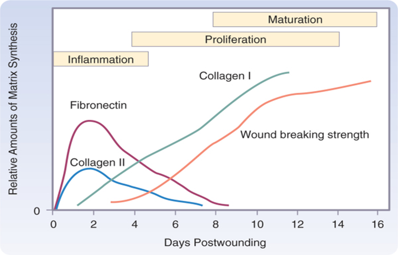
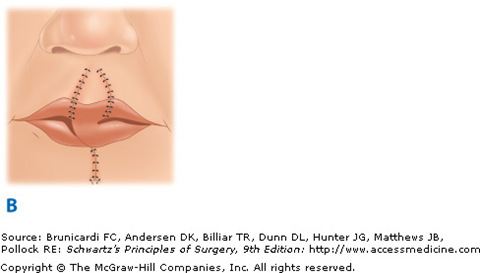
Sabiston:- Intralesional injection of steroids into a keloid scar can inactivate and shrink the scar; such therapy is not indicated for hypertrophic scars.

Scars that are perpendicular to the underlying muscle fibers tend to be flatter and narrower, with less collagen formation than when they are parallel to the underlying muscle fibers. The position of an elective scar can be chosen in such a way to make a narrower and less obvious scar in the distant future. As muscle fibers contract, the wound edges become reapproximated if they are perpendicular to the underlying muscle. If, however, the scar is parallel to the underlying muscle, contraction of that muscle tends to cause gaping of the wound edges and leads to more tension and scar formation.

____________________________________________________________________________________________________________________________________________________________________________

Wounds

Primary closure of incised wounds must be done in –
a) 2 hrs b) 4 hrs
c) 6 hrs d) 12 hrs
e) 16 hrs

(Because of the fear of bacterial invasion, primary wound closure beyond 6 to 8 hours after injury was historically proscribed. However, several scientific studies have since shown that when blood supply to a wound is adequate and bacterial invasion is absent, wounds can be safely closed at any time after proper débridement and irrigation)

The tensile strength of wound reaches that of normal tissue by – (PGI 88)
) 6 weeks
c) 4 months
b) 2 months
d) 6 months

NEVER

In the healing of a clean wound the maximum immediate strength of the wound is reached by –
a) 2 – 3 days b) 4 – 7 days
10 – 12 days d) 13 – 18 days

21 days is ans

The tensile strength of the wound starts and increases after – (MAHE 05)
a)Immediate suture of the wound
b)3 to 4 days
c)7-10 days
d 6 months

see figure

When is the maximum collagen content of wound
tissue – (PGI 81, ROHTAK 87)
a)Between 3rd to 5th day
b)Between 6th to 17th day
C) Between 17th to 21st day d) None of the above

In a sutured surgical wound, the process of epithelialization is completed within – (UPSC 07)
a) 24 hours b) 48 hours
c) 72 hours d) 96 hours

Ref schwartz Epithelialization:- While tissue integrity and strength are being re-established, the external barrier must also be restored. This process is characterized primarily by proliferation and migration of epithelial cells adjacent to the wound The process begins within 1 day of injury and is seen as thickening of the epidermis at the wound edge.Re-epithelialization is complete in less than 48 hours in the case of approximated incised wounds, but may take substantially longer in the case of larger wounds, in which there is a significant epidermal/dermal defect.

Sabiston : – Finally, adequate dressing of the closed wound isolates it from the outside environment. Providing an appropriate dressing for 48 to 72 hours can decrease wound contamination. However, dressings after this period increase the subsequent bacterial count on adjacent skin by altering the microenvironment underneath the dressing.

Following are required for wound healing except – a) Zinc
b) Copper c) Vitamin C d) Calcium

Copper is also a component of ferroprotein, a transport protein involved in the basolateral transfer of iron during absorption from the enterocyte. As such, copper plays a role in iron metabolism, melanin synthesis, energy production, neurotransmitter synthesis, and CNS function; the synthesis and cross-linking of elastin and collagen :- Harrison

Copper Deficiency:- Anemia, growth retardation, defective keratinization and pigmentation of hair, hypothermia, degenerative changes in aortic elastin, osteopenia, mental deterioration.

Patient has lacerated untidy wound of the leg and attended the casualty after 2 ‘hours. His wound (AIIMS 84)should be –
a) Sutured immediately  b) Debrided and sutured immediately  c) debrided and sutured secondarily d) Cleaned and dressed

Wound healing is worst at –
(ALL INDIA 93) a) Sternum b) Anterior neck
c) Eyelid d) Lips

After closing deep tissues and replacing significant tissue deficits, skin edges should be reapproximated for cosmesis and to aid in rapid wound healing. Skin edges may be quickly reapproximated with stainless steel staples or nonabsorbable monofilament sutures. Care must be taken to remove these from the wound before epithelialization of the skin tracts where sutures or staples penetrate the dermal layer. Failure to remove the sutures or staples by 7 to 10 days after repair will result in a cosmetically inferior wound

(Anatomic areas where tension is excessive are avoided if possible. The shoulders, back, and anterior chest are high tension and mobile areas where wide scarring is difficult to avoid. Patients are also questioned as to propensity for development of hypertrophic scars or keloid formation. Ears, anterior chest, and shoulders are areas prone to these problematic scars)

Sabiston :-Wound strength increases rapidly within 1 to 6 weeks and then appears to plateau up to 1 year after the injury .When compared with unwounded skin, tensile strength is only 30% in the scar. An increase in breaking strength occurs after approximately 21 days, mostly as a result of cross-linking.The rate of collagen synthesis declines after 4 weeks and eventually balances the rate of collagen destruction by collagenase (MMP-1). At this point the wound enters a phase of collagen maturation.

Taylor:-The tensile strength of the young scar is only about 10% that of normal skin. Scar strength increases to about 30–50% of normal skin by 4 weeks and to 80% after several months.

Robbins:-We now turn to the questions of how long it takes for a skin wound to achieve its maximal strength, and which substances contribute to this strength. When sutures are removed, usually at the end of the first week, wound strength is approximately 10% of the strength of unwounded skin, but it increases rapidly over the next 4 weeks. This rate of increase then slows at approximately the third month after the original incision and then reaches a plateau at about 70 to 80% of the tensile strength of unwounded skin, which may persist for life.

Schwartz:-Wound strength and mechanical integrity in the fresh wound are determined by both the quantity and quality of the newly deposited collagen. The deposition of matrix at the wound site follows a characteristic pattern: Fibronectin and collagen type III constitute the early matrix scaffolding, glycosaminoglycans and proteoglycans represent the next significant matrix components, and collagen type I is the final matrix. By several weeks postinjury the amount of collagen in the wound reaches a plateau, but the tensile strength continues to increase for several more months.20 Fibril formation and fibril cross-linking result in decreased collagen solubility, increased strength, and increased resistance to enzymatic degradation of the collagen matrix. Scar remodeling continues for many (6 to 12) months postinjury, gradually resulting in a mature, avascular, and acellular scar. The mechanical strength of the scar never achieves that of the uninjured tissue.

image


Factors That Inhibit Wound Healing

Infection

Ischemia

Circulation

Respiration

Local tension

Diabetes mellitus

Ionizing radiation

Advanced age

Malnutrition

Vitamin deficiencies:- Vitamin C Vitamin A

Mineral deficiencies:-Zinc Iron

Exogenous drugs:-Doxorubicin (Adriamycin) Glucocorticosteroids

suture marks are to be avoided, skin sutures should be removed by -  a) hours b) 1 week
2 weeks d) 3 weeks

Epidermal skin sutures function for fine alignment of skin edges. Interrupted sutures are less constrictive than running sutures. The needle enters and exits the skin at 90 degrees in order to evert the skin edges. These skin sutures are removed as soon as adequate intrinsic bonding strength is sufficient. Skin sutures left in place too long result in an unsightly track pattern. On the other hand, sutures removed prematurely risk wound dehiscence. Nonabsorbable sutures on the face are typically removed after 5 days. Sutures in the hand, foot, or across areas that are acted on by motion are left for 14 days or longer .Alternatively, by employing the running intradermal suturing technique, the time constraints of suture removal may be disregarded, and these sutures may be left in place for a longer time without risking a track pattern scar. Finally, epidermal approximation can be achieved without suture using a medical-grade cyanoacrylate adhesive such as Dermabond. Such adhesives are applied across the coapted skin edges only and contribute no tensile strength. Tape closure strips such as Steri-Strips can be applied at the completion of wound closure to help splint the coapted skin edges.


 Guidelines for Day of Suture Removal by Area
BODY REGION REMOVAL (DAYS)
Scalp 6-8
Ear 10-14
Eyelid 3-4
Eyebrow 3-5
Nose 3-5
Lip 3-4
Face (other) 3-4
Chest, abdomen 8-10
Back 12-14
Extremities 12-14
Hand 10-14
Foot, sole 12-14

A patient with grossly contaminated wound presents 12 hours after an accident. His wound should be managed by – (UPSC 96)
a)Thorough cleaning and primary repair
b)Thorough cleaning with debridement of all dead and devitalised tissue without primary closure
c)Primary closure over a drain
d)Covering the defect with split skin graft after cleaning

Management of an open wound seen 12 hrs. after
the injury – (AIIMS 87)
a)Suturing
b)Debridement and suture
c)Secondary suturing
d)Heal by granulation

Delayed wound healing is seen in all except-(AP 96)
a) Malignancy b) Hypertension
c) Diabetes d) Infection

All of the following favour postoperative wound dehiscence except – (Karnat 05)
a)Malignancy
b)Vitamin B complex deficiency
c)Hypoproteinaemia
d)Jaundice

Fibroblast in healing wound derived from –
a) Local mesenchyme b) Epithelium (PGI 98)
c) Endothelial d) Vascular fibrosis

(Sabiston) Fibroplasia:- Fibroblasts are specialized cells that differentiate from resting mesenchymal cells in connective tissue; they do not arrive in the wound cleft by diapedesis from circulating cells. After injury, the normally quiescent and sparse fibroblasts are chemoattracted to the inflammatory site, where they divide and produce the components of the ECM.The primary function of fibroblasts is to synthesize collagen, which they begin to produce during the cellular phase of inflammation. The time required for undifferentiated mesenchymal cells to differentiate into highly specialized fibroblasts accounts for the delay between injury and the appearance of collagen in a healing wound. This period, generally 3 to 5 days, depending on the type of tissue injured, is called the lag phase of wound healing.The rate of collagen synthesis declines after 4 weeks and eventually balances the rate of collagen destruction by collagenase (MMP-1). At this point the wound enters a phase of collagen maturation. The maturation phase continues for months or even years. Glycoprotein and mucopolysaccharide levels decrease during the maturation phase, and new capillaries regress and disappear. These changes alter the appearance of the wound and increase its strength.

Degloving injury is – (KERALA 2K)
a) Surgeon made wound b) Lacerated wound
c) Blunt injury d) Avulsion injury
e)Abrasive wound

Avulsion injuries are open injuries where there has been a severe degree of tissue damage. Such injuries occur when hands or limbs are trapped in moving machinery, such as in rollers, producing a degloving injury. Degloving is caused by shearing forces that separate tissue planes, rupturing their vascular interconnections and causing tissue ischaemia. This most frequently occurs between the subcutaneous fat and deep fascia. Degloving injuries can be open or closed. Degloving can be localised or circumferential. It can occur only in the single, subcutaneous plane, but where present in multiple planes, such as between muscles and fascia and between muscles and bone, is an indication of a severe high-energy injury with a limited potential for primary healing. Similar injuries occur as a result of runover road traffic accident injuries where friction from rubber tyres will avulse skin and subcutaneous tissue from the underlying deep fascia (Fig. 3.11). The history should raise the examiner’s suspicion and it is often possible to pinch the skin and lift it upwards revealing its detachment from the normal anchorage. The danger of degloving or avulsion injuries is that there is devascularisation of tissue and skin necrosis may become slowly apparent in the following few days. Even tissue that initially demonstrates venous bleeding may subsequently undergo necrosis if the circulation is insufficient. Treatment of such injuries is to identify the area of devitalised skin and to remove the skin, defat it and reapply it as a full-thickness skin graft. Avulsion injuries of hands or feet may require immediate flap cover using a one-stage microvascular tissue transfer of skin and/or muscle.

In treatment of hand injuries, the greatest priority is – (A1 96)
a)Repair of tendons
b)Restoration of skin cover
c)Repair of nerves
d) Repair of blood vessels

During the surgical procedure – (AIIMS 83)
a)Tendons should be repaired before nerves
b)Nerves should be repaired before tendons
c)Tendons should not be repaired at the same time
d)None is true

In hand injuries first to be repaired is – (A195)
a) Bone b) Tendon
c) Muscle d) Nerve

In the case of injuries, treatment is directed at the specific structures damaged: skeletal, tendon, nerve, vessel, and integument. In emergency situations, the goals of treatment are to maintain or restore distal circulation, obtain a healed wound, preserve motion, and retain distal sensation. Stable skeletal architecture is established in the primary phase of care because skeletal stability is essential for effective motion and function of the extremity. This also results in reestablishing skeletal length, straightening deformities, and correction of compression or kinking of nerves and vessels. Arteries are also repaired in the acute phase of treatment to maintain distal tissue viability. Additionally, extrinsic compression on arteries must be released emergently such as in compartment pressure problems. In clean-cut injuries, tendons can be repaired primarily. In situations in which there is a chance that tendon adhesions may form, such as when there are associated fractures, it is nonetheless better to repair tendons primarily with preservation of their length and if necessary at a later date to perform tenolysis. However, when there are open and contaminated wounds or a severe crushing injury, it is best to delay repair of both tendon and nerve injuries

Prevention of wound infection done by –
a)Pre-op shaving (PGI  05)
b)Pre-op antibiotic therapy
c)Monofilament sutures
d)Wound apposition

SSIs are the most common nosocomial infection in our population and constitute 38% of all infections in surgical patients. By definition, they can occur anytime from 0 to 30 days after the operation or up to 1 year after a procedure that has involved the implantation of a foreign material (mesh, vascular graft, prosthetic joint, and so on). Incisional infections are the most common; they account for 60% to 80% of all SSIs and have a better prognosis than organ/space-related SSIs do, with the latter accounting for 93% of SSI-related mortalities.

Preoperative shaving has been shown to increase the incidence of SSI after clean procedures as well. This practice increases the infection rate about 100% as compared with removing the hair by clippers at the time of the procedure or not removing it at all, probably secondary to bacterial growth in microscopic cuts. Therefore, the patient is not shaved before an operation. Extensive removal of hair is not needed, and any hair removal that is done is performed by electric clippers with disposable heads at the time of the procedure and in a manner that does not traumatize the skin

1.Basic principles include size of the OR, air management (filtered flow, positive pressure toward the outside, and air cycles per hour), equipment handling (disinfection and cleansing), and traffic rules. All OR personnel wear clean scrubs, caps, and masks, and traffic in and out of the OR is minimized.

2.The CDC recommends the use of chlorhexidine showers, and it is reasonable to implement such a policy, particularly in patients who have been in the hospital for a few days and in those in whom an SSI will cause significant morbidity (cardiac, vascular, and prosthetic procedures). Skin preparation of the surgical site is done with a germicidal antiseptic such as tincture of iodine, povidone-iodine, or chlorhexidine. An alternative preparation is the use of antimicrobial incise drapes applied to the entire operative area. Traditionally, the surgical team has scrubbed their hands and forearms for at least 5 minutes the first time in the day and for 3 minutes every consecutive time.

3.As many as 90% of an operative team puncture their gloves during a prolonged operation. The risk increases with time, as does the risk for contamination of the surgical site if the glove is not changed at the moment of puncture. The use of double gloving is becoming a popular practice to avoid contamination of the wound, as well as exposure to blood by the surgical team. Double gloving is recommended for all surgical procedures.Instruments that will be in contact with the surgical site are sterilized in standard fashion, and protocols for flash sterilization or emergency sterilization, or both, must be well established to ensure the sterility of instruments and implants.

Local Wound Related:-Intraoperative measures include appropriate handling of tissue and assurance of satisfactory final vascular supply, but with adequate control of bleeding to prevent hematomas/seromas. Complete débridement of necrotic tissue plus removal of unnecessary foreign bodies is recommended, as well as avoiding the placement of foreign bodies in clean-contaminated, contaminated, or dirty cases. Monofilament sutures have proved in experimental studies to be associated with a lower rate of SSI. Sutures are foreign bodies that are used only when required. Suture closure of dead space has not been shown to prevent SSI. Large potential dead spaces can be treated with the use of closed-suction systems for short periods, but these systems provide a route for bacteria to reach the wounds and may cause SSI. Open drainage systems (e.g., Penrose) increase rather than decrease infections in surgical wounds and are avoided unless used to drain wounds that are already infected.

In heavily contaminated wounds or wounds in which all the foreign bodies or devitalized tissue cannot be satisfactorily removed, delayed primary closure minimizes the development of serious infection in most instances. With this technique, the subcutaneous tissue and skin are left open and dressed loosely with gauze after fascial closure. The number of phagocytic cells at the wound edges progressively increases to a peak about 5 days after the injury. Capillary budding is intense at this time, and closure can usually be accomplished successfully even with heavy bacterial contamination because phagocytic cells can be delivered to the site in large numbers. Experiments have shown that the number of organisms required to initiate an infection in a surgical incision progressively increases as the interval of healing increases, up to the fifth postoperative day.

Finally, adequate dressing of the closed wound isolates it from the outside environment. Providing an appropriate dressing for 48 to 72 hours can decrease wound contamination. However, dressings after this period increase the subsequent bacterial count on adjacent skin by altering the microenvironment underneath the dressing.

Elective cholecystectomy is – (APPG 06)
a) Clean contaminated b) Clean
Dirty d) Contaminated

Which one of the following surgical procedures is considered to have a clean-contaminated wound ?

a),Elective open cholecystectomy for cholelithiasis
b)Hemiorrhaphy with mesh repair
c)Lumpectomy with axillary node dissection
d)Appendectomy with walled off abscess

The accepted range of infection rates has been 1% to 5% for clean, 3% to 11% for clean-contaminated, 10% to 17% for contaminated, and greater than 27% for dirty wounds.

Table 14-2 Surgical Wound Classification According to Degree of Contamination
WOUND CLASS DEFINITION
Clean An uninfected operative wound in which no inflammation is encountered and the respiratory, alimentary, genital, or infected urinary tract is not entered. Wounds are closed primarily and, if necessary, drained with closed drainage. Surgical wounds after blunt trauma should be included in this category if they meet the criteria
Clean-contaminated An operative wound in which the respiratory, alimentary, genital, or urinary tract is entered under controlled conditions and without unusual contamination
Contaminated Open, fresh, accidental wounds. In addition, operations with major breaks in sterile technique or gross spillage from the gastrointestinal tract and incisions in which acute, nonpurulent inflammation is encountered are included in this category
Dirty Old traumatic wounds with retained devitalized tissue and those that involve existing clinical infection or perforated viscera. This definition suggests that the organisms causing postoperative infection were present in the operative field before the operation

Staphylococcus aureus remains the most common pathogen in SSIs, followed by coagulase-negative staphylococci, enterococci, and Escherichia coli. However, for clean-contaminated and contaminated procedures, E. coli and other Enterobacteriaceae are the most common cause of SSI.


The Vitamin which has inhibitory effect on wound healing is – (MAHE 05)
a) Vitamin-A b) Vitamin-E
c) Vitamin-C d) Vitamin B-complex

Golden period for treatment of open wounds is
….hours – (AIIMS 86, 88)
a) 4 b) 6
c) 12 d) 24

In the first 4 hours after a breach in an epithelial surface and underlying connective tissues made during surgery or trauma, there is a delay before host defences can become mobilised through acute inflammatory, humoral and cellular processes. This period is called the ‘decisive period’ and it is during these first 4 hours after incision that bacterial colonisation and established infection can begin. It is logical that prophylactic antibiotics will be most effective during this time.

Abbey-Estlander flap is used in the reconstruction
of- (AI 05)
a) Buccal mucosa b) Lip
c) Tongue d) Palate

In defects of less than one third the horizontal length, enough redundancy is present to allow primary closure. More complex decisions must be made for defects that are between one third and two thirds of the total lip length. The two categories of lip flap technique are transoral cross-lip flaps and circumoral advancements flaps. Cross-lip flaps include the Abbé flap and the Estlander flap. The Abbé flap was originally designed to reconstruct central upper lip (tubercle) defects with lower lip full-thickness tissue vascularized by one of the labial arteries.The technique requires a second-stage procedure for division of the pedicle. The Estlander flap is similar in principle but is based laterally at the oral commissure and is used to reconstruct lateral upper or lower lip lesions. Both the Estlander and Abbé flaps are denervated, but sensation and perhaps even motor function return over months.The Karapandzic technique is an advancement-rotation flap technique designed for central lower lip defects. Although good function, sensation, and mobility are preserved, a side effect is reduction in the size of the oral aperture. The Webster-Bernard technique uses cheek tissue advancement flaps to replace defects with full-thickness or partial-thickness cheek incisions extended laterally from the commissure (Fig. 45-34). When performed bilaterally, both the Karapandzic and the Webster-Bernard methods can be used to reconstruct a complete upper or lower lip.

imageimageimage


Abbé flap upper lip reconstruction. A. Defect and flap design.                     B. Rotation of the flap and primary closure of the donor site.                                     C. Division of the pedicle (after 2 to 3 weeks) and final insetting.

Cock’s peculiar tumour is-(UPSC 86,NIMHANS 87,
a)Papilloma Kerala 87, TN 90 )
b)Infected sebaceous cyst
c)Cylindroma
d)Sqaumous cell carcinoma (RESEMBLES SCC but it’s not SCC)

COCK

Epidermoid cyst(syn. sebaceous cyst, wen)
These cysts contain keratin and its breakdown products, surrounded by a wall of stratified squamous keratinising epithelium (the commonly used term sebaceous cyst is incorrect — these cysts only rarely have associated sebaceous glands and do not contain sebum). Epidermoid cysts often have a punctum. They are inherited in an autosomal dominant fashion. The common sites are the face, neck, shoulders and chest, areas favoured by acne vulgaris. Lesions may be solitary but are commonly multiple. They enlarge slowly and may become inflamed and tender from time to time. Suppuration may occur. The contents of an infected cyst become semiliquid and usually very foetid. Recurrent infective episodes cause the cyst wall to become adherent to surrounding subcutaneous tissue, and consequently more difficult to remove. If ulceration occurs it can resemble squamous cell carcinoma to which the term ‘Cock’s peculiar tumour’ may be applied .The contents of a cyst sometimes escape slowly from the duct orifice and dry in successive layers on the skin, forming a ‘sebaceous horn’.Treatment is by surgical excision (except if inflamed, when it is better incised and drained). This can be performed under local anaesthesia; an ellipse of skin including the punctum is removed with the cyst. Unless the wall is completely removed, recurrence is likely.

Cause of persistance of a sinus or fistulae includes-
a)Foreign body (JIPMER 86)
b)Non dependentt drainage
c)Unrelieved Obstruction
d)Presence of malignancy
e)All of the above

FISTULA

Sinuses and fistulas
A sinus is a blind track (usually lined with granulation tissue) leading from an epithelial surface into the surrounding tissues. Pathological sinuses must be distinguished from normal anatomical sinuses (e.g. the frontal and nasal sinuses). A fistula  is an abnormal communication between the lumen or surface of one organ and the lumen or surface of another, or between vessels. Most fistulas connect epithelial­lined surfaces .Sinuses and fistulas may be congenital or acquired. Forms which have a congenital origin include preauricular sinuses, branchial fistulas , tracheo-oesophageal fistulas  and arteriovenous fistulas.The acquired forms often follow inadequate drainage of an abscess. Thus, a perianal abscess may burst on the surface and lead to a sinus (erroneously termed a blind external ‘fistula’). In other cases, the abscess opens both into the anal canal and on to the surface of the perineal stem resulting in a true fistula-in-ano .Acquired arteriovenous fistulas are caused by trauma or operation (for renal dialysis).
  Persistence of a sinus or fistula
  The reason for this will be found among the following:
•  a foreign body or necrotic tissue is present, e.g. a suture, hairs, a sequestrum, a faecolith or even a worm (see below);
•  inefficient or nondependent drainage: long, narrow, tortuous track predisposes to inefficient drainage;
•  unrelieved obstruction of the lumen of a viscus or tube distal to the fistula;
•  high pressure, such as occurs in fistula-in-ano due to the normal contractions of the sphincter which force faecal material through the fistula;
•  the walls have become lined with epithelium or endothelium (arteriovenous fistula);
•  dense fibrosis prevents contraction and healing;
•  type of infection, e.g. tuberculosis or actinomycosis;
•  the presence of malignant disease
•  ischaemia;
•  drugs, e.g. steroids, cytotoxics;
•  malnutrition;
•  interference, e.g. artefacta;
•  irradiation, e.g. rectovaginal fistula after treatment for a carcinoma of the cervix;
•  Crohn’s disease;
• high-output fistula, e.g. duodenocutaneous fistula.

Premalignant conditions of the skin include –
a)Bowen disease (JIPMER 86)
b)Pagel’s disease of nipple
c)Leukoplakia
d)Solar keratosis
e)All of the above

Premalignant lesions :-Actinic keratoses
Bowen’s disease
Erythroplasia of Querat
Radiodermatitis
Chronic scars(A carcinoma which develops in a scar (Marjolin’s ulcer) )
Sebaceous epidermal naevus
Porokeratosis


Melanoma should be excised with a margin of –
a) 2 cm b) 5 cm (UPSC 88)
c) 7 cm d) 10 cm

image


Harrison also recommends same treatment for Melanoma as described in above figure.

Hidradenitis suppurativa. is found to occur in – (JIPMER 86, AIMS 87)
a) Axilla b) Circumanal
c) Scalp d) Groin

Hidradenitis suppurativa. :- This is a chronic infection of apocrine glands around the anal margin giving rise to numerous sinuses. The mons pubis and groin can also be affected. After excision of the area, granulation and healing ate accelerated by using Silastic foam dressing (Hughes).

The term universal tumour refers to – (PGI 88)
a) Adenoma b) Papilloma
c) Fibroma d) Lipoma

Lipoma
A lipoma is a slowly growing tumour composed of fat cells adult type. Lipomas may be encapsulated or diffuse. It occur anywhere in the body where fat is found and earn tl titles of the ‘universal tumour’ or the ‘ubiquitous tumour The head and neck area, abdominal wall and thighs are particularly favoured sites.

Hydrocele is a type of ….cyst – (PGI 88)
a) Retention b) Distension
c) Exudation d) Traumatic

Acquired cysts
Retention cysts are due to the accumulated secretion of a gland behind an obstruction of a duct. Examples are seen in the pancreas, the parotid, the breast, the epididymis and Bartholin’s gland. A sebaceous cyst starts with the obstruction of a sebaceous gland, but this is followed by the down-growth and the accumulation of desquamated epidermal cells, thus turning it into an epidermoid cyst. In the epididymis, if the retention cyst contains sperms, it is known as a ‘spermatocele’.

Distension cysts occur in the thyroid from dilatation of the acini, or in the ovary from a follicle. Lymphatic cysts and cystic hygromas are distension cysts.

Exudation cysts occur when fluid exudes into an anatomical space already lined by endothelium, e.g. hydrocele, a bursa, or when a collection of exudate becomes encrusted.

Cystic tumours. Examples are cystic teratomas (dermoid cyst of the ovary) and cystadenomas (pseudomucinous and serous cystadenoma of the ovary).
Ganglia. Implantation dermoids arise from squamous epithelium which has been driven beneath the skin by a penetrating wound. They are classically found in the fingers of women who sew assiduously and metal workers.


Sebaceous cyst does not occur in the …. – (PGI 88)
a) Scalp b) Scrotum
c) Back d) Sole

Sebaceous cysts are common in the scrotal skin. They are usually small and multiple.

Fordyce spots are – (All India 95)
a)Ectopic sebaceous glands
b)Ectopic eccrine
c)Ectopic apocrine
d)Ectopic mucossal glands

Broke’s tumor is a tumor of–
a)Superficial dermal vesels
b)Sweat glands
c)Hair follicles
d)Sebaceous glands


Commonest site for rodent ulcer is – (PGI 88)
a) Inner canthus b) Outer canthus
c) Angle of mouth d) Cheek

Squamous cell carcinoma can arise from-(PGi88)
a)Long standing venous ulcers
b)chronic lupus vulgaris
c)Rodent ulcer
d)All of the above

There is a strong correlation with damage to the skin by the sun , and can be experimentally produced by ultraviolet light. Occa­sionally it arises as a complication of long-standing chronic granulomas, such as syphilis, lupus vulgaris and leprosy, chronic ulcers, osteomyelitis, Hydradenitis suppurativa, long-standing venous ulcers or old burn scars

The best results in treatment of capillary nevus have been achieved by – (AIIMS 84)
a)Full thickness skin graft
b)Dermabrasion
c)Tatooing
d)Argon laser treatment

Capillary malformation, usually referred to as a port-wine stain or nevus flammeus, is the most common type of vascular malformation

Vascular malformations:-These are structural and morphological anomalies due to faulty embryological morphogenesis. The lesions are present at birth, grow commensurate with the child and do nor regress. They can lead to underlying soft tissue or bony hypertrophy, nodular development and discoloration as a consequence of blood vessel ectasia with age. The natural history of these lesions is determined by their haemodynamic and Iymphodynamic characteristics.
• High-flow lesions include arterial malformations and arteriovenous malformations (arterial plexiform angiomas, cirsoid aneurysm).
• Low-flow lesions include lymphatic (LM) venous (VM) and capillary (CM — port-wine stain). Frequently these lesions combine arterial, venous and lymphatic elements.
Port-wine stains:-Port-wine stains are intradermal capillary mal­formations that change very little throughout life, although the colour may alter a little and they may become nodular in some areas. Treatment is for reason of appearance. Treatment of choice for these lesions is the use of the pulsed tunable dye laser.

Eleven month old child presents with erythematous lesion with central clearing which has been decreasing in size – (Al 97)
a)Strawberry angioma
b)Nevus
c)Portwine stain
d)Cavernous haemangioma

Malignant melanoma most often develops from –
a)Hairy naevus (SGPGI 05)
b)Junctional naevus
c)Intradermal naevus
d)Blue naevus


The aim of differential diagnosis is to distinguish benign pigmented lesions from melanoma and its precursor. If melanoma is a consideration, then biopsy is appropriate. Some benign look-alikes may be removed in the process of trying to detect authentic melanoma. Table 83-5 summarizes the distinguishing features of benign lesions that may be confused with melanoma.

nevus

Full thickness skin graft can be taken from the following sites except – (AIIMS 87)
a) Elbow b) Back to neck
c) Supraclavicular area d) Upper eyelids

Posted in Medical | Tagged: , , , , , , , , | 6 Comments »

Physiology

Posted by Dr KAMAL DEEP on February 14, 2011

1.Body Composition

image

Note :- Proteins are not in interstitial fluid (because there are more proteins in plasma than in interstitial fluid, there is a Donnan effect on ion movement across the capillary wall also.)

60% Water (40%ICF;20%ECF(15%Interstitial plus 5% Plasma Volume) 18% Protein Fat 15% Minerals 7%

TBW 60%—measured by D20,HEAVY WATER.It is ECF PLUS ICF.

ICF (40%)—Measured by TBW minus ECF.It cannot be measured directly just like interstitial fluid.

ECF (20%) –Measured by Inulin (Mannitol and sucrose have also been used to measure this space though not as much accurate as inulin)

ECF=Interstitial Fluid(15%) Plus Plasma Volume(5%).

Interstitial Fluid is measured by ECF minus Plasma Volume.It cannot be measured directly just like intracellular fluid.

Plasma Volume(3.5 liters ;5% of body weight) can be measured using dyes Evans Blue(T-1824).It can also be measured by serum albumin labeled with radioactive iodine.

In short TBW(D20,Titanium oxide and aminopyrine),ECF(INULIN,MANNITOL,SUCROSE),PLASMA(T-1824,ALBUMIN) can be measured directly by bracketed methods while Intracellular fluid(TBW-ECF)and interstitial fluid(ECF –PLASMA) can be derived from them indirectly by subtraction methods.

TBW= 40-45 LITRES;60% of body weight; (Intracellular Fluid=28.8Litres Plus Interstitial Fluid =10.5 Liters Plus Blood plasma=3.5 Liters) where ECF(interstitial plus blood plasma) is 14 L or another name is sucrose space in a 70 KG MAN.

Total Blood volume = 3500 Multiply 100/100-38 where 38 is hematocrit.Hematocrit is percentage of blood volume that is made up of red blood cells..So Total blood volume equals 5645 ml.In other words TTAL  is about 8% of body weight in which 5% part is plasma volume and 3 % is red cell volume.

The water content of lean body tissue is constant at 71-72 ml/100g of tissue.

Q:-Most accurate measurement of extracellular fluid
volume (ECF) can be done by using – (AIIMS 03)
a) Sucrose b) Mannitol
c) Inulin d) Aminopyrine

Ans is C i.e. INULIN

Q:- In an adult man 70kgs, the extracellular
fluid volume will be about – (Karn PG MEE 2006)
a) 42L b) 25L
c) 15L d) 121L  Ans is not A as given in guides…Thanks to 
drirfan_ahmad@hotmail.com

Q:-Which of the following methods is not used for measurement of body fluid volumes –
a) Aminopyrine for total body water-true (AIIMS May 05)
b)Inulin for extracellular fluid-true
c)Evans blue for plasma volume-true
d)albumin for blood volume-false it is used for plasma volume   Ans is D

Note:- Red cell volume can be determined by subtracting the plasma volume from the total blood volume..Total blood volume was determined by plasma volume and hematocrit.A commonly used tag is 15Cr,a radioactive isotope of chromium that is attached to the cells by incubating them in a suitable chromium solutions.Isotopes of iron and phosphorus and antigenic tagging have also been employed.

Heavy water (D20) or Deuterium Oxide is most frequently used for measuring the TBW.Titanium oxide and aminopyrine have also been used for this purpose.

Solvent Drag:- When solvent is moving along one direction,it tends to drag along some molecules of solute.

Q:- The percentage of the circulating blood volume in the venous system and splanchnic vessels is
normally between – (JIPMER 70, AMU 88)
a) 20-30% b) 40-50%
c) 60-70% d) None of these

Ans is c

Q:-Splanchnic vessels and venules contain what percentage of blood volume – (AMC 86,87)
a) 10-20 %
b) 20-30 %
c) 40-50 % d) 60-70 %

Ans is b given…..

Two different answers

Because different questions.

Veins are also called “capacitance vessels” because most of the blood volume (60%) is contained within veins.the splanchnic circulation contains about 25—30% of the total blood volume.

Q:-Osmolarity is – (JIPMER 98)
a)Osmolarity per kg of solvent
b)Osmolarity per litre of solvent
c)Moles per kg of solvent
d)Moles per litre of solvent

The osmolal concentration of a substance in a fluid is measured by the degree to which it depresses the freezing point, with 1 mol of an ideal solution depressing the freezing point 1.86 °C. The number of milliosmoles per liter in a solution equals the freezing point depression divided by 0.00186

Osmolarity is the number of osmoles per litre of the solution.

Osmolality is the number of osmoles per kg of the solvent.

Therefore, osmolarity is affected by the volume of the various solutes in the solution and the temperature, while the osmolality is not.

Osmotically active substances in the body are dissolved in water, and the density of water is 1, so osmolal concentrations can be expressed as osmoles per liter (Osm/L) of water. In this book, osmolal (rather than osmolar) concentrations are considered, and osmolality is expressed in milliosmoles per liter (of water).

Note that although a homogeneous solution contains osmotically active particles and can be said to have an osmotic pressure, it can exert an osmotic pressure only when it is in contact with another solution across a membrane permeable to the solvent but not to the solute.

Q:-A solution contains 1 gram-mole of magnesium sulfate per liter. Assuming full ionization of this compound , calculate the osmotic pressure of the solution (1 mosmole/liter concentration is equivalent to 19.3 mm.IIg osmotic pressure) – (AI 86)
a) 19.3 mm Hg b) 3.86 mm Hg
c) 19.300 mm Hg d) 38.600 mm Hg
e) 57.900 mm Hg

Ans is D  19.3 MULTIPLY BY 2

Magnesium sulfate (or magnesium sulphate) is a chemical compound containing magnesium, sulfur and oxygen, with the formula MgSO4 SUPPLYING 2 Osm

If a solute is a nonionizing compound such as glucose, the osmotic pressure is a function of the number of glucose molecules present. If the solute ionizes and forms an ideal solution, each ion is an osmotically active particle. For example, NaCl would dissociate into Na+ and Cl ions, so that each mole in solution would supply 2 Osm. One mole of Na2SO4 would dissociate into Na+, Na+, and SO42– supplying 3 Osm..

Q:-When solvent is moving in one direction, the
solvent tends to drag along some molecules of solute. This is called – (PGI 81„AMU 86)
a) Filtration b) Osmosis
c) Dorman effect d) Solvent drag

D is ans

Q:-Osmolality of plasma in a normal adult (in m
Osm/L) is – (Delhi 87)
a) 250 -270
b) 270 – 290
c) 300 -310 d)310-330

The freezing point of normal human plasma averages –0.54 °C, which corresponds to an osmolal concentration in plasma of 290 mOsm/L

It is important to note the relative contributions of the various plasma components to the total osmolal concentration of plasma. All but about 20 of the 290 mOsm in each liter of normal plasma are contributed by Na+ and its accompanying anions, principally Cl and HCO3. Other cations and anions make a relatively small contribution. Although the concentration of the plasma proteins is large when expressed in grams per liter, they normally contribute less than 2 mOsm/L because of their very high molecular weights. The major nonelectrolytes of plasma are glucose and urea, which in the steady state are in equilibrium with cells. Their contributions to osmolality are normally about 5 mOsm/L each but can become quite large in hyperglycemia or uremia.

Gibbs-donnan Equation

Donnan Effect

When an ion on one side of a membrane cannot diffuse through the membrane, the distribution of other ions to which the membrane is permeable is affected in a predictable way. For example, the negative charge of a nondiffusible anion hinders diffusion of the diffusible cations and favors diffusion of the diffusible anions. Consider the following situation,

image

in which the membrane (m) between compartments X and Y is impermeable to charged proteins (Prot–) but freely permeable to K+ and Cl–. Assume that the concentrations of the anions and of the cations on the two sides are initially equal. Cl– diffuses down its concentration gradient from Y to X, and some K+ moves with the negatively charged Cl– because of its opposite charge. Therefore

image

Furthermore,

image

that is, more osmotically active particles are on side X than on side Y.

Donnan and Gibbs showed that in the presence of a nondiffusible ion, the diffusible ions distribute themselves so that at equilibrium their concentration ratios are equal:

image

Cross-multiplying,

image

This is the Gibbs–Donnan equation. It holds for any pair of cations and anions of the same valence.

The Donnan effect on the distribution of ions has three effects in the body introduced here and discussed below. First, because of charged proteins (Prot–) in cells, there are more osmotically active particles in cells than in interstitial fluid, and because animal cells have flexible walls, osmosis would make them swell and eventually rupture if it were not for Na, K ATPase pumping ions back out of cells. Thus, normal cell volume and pressure depend on Na, K ATPase. Second, because at equilibrium the distribution of permeant ions across the membrane (m in the example used here) is asymmetric, an electrical difference exists across the membrane whose magnitude can be determined by the Nernst equation. In the example used here, side X will be negative relative to side Y. The charges line up along the membrane, with the concentration gradient for Cl– exactly balanced by the oppositely directed electrical gradient, and the same holds true for K+. Third, because there are more proteins in plasma than in interstitial fluid, there is a Donnan effect on ion movement across the capillary wall.

Nernst equation deals with – (JIPMER 92)
a)Oxygen uptake
b)Chloride shift
c)Cellular ATP levels
d)Plasma bicarbonate level

The forces acting across the cell membrane on each ion can be analyzed mathematically. Chloride ions (Cl) are present in higher concentration in the ECF than in the cell interior, and they tend to diffuse along this concentration gradient into the cell. The interior of the cell is negative relative to the exterior, and chloride ions are pushed out of the cell along this electrical gradient. An equilibrium is reached between Cl influx and Cl efflux. The membrane potential at which this equilibrium exists is the equilibrium potential. Its magnitude can be calculated from the Nernst equation, as follows:

image

where

ECl = equilibrium potential for Cl
R = gas constant
T = absolute temperature
F = the faraday (number of coulombs per mole of charge)
ZCl = valence of Cl (–1)
[Clo] = Cl concentration outside the cell
[Cli] = Cl concentration inside the cell

The equilibrium potential for Cl (ECl), calculated from the standard values listed in Table 1–1, is –70 mV, a value identical to the measured resting membrane potential of –70 mV. Therefore, no forces other than those represented by the chemical and electrical gradients need be invoked to explain the distribution of Cl across the membrane

Table 1–1 Concentration of Some Ions Inside and Outside Mammalian Spinal Motor Neurons.


Ion Concentration (mmol/L of H2O) Equilibrium Potential (mV)
Inside Cell Outside Cell
Na+ 15.0 150.0 +60
K+ 150.0 5.5 –90
Cl 9.0 125.0 –70

The magnitude of membrane potential at any given time depends,of course,upon the distribution of Na+,K+ and CL-

Table 4–1 Nerve Fiber Types in Mammalian Nerve.a


Fiber Type Function Fiber Diameter (micronm) Conduction Velocity (m/s) Spike Duration (ms) Absolute Refractory Period (ms)
A
Alpha Proprioception; somatic motor 12–20 70–120
Beta Touch, pressure 5–12 30–70 0.4–0.5 0.4–1
Gamma Motor to muscle spindles 3–6 15–30
Delta Pain, cold, touch 2–5 12–30
B Preganglionic autonomic 3–15 1.2 1.2
C
Dorsal root Pain, temperature, some mechano-reception 0.4–1.2 0.5–2 2 2
Sympathetic Postganglionic sympathetic 0.3–1.3 0.7–2.3 2 2

aA and B fibers are myelinated; C fibers are unmyelinated.

Table 4–3 Relative Susceptibility of Mammalian A, B, and C Nerve Fibers to Conduction Block Produced by Various Agents.


Susceptibility to: Most Susceptible Intermediate Least Susceptible
Hypoxia B A C
Pressure A B C
Local anesthetics C B A

image

A) Arrangement of thin (actin) and thick (myosin) filaments in skeletal muscle (compare to Figure 5–2). B) Sliding of actin on myosin during contraction so that Z lines move closer together. C) Detail of relation of myosin to actin in an individual sarcomere, the functional unit of the muscle. D) Diagrammatic representation of the arrangement of actin, tropomyosin, and troponin of the thin filaments in relation to a myosin thick filament. The globular heads of myosin interact with the thin filaments to create the contraction. Note that myosin thick filaments reverse polarity at the M line in the middle of the sarcomere, allowing for contraction.

The width of the A bands is constant

image

image


During phases 0 to 2 and about half of phase 3 (until the membrane potential reaches approximately –50 mV during repolarization), cardiac muscle cannot be excited again; that is, it is in its absolute refractory period. It remains relatively refractory until phase 4. Therefore, tetanus of the type seen in skeletal muscle cannot occur. Of course, tetanization of cardiac muscle for any length of time would have lethal consequences, and in this sense, the fact that cardiac muscle cannot be tetanized is a safety feature.

Cardiac muscle is a collection of individual cells (cardiomyocytes) that are linked as a syncytium by gap junctional communication.

  • Smooth muscle cells are largely under control of the autonomic nervous system.
  • There are two broad categories of smooth muscle cells: unitary and multiunit. Unitary smooth muscle contraction is synchronized by gap junctional communication to coordinate contraction among many cells. Multiunit smooth muscle contraction is coordinated by motor units, functionally similar to skeletal muscle.
  • Smooth muscle cells contract through an actomyosin system, but do not have well-organized striations. Unlike skeletal and cardiac muscle, Ca2+ regulation of contraction is primarily through phosphorylation–dephosphorylation reactions
  • the excitation–contraction coupling in unitary smooth muscle can occur with as much as a 500-ms delay. Thus, it is a very slow process compared with that in skeletal and cardiac muscle, in which the time from initial depolarization to initiation of contraction is less than 10 ms
  • Unitary smooth muscle is characterized by the instability of its membrane potential and by the fact that it shows continuous, irregular contractions that are independent of its nerve supply. This maintained state of partial contraction is called tonus, or tone. The membrane potential has no true “resting” value, being relatively low when the tissue is active and higher when it is inhibited, but in periods of relative quiescence values for resting potential are on the order of –20 to –65 mV. Smooth muscle cells can display divergent electrical activity

image

image

EEG records showing the alpha and beta rhythms. When attention is focused on something, the 8–13 Hz alpha rhythm is replaced by an irregular 13–30 Hz low-voltage activity, the beta rhythm.EEG records showing the alpha and beta rhythms. When attention is focused on something, the 8–13 Hz alpha rhythm is replaced by an irregular 13–30 Hz low-voltage activity, the beta rhythm.

1.A person falling asleep first enters stage 1, the EEG begins to show a low-voltage, mixed frequency pattern. A theta rhythm (4–7 Hz) can be seen at this early stage of slow-wave sleep. Throughout NREM sleep, there is some activity of skeletal muscle but no eye movements occur.

2.Stage 2 is marked by the appearance of sinusoidal waves called sleep spindles (12–14 Hz) and occasional high voltage biphasic waves called K complexes.

3.In stage 3, a high-amplitude delta rhythm (0.5–4 Hz) dominates the EEG waves.

4.Maximum slowing with large waves is seen in stage 4. Thus, the characteristic of deep sleep is a pattern of rhythmic slow waves, indicating marked synchronization; it is sometimes referred to as slow-wave sleep. Whereas theta and delta rhythms are normal during sleep, their appearance during wakefulness is a sign of brain dysfunction

Posted in Medical | Tagged: , , , , , , , , | 5 Comments »

Forensic Medicine Notes & MCQs Part 1 (IPCs and Courts)

Posted by Dr KAMAL DEEP on February 1, 2011

1.In case of death in lock up, the inquest is held by –
a) A Police officer b) Magistrate (PGI 86)
c) Panchayat officer d) District Attorney

2.In India, inquest is carried out by all except –
a) Police b) Coroner (AIIMS 91)
c) Doctor d) Magistrate

3.The enquiry into the circumstances of death is
called – (TN 91)
a) Homicide enquiry b) Inquest report
c) Open verdict d) Adjounred verdict

4.The common type of inquest in India is –
a) Coroner’s inquest – (Karn 94)
b) Police inquest
c) Judicial magistrate inquest
d) Medical examiner’s inquest

5.In India, magrstrate inquest is done in the following
cases except – (A1 05)
a) Exhumation cases
b) Dowry deaths within 5 years of marriage
c) Murder cases
d) Death of a person in police custody

U/S 174 Cr.P.C-Police to enquire -Under section 174(subsection 3) Cr.P.C, it’s mandatory provision which requires that the body of a woman, who has committed suicide within seven years of her marriage, has to be subjected to postmortem examination and the inquest also has to be held.

U/S 176 Cr.P.C.-Inquiry by Magistrate into cause of death:-[When any person dies while in the custody of the police or when the case is of the nature referred to in clause (i) o or clause (ii) of sub-section (3) of section 174] the nearest Magistrate empowered to hold inquests shall, and in any other case mentioned in sub-section (1) of section 174, any Magistrate so empowered may hold an inquiry into the cause of death either instead of, or in additional to, the investigation held by the police officer; and if he does so, he shall have all the powers in conducting it which he would have in holding an inquiry into an offence.

Magistrate’s inquest means an inquiry conducted by a magistrate to ascertain matters of fact. It is commonly held in the following cases: Admission of a mentally ill person in a psychiatric hospital or a psychiatric nursing home under certain provisions of the Mental Health Act, 1987; Death of a convict in jail; Death of a person in police custody or during police interrogation; Death as a result of police shooting killing; Exhumation cases and Dowry deaths under section Sec 176 Criminal Procedure Code, 1973.


Important Crpc Sections:-

193 crpc–Perjury means giving willful false evidence by a witness while under oath, the witness is liable to be prosecuted for perjury and the imprisonment may extend to seven years. This falls under 193 of Penal Code

Section 53:- Examination at the request of police

Section 54:- Examination at the request of accused (imp:-NOT victim)

                                                                 IPCs:-

44-Injury Body Mind Reputation Property
84-Criminal responsibility of insane

85-Involuntary Drunkeness

86-Voluntary Drunkensess


312-316 Deals with criminal abortion

317- Abandoning of child by parents

318- Concealment of death of infant


definition of rape is sec 375
punishment for rape is sec 376
sec 377
is about unnatural sexual offences
Sections 354 (molestation),Section 354 of the IPC considers the assault or criminal force to woman with the intention to outrage her modesty. This offense is considered less serious than Rape.

Section 509. Word, gesture or act intended to insult the modesty of a woman.– Whoever, intending to insult the modesty of any woman, utters any word, makes any sound or gesture, or exhibits any object, intending that such word or sound shall be heard, or that such gesture or object shall be seen, by such woman, or intrudes upon the privacy of such woman, shall be punished with simple imprisonment for a term which may extend to one year, or with fine, or with both.

506 (2) (criminal intimidation)
The India Penal Code 1860 does not recognise child abuse. Only rape and sodomy can lead to criminal conviction
Section 5 of the Immoral Traffic Prevention Act 1956 prescribes punishment of not less than 7 years for inducing a child into prostitution, but does not directly address child abuse.
Most of these forms of child abuse are sought to be covered under sec. 354 of the Indian Penal Code as a violation of a woman’s modesty. Though offences under Sec. 354 of the IPC are cognizable, they are also bailable, allowing the perpetrator to abscond before the case comes up in court.


300-Defines MURDER
299-Defines CUL. HOMIICIDE NOT AMOUNTIN TO MURDER
Section 302. Punishment for murder

Section 304:- Punishment for 299

Section 304. Punishment for culpable homicide not amounting to murder

Whoever commits culpable homicide not amounting to murder shall be punished with 1 [imprisonment for life ],or imprisonment of either description for a term which may extend to ten years, and shall also be liable to fine, if the act by which the death is caused is done with the intention of causing death, or of causing such bodily injury as is likely to cause death,Or with imprisonment of either description for a term which may extend to ten years, or with fine, or with both, if the act is done with the knowledge that it is likely to cause death ,but without any intention to cause death, or to cause such bodily injury as is likely to cause death


Sec 304 A Death due to negligent act (Medical or any)-2yrs punishment:-

Section 304A. Causing death by negligence
1[304A. Causing death by negligence.

Whoever causes the death of any person by doing any rash or negligent act not amounting to culpable homicide, shall be punished with imprisonment of either description for a term which may extend to two years, or with fine, or with both.]

Medical negligence falls under section 304A

Sec 304 b is dowry death- 7 yrs to Life  imprisonment


Section 320. Grievous hurt.

Section 312. Causing miscarriage
Section 37. Co-operation by doing one of several acts constituting an offence–A and B agree to murder Z by severally and at different times giving him small doses of poison. A and B administer the poison according to the agreement with intent to murder Z. Z dies from the effects of the several doses of poison so administered to him. Here A and B intentionally co-operates in the commission of murder and as each of them does an act by which the death is caused, they are both guilty of the offence though their acts are separate.


IPC Crimes
(i) Procuration of minor girls (section 366-A IPC)
(ii) Importation of girls ((Sec. 366-B IPC) 

SLL Crimes
(i) Immoral Trafficking (Prevention) Act 1956
(ii) Child Marriage Restraint Act, 1929.
Section 366B. Importation of girl from foreign country
1[366B. Importation of girl from foreign country. Whoever imports into 2[India] from any country outside India 3[or from the State of Jammu and Kashmir] any girl under the age of twenty-one years with intent that she may be, or knowing it to be likely that she will be, forced or seduced to illicit intercourse with another person, 4[***] shall be punishable with imprisonment which may extend to ten years and shall also be liable to fine.]
Section 366A. Procreation of minor girl
1[366A. procreation of minor girl.:–Whoever, by any means whatsoever, induces any minor girl under the age of eighteen years to go from any place or to do any act with intent that such girl may be, or knowing that it is likely that she will be, forced or seduced to illicit intercourse with another person shall be punishable with imprisonment which may extend to ten years, and shall also be liable to fine.]

Section 372. Selling minor for purposes of prostitution, etc
Section 373. Buying minor for purposes of prostitution, etc.


Section 299. Culpable homicide

Who ever causes death by doing an act with the intention of causing death, or with the intention of causing such bodily injury as is likely to cause death, or with the knowledge that he is likely by such act to cause death, commits the offence of culpable homicide.

Section 300. Murder

Except in the cases hereinafter excepted, culpable homicide is murder, if the act by which the death is caused is done with the intention of causing death, or-2ndly If it is done with the intention of causing such bodily injury as the offender knows to be likely to cause the death of the person to whom the harm is caused, or-3rdly If it is done with the intention of causing bodily injury to any person and the bodily injury intended to be inflicted is sufficient in the ordinary course of nature to cause death, or-4thly If the person committing the act knows that it is so imminently dangerous that it must, in all probability, cause death or such bodily injury as is likely to cause death, and commits such act without any excuse for incurring the risk of causing death or such injury as aforesaid.

Illustrations

(a) A shoots Z with the intention of killing him. Z dies in consequence. A commits murder.
(b) A, knowing that Z is labouring under such a disease that a blow is likely to cause his death, strikes him with the intention of causing bodily injury. Z dies in consequence of the blow. A is guilty of murder, although the blow might not have been sufficient in the ordinary course of nature to cause the death of a person in a sound state of health. But if A, not knowing that Z is labouring under any disease, gives him such a blow as would not in the ordinary course of nature kill a person in a sound state of heath, here A, although he may intend to cause bodily injury, is not guilty of murder, if he d8id not intend to cause death, or such bodily injury as in the ordinary course of nature would cause death.
(c) A intentionally gives Z a sword-cut or club-wound sufficient to cause the death of a man in the ordinary course of nature. Z dies in consequence. Here, A is guilty of murder, although he may not have intended to cause Z’s death.
(d) A without any excuse fires a loaded connon into a crowd of persons and kills one of them. A is guilty of murder, although he may not have had a premeditated design to kill any particular individual.

Exception I-When culpable homicide is not murder-Culpable homicide is not murder if the offender, whilst deprived of the power of self-control by grave and sudden provocation, causes the death of the person who gave the provocation or causes the death of any other person by mistake or accident.
The above exception is subject to the following provisos :–
First-That the provocations not sought or voluntarily provoked by the offender as an excuse for killing or doing harm to any person.
Secondly-That the provocation is not given by anything done in obedience to the law, or by a public servant in the lawful exercise of the powers of such public servant.
Thirdly-That the provocations not given by anything done in the lawful exercise of the right of private defence.
Explanation-Whether the provocation was grave and sudden enough to prevent the offence from amounting to murder is a question of fact.

Culpable Homicide not Amounting to Murder is an offence under s.304 of the Indian Penal Code. It applies to an event where the death is intentional but does not come within the IPC definition of Murder.

Murder versus Culpable Homicide:

Murder (defined under Section 300) and culpable homicide (defined under Section 299) are two offences under the Indian Penal Code the distinction between which has always been perplexing to the law students.

Section 299 and Section 300 IPC deals with the definition of culpable homicide and murder respectively. Section 299 defines culpable homicide as the act of causing death; (i) with the intention of causing death or (ii) with the intention of causing such bodily injury as is likely to cause death or (iii) with the knowledge that such act is likely to cause death. The bare reading of the section makes it crystal clear that the first and the second clause of the section refer to intention apart from the knowledge and the third clause refers to knowledge alone and not intention. Both the expression “intent” and “knowledge” postulate the existence of a positive mental attitude which is of different degrees. The mental element in culpable homicide i.e. mental attitude towards the consequences of conduct is one of intention and knowledge. If that is caused in any of the aforesaid three circumstances, the offence of culpable homicide is said to have been committed. Section 300 IPC, however, deals with murder although there is no clear definition of murder provided in Section 300 IPC. It has been repeatedly held by this Court that culpable homicide is the genus and murder is species and that all murders are culpable homicide but not vice versa. Section 300 IPC further provides for the exceptions which will constitute culpable homicide not amounting to murder and punishable under Section 304. When and if there is intent and knowledge then the same would be a case of Section 304 Part I and if it is only a case of knowledge and not the intention to cause murder and bodily injury, then the same would be a case of Section 304 Part II. The aforesaid distinction between an act amounting to murder and an act not amounting to murder has been brought out in the numerous decisions.

————(a) 300 (amounting to murder).Definition

Section 299(culpable homicide)

————b) 304  (not amounting to murder).Punishment for 299 if not (a)

_______c) 302 Punishment if (a)

Which carries more weight in a court of law –
a) Dying declaration (PGI 87)
b) Dying deposition
c) Both carry the-same weight
d) Both are not significant

Dying declaration is very important documentary evidence. It is hearsay evidence but even then it is given a lot of weightage in the court proceedings. Recording of dying declaration is very important. If it is recorded properly by the proper person keeping in mind the essential ingredients of the dying declaration it retains its full value. Missing any single ingredients of dying declaration makes it suspicious and offenders are likely to get the benefits of its shortcomings.

Its admissibility is explained in the section 32 (1) of Indian Evidence Act. According to this section when the statement is made by a person as to the cause of his death, or any of the circumstances of the transaction which resulted in his death, in cases in which the cause of that person’s death comes into question. Such statements are relevant whether the person who made this was expecting death or not [1]. In English law he must be under expectation of death only then this declaration is valid.

This can be best certified by the doctor who knows best about the condition of the patient. But even in conditions where it was not possible to take fitness from the doctor, dying declarations have retained their full sanctity if there are other witnesses to testify that victim was in such a condition of the mind which did not prevent him from making statement. Medical opinion cannot wipe out the direct testimony of the eyewitness stating that the deceased was in fit and conscious state to make the dying declaration.

It is best that it is recorded by the magistrate but if there is no time to call the magistrate due to the deteriorating condition of the victim it can be recorded by anybody e.g. public servant like doctor or any other person. Courts discourage the recording of dying declaration by the police officers but if there is no body else to record it dying declarations written by the police officers are also considered by the courts. If these are not recorded by the magistrate it is better that signatures of the witnesses are taken who are present at the time of recording it.

Ideally it should be recorded by executive magistrate. [But if magistrate is not available, it can be recorded by doctor himself or if the patient is not in the hospital it can also be recorded by any person present near the patient.] Doctor has to certify pt.to be ‘compos mentis’ .During recording police, relatives should not be present there. .It should be recorded in the presence of magistrate, doctor & 2 disinterested witness. .No oath is administered & leading questions are not permitted. .It is recorded in Question & answer form in the vernacular of patient.

At the end it is read over to the patient & is signed by the magistrate, doctor & 2 disinterested witness. If the patient dies during recording Dr. should certify that pt. is dead & incomplete declaration is signed by all the concerned. The date, time & place of recording is noted & report is sent to magistrate (if not present) in a sealed cover. If the pt. survives after recording, the declaration looses its value, because now he can be called to the court for evidence where cross examination will be possible.However under section 157 IEA,the declaration may still be relied upon to corroborate the statement of the complainant at the time of oral examination.

Ref:-JIAFM, 2004; 26(1). ISSN 0971-0973
CRITICAL APPRAISAL OF DYING DECLARATION
Dr. R.K.Gorea, Professor and Head, Forensic Medicine, GMC, Patiala
Dr. O.P.Aggarwal, Professor, Forensic Medicine, M.M. Medical College, Mullana, Ambala

DYING DEPOSITION It is defined as the deposition (statement on oath) made by a person likely to die bcoz of some unnatural act done on his body,narrating the cause of his likely unnatural death to the magistrate, in the +nse of accused & lawyers of both parties who can cross examine the pt. Precautions- It should by magistrate, in the +nse of accused & lawyers of both parties. Dr. has to be +ntthrough out the procedure & has to certify the pt. in compos mentis. Oath is administered to the pt. Cross examination includes leading questions.Any person may be present during recording .No witness required. Recording is done as per the procedure of court & hence almost it seems that a court is +nt at bedside of pt.

Importance-Dying deposition is having more value than that of dying declaration bcoz, It is recorded by magistrate. In the +nseof accused & lawyers of both parties. It is recorded after cross examination. It retains its value even if pt. survives bcozcross examination has already been done.

The power of Amnesty for capital punishment
is vested with – . (AMU 88)
a) The president b) Supreme court
c) High court d) The governer

Amnesty is a legislative or executive act by which a state restores those who may have been guilty of an offense against it to the positions of innocent people. It includes more than pardon, in as much as it obliterates all legal remembrance of the offense.

Courts can only give stay order pending hearing for a lower court order.

Conduct money is paid to expert witness with
summons from – (AI 90)
a) Civil court b) Criminal court
c) High court d) Sub magistrate

Conduct money is the money paid by the court of law to the witness who is under summon or subpoena to meet his travelling expenses from his place of residence to the court, and back . In CIVIL cases , this is paid to witness at the time the summon is served on him . If this amount is not paid to him/her, he/she may ignore the summons, if he/she so desire. If he/she feels the sum is inadequate can inform the court accordingly & get it enhanced . In CRIMINAL cases however, no such fee is given usually, & it is bounden duty of every citizen to attend the court whenever summoned.(It may however be noted that provision is made by government under section 312 of Crpc for payment of reasonable expenses of an expert witness attending before any criminal court.Payment of reasonable expenses of an expert witness in magistrate courts in summons cases is provided for under section 254(3) and in warrant cases under section 243 (3) CrPC.)


Death sentence can be awarded by – (Kerala 91)
a) First class magistrate
b) Second class magistrate
c) Session court
d) Chief judicial magistrate

Important POINTS:-

1.A session court cannot commute death sentence.How can it commute a death sentence when it is the lowest court to announce the death sentence.

2.President cant Give death sentence It can give amnesty/mercy plea.

3.Additional session court have same power as session court

4.Assistant session court has lower power.

5.None of the magistrate can give death sentence or Life imprisonment

6.CJM power:- 7 years,fine without limit

1st class Judicial magistrate power:- 3 years,upto Rs 5000

2nd class Jud Mag:- 1 Year Max, upto 1000

  1. According to Indian Majority Act 1875, a person attains majority when he/she attains the age of 18 years.
  2. According to Section 4(a) of the TheHindu Minority and Guardianship Act 1956, a person who is below 18 years of age is a minor.

Age

According to Section 363-A of I.P.C, kidnapping or maiming of a minor for the purpose of begging is an offence. If the person merely kidnaps, the sentence is 10 years and fine. If he maims, the sentence is life imprisonment and fine. For the purposes of this section a minor is a female below 18 years (or a male below the age of 16 years). This is an interesting section, as the ages for minors are different for males and females.

However Sections 87 IPC mentions eighteen years as the age for giving consent for acts not intended and not known to be likely to cause death or grievous hurt. These acts are not necessarily for the benefit of the person . Hence Section 87 IPC is not applicable to the medical profession as here (in Section 87 IPC), the acts are NOT done for the person’s benefit. .

According to Section 87 of I.P.C, a person under the age of 18 years can not give valid consent whether express or implied to suffer any harm which may result from an act not intended or not known to cause death or grievous hurt (example: fencing)’

According to Section 89 of I.P.C, a child below 12 years can not give valid consent to suffer any harm which may occur from an act done in good faith and for its benefit(as a general physical examination by a doctor,surgical operations).


The age of a 16 years old female is best determined
by the radiograph of – (PGI 87)
a) Lower end of radius and Ulna
b) Upper end of humerus
c) Upper end of radius and Ulna
d) Xiphisternum

Female                       Male

Elbow     13-14 yrs                   15-17 yrs

Wrist:-    16-17 years                18-19 yrs

Shoulder      17-18                   19-20 yrs

Crest of Ilium 18-19                  20-21yrs

Ref Parikh

Elbow(13-14)—Wrist(16-17)—Shoulder(17-18)—Crest of ilium(18-19) Add plus 2 for Males

By 13-14 years the epiphysis at the elbow join their respective shafts in Females. In males by 16-18 yrs.,all the epiphysis at the elbow (except the medial epicondyle),head of the femur,and lower end of tibia join the respective shafts.

The age of a 15 year old female is best determined
by the radiograph of –

a) Lower end of Radius and Ulna
b) Upper end of humerus
c) Upper end of Radius and Ulna??? NOT SURE
d) Xiphisternum

Ossification centre appearing just before birth is—LOWER END OF FEMUR

Capitate and hamate ossify at the age of 4 months,so before 4 months of age,no bone is seen on radiography

Pre-auricular sulcus is used for – (AMJ 88)
a) Determination of Race
b) Determination of age
c) Determination of sex
d) None of the above

The narrow part of the pelvic surface, between the auricular surface and the upper rim of the greater sciatic notch, often shows a rough preauricular sulcus for the lower fibers of the anterior sacroiliac ligament, more apparent in females.

In forensic practice, identification of human skeletal remains (which are sometimes fragmentary) usually involves diagnosis of sex, and this is most certainly established from the pelvis. Even parts of the pelvis may be useful. Several studies of metrical characteristics in various pelvic regions have been made, leading to the production of various indices. The ilium has received particular attention, e.g. one index compares the pelvic and sacroiliac parts of the bone. A line is extended back from the iliopectineal eminence to the nearest point on the anterior auricular margin and thence to the iliac crest. The auricular point divides this chilotic line into anterior (pelvic) and posterior (sacral) segments, each expressed as a percentage of the other. Chilotic indices display reciprocal values in the sexes: the pelvic part of the chilotic line is predominant in females, and the sacral part in males. Detailed metrical studies of the ilium have indicated its limited reliability in ‘sexing’ pelvis. However, the higher incidence and definition of the female preauricular sulcus is recognized.The desirability of correlating all available metrical data is to be emphasized; when a range of pelvic data can be combined, especially if they are metrical, 95% accuracy should be achieved. Complete accuracy has been claimed when the rest of the skeleton is available. Assessment of sex from isolated and often incomplete human remains is less reliable.

image

image

image

Males:-Suprapubic arch narrow,V shaped,angle not more than about 70 degrees and hence less Distance between ischia,Greater sciatic notch is narrow,deep,and less than right angle.Obturator foramina ovoid.Ischial Tuberosities inverted

Females:- Suprapubic arch wide,U shaped,angle more than a right angle and hence more distance between ischia.Greater sciatic notch is wide shallow and almost a right angle or more.Obturator foramina triangular.Ischial Tuberosity everted.

image

Structure of the bony pelvis. A. In women. B. In men. The angle formed by the pubic arch can be approximated by the angle between the thumb and index finger for women and the angle between the index finger and middle finger for men as shown in the insets.

Skull:- Orbital opening big and rectangular in males while it is rounded and small in females.Muscle attachments are more pronounced in males.

Cephalic index(for the determination of race) is obtained by multiplying the maximum transverse breadth by 100 and dividing it by maximum AP Length. A skull is dolichocephalic(long headed)—70-74.9 seen in Aryans,aborigines,and blacks.

Mesaticephalic is medium long headed—75—79.9—seen in European,, Indians and Chinese.

Mongolian- is Brachycephalic——index is more than 80or above..(transverse diameter approaches 80 per of AP length).

100 % sex differentiation is not possible by pelvis alone.Entire skeleton is needed.

According to Krogman:- Pelvis alone—95%

                                   Skull alone—90%

                                   Long bones alone—80%

                                   Pelvis and Skull—98%, Entire skeleton—100%

A method of sexing of bones by the use of medullary index has also been described.The humerus,radius,ulna,and tibia are the most reliable bones for this purpose.

Medullary index:- Diameter of medulla/diameter of whole bone multiply by 100

There is thinning of cortex in old age so it may also give some idea about age.

Medullary index of Hair is used to distinguish between animal or human hair.In animals it is more than 0.5 while in humans it is less than 0.3.

In human hair,only the cortex is pigmented;the medulla is narrow ,absent or pigmented.In Negros’s fragmentation of medulla of scalp hair is seen.

Teeth:-

Temporary—Lower central incisors,2(6-8 months)—-Upper central incisors,2(7-9 months)———Upper Lateral incisors,2(7-9 months)———Lower lateral incisors,2(10-12 months)——-First molars,4(12-14 months)———Canines,4(17-18 months)——-Second Molars,4(20-30 months)

Permanent—-First Molars,4(6-7 years)—At new place

                   Central incisors,4(6-8 years)—Replacement

                   Lateral incisors,4(8-9 years)—Replacement

                  First bicuspids/premolars,4(9-11 years)—-Replaces 1st temp molar

                  Second bicuspids/premolars,4(10-12 years)-Replaces second temp molars

                  Canines,4(11-12 years)—-Replaces temp canines

                   Second molars,4(12-14 years)—-At new place

                  Third Molars,4(17-21 years)—— At new place

All permanent molars are additions at new place not replacements behind the second temporary molars.

There is no temporary premolars or bicuspids.Only permanent.

Premolars replaces first and second temporary molars.

The best method to determine age up to 14 years is DENTITION.

20-Temporary teeths-TEMP DENTITION COMPLETES AT ABOUT 2 AND HALF YEARS

The temporary teeth begin to shed from the 6th to 7th years after the eruption of first molar behind the second “temporary” molar teeth and all temporary teeths have shed when permanent canines appears.Period of mixed dentition is from 6-11 years.

MOST IMPORTANT POINT:-From the age of 6 years till the age of 12 years-The total number of teeth remains 24 in number as 4 permanent molars in 6 years are added at new place other permanent teeth’s replaces temporary teeths; so total number doesn’t changes after addition of first permanent molars till the appearance of second molars at the age of 12-14 years.

A girl of 10 years will have 24 teeth—16 permanent and 8 temporary teeths

TEMP TEETH APPEARANCE-MIB  CM—-MOLAR INCISORS BICUSPIDS CANINES MOLARS

M      -7 yrs—-NEW PLACE

I      —CENTRAL—8yrs LATERAL—9yrs—REPLACEMENT

B    -BICUSPIDS—FIRST—10yrs  SECOND—11yrs—REPLACEMENT

C    —CANINES—12yrs—REPLACEMENT

M    OLARS—SEC—12-14 YRS,    WISDOM/THIRD—17-21 YEARS—NEW PLACE

FIRST PERMANENT MOLARS AT 6 YEARS,SECOND AT 12 YRS.

MOLARS ARE TRICUSPIDS.

Q:-Second molar erupts at – (A1 91)
a) 6 years b) 12 years
c) 18 to 22 years d) 25 to 28 years

20 permanent teeth and 8 temporary teeth are
seen at the age of – (Kerala 2001)
a)10yrs  b) 11 yrs
c) 9 v s dl 12 yrs

First permanent tooth to arise- (JIPMER 02)
a) Incisor b) Canine
c) Premolar d) Molar

Among the secondary changes in tooth the most
useful one for age determination is – (Corned 08)
a) Attrition
b) Secondary dentine deposition
c) Root resorption
d) Root transparency

On tenth day of a tooth being knocked out, the local
clinical finding will be – (AIIMS 81, Kerala 89)
a) Tooth socket being filled up by tissue
b) Blood clots in the socket
c) Alvoelar process smooth
d) Fluid blood in the socket

In antemortem tooth loss or extraction of the
alveolus is – (Karnataka 02)
a) Smooth
b) Sharp and feathered
c) Does not show any injury
d) sMay have a regular or an irregular appearance

When a tooth has been knocked out,bleeding from its socket stops in about one to two days and a clot is formed.The clot is obliterated by fibrous tissue in about 14 days.The socket is completely filled with gradual new bone in about a  year,as seen on X-RAY examination.

Eruption of temporary teeth will be completed by

a)One to one and half year (JIPMER 81,
b) Two to two and half years Kerala 90)
c) Three to four years
d) Four to five years

A girl of 10 years wilt have —- permanent and
–temporary teeth – (PGI 79, DNB 90)
a) 8,12 b)8,16
c) 12,12 d) 16,8

A child at the age of 7 years has how many teetha)
16 b) 20 (PGI 83, UPSC 81)
C) 24 d) 28

IMPORTANT:- A boy has 20 permanent and 8 temporary teeth
his age would be –
‘ (APPPGE 04)
a) 9 years b) 10 years
c) 11 years d) 12 years

Answer in guides is 10 years.But answer is d i.e.12 years.

The total number of teeths is 28 in this Q

“In question there should have been 16 permanent and 8 temp teeth.If there are 20 permanent teeth there should be 4 temp teeth. But since there is total number of 28 teeth,A second molars must have got erupted also so age must be 12 years.8 temporary teeth i.e canines and second temp molars also get replaced as late as 12 years.So period of mixed dentition persists till about 12 to 13 years.The bicuspids are most irregular and are of little value in fixing age.”

8 TEMP TEETH are still here so 4 temporary canines and 4  temporary second molars are not replaced by 4 permanent canines and  4 permanent second premolars respectively but 4 second permanent molars have got erupted at new place making total number to 28 .since 24 teeth are till 12 years of age only. so age is 12 years.

B    -BICUSPIDS—FIRST—10yrs  SECOND—11yrs—REPLACEMENT

C    —CANINES—12yrs—REPLACEMENT

Number of deciduous teeth is – (JIPMER 95)
a) 20 b) 24
C) 28 d) 32

Tattoo is useful in identifying body – (A1 91)
a) Living b) Dead
c) Decomposed d) Burnt

Tattoo marks on unidentified putrefied bodies may be photographed with a sharp definition if the loose epidermis is first removed and the design on the dermis recorded.

Blackening and tattooing of skin and clothing can
be best demonstrated by –  (A1 03)
a) LuminoI spray
b) Infra red photography
c) Ultra violet light
d) Magnifying lens

To reveal latent tattoo marks,the use of high-contrast photography,computer image enhancement,ultraviolet lamp or infra-red photography is helpful.

Tattooing is seen surrounding the wound of
entrance from a revolver or pistol, if the weapon
is discharged up to distance of – (COMEDK 05)
a) 10 cm b) 30 cm
c) 50 cm d) 90 cm

Rigor mortis

Rigor mortis is simulated by – (AIIMS 92)
a) Mummification b) Algor mortis
c) Cadaveric spasm d) All of the above

Stiffening and shortening of muscles.

After death ATP is resynthesized for a short period of time depending upon the glycogen available locally,but after this glycogen is used up,ATP cannot be resynthesized.This leads to fusion of myosin and actin filaments into a dehydrated stiff gel resulting in condition known as RIGOR MORTIS.

imp-it is due to decreased ATP not due to increase.

It can be broken down by mechanical force.

In the involuntary muscles,rigor mortis appear in the heart within an hour after death.

In the voluntary muscles sequence is as follows:-

EYELIDS(3-4 HRS)—->FACE(4-5 HRS)—->NECK AND TRUNK—->(5-7 HRS)——>MUSCLES OF UPPER EXTREMITIES—->(7-9 HRS),—-> of the legs(9-11hrs)—–>FINGERS AND TOES(11-12 HRS)

In india rigor mortis commences within 2-3 hrs,takes about 12 hrs to develop from head to toes,persist for another 12 hrs and takes about 12 hrs to pass off.

So if rigor mortis has not set in,the time since death would be within 2 hours and if it has affected the whole body,the time since death would be within about 12- 24 hrs.

Rigor mortis in fetus develops after attaining 7 months of age.

As a general rule,the longer it takes to appear for rigor,the longer it lasts and vive  versa.

In chronic diseases and convulsive disorders,rigor appears early and passes off quickly due to depletion of  glycogen stores.In strychnine poisoning,rigor sets in almost immediately and passes off early.

In cases of sudden death,in healthy adults,a late onset and long duration is usual.

In death from drowning,rigor appears early due to muscular exhaustion but lasts longer due to coldness of water.

Rigor is frequently absent in septicemic conditions.

Surroundings effect:-Rigor is delayed by cold and accelerated by heat.

It should be differentiated from other causes of postmortem muscular stiffening or condition simulating rigor mortis such as:
a)Cadaveric spasm.

Cadaveric spasm OR Instantaneous rigor
It is the muscular stiffening in special muscles and special type of death.
* It occurs in voluntary muscles and in cases of deaths accompanied with violence (violent death).
*Accompanied by severe mental and nervous excitation.

-It occurs as a continuation of the antemortem contraction of muscles Justbefore death without passing the stage of primary flaccidity.
-Cadaveric spasm records the last act of life Justbefore death.

Cadaveric spasm cannot be fabricated.
-Cadaveric spasm occurs in injuries of the CNS, drowning, suicide, struggling homicide and such cases of death with nervous over stimulation.

Medicolegal importance
of
cadaveric spasm
1-In cases of drowning, the dead animal keeps the mouth grasping some aquatic plants, weeds, and mud which is a sure sign of drowning.
2-In human forensic medicine, the hand of the victim is seen firmly grasping the weapon in suicide or the cloths or hairs of the assailant in homicide.

When a group of muscles of a dead body were in
state of strong contraction immediately prior to
death and remain so even after death, the condition
is termed as – (AIIMS May 05)
a) Gas stiffening b) Rigor mortis
c) Cadaveric spasm d) Cold stiffening

b)Cold stiffening.

Cold stiffening of muscles is due to the effect of very low temperature or freezing. It is reversible if the dead body warmed, the dead body then passes the primary flaccidity and rigor mortis as soon as thawing takes place.

c)Heat stiffening.Exposure of a body to intense heat results in heat stiffening due to coagulation of the muscle protein. Shortening of the muscles due to the effect of heat or fire of hot liquids. Heat coagulation or stiffening persists till the beginning of secondary flaccidity.

Cold stiffening may be associated with –
a) Trench foot (AIIMS 80, AMC 85)
b) Immersion foot
c) Body rigid, heavy and stiff
d) .Any of the above

It’s a POST-MORTEM finding

Rigor mortis is simulated by – (AIIMS 92)
a) Mummification b) Algor mortis
c) Cadaveric spasm d) All of the above

Rigor moreis in a foetus develops after
attaining – (TN 02)
a) 3 months of age b) 4 months of age
c) 7 months of age d) 10 months of age

Rigor mortis develops …. after death-(PGI 86,87)
a) 1/2- 1 hr. b) 1 to 2 hrs
c) 3 to 6 hrs. d) 12 hrs.

Rigor mortis first starts in – (PGI 81, AIIMS82)
a) Upper eyelids b) Lower eyelids
c) Lower limbs d) Fingers

Prolongation of Rigor mortis is seen in – (PGI 03)
a) Lead b) Arsenic
c) Mercury d) Copper

Delayed rigor mortis occurs with – (UP 07)
a) Vegetables poisons b) Strychnine
c) Opium d) Septicemia

Pugilistic attitude is due to – (PGI 99)
a) Coagulation of proteins b) Cadaveric spasm
c) Rigor mortis d) Coagulation of fats

Seen in heat stiffening

Posted in Medical | Tagged: , , , , , , , , | 18 Comments »

Gynaecology & Obstetrics Notes

Posted by Dr KAMAL DEEP on January 21, 2011

1. Feto maternal transfusion is demonstrated in the mother by: a) Coombs test b) Kleihauer count c) Electrophoretic methods, d) reticulocyte count

Fetal red cells in the maternal circulation can be identified by use of the acid elution principle first described by Kleihauer, Brown, and Betke, or any of several modifications. Fetal erythrocytes contain hemoglobin F, which is more resistant to acid elution than hemoglobin A. After exposure to acid, only fetal hemoglobin remains. Fetal red cells can then be identified by uptake of a special stain and quantified on a peripheral smear (Fig. 29–7). This test is very accurate unless the maternal red cells carry excess fetal hemoglobin as the result of a hemoglobinopathy.

During all pregnancies, very small volumes of blood cells escape from the fetal intravascular compartment across the placental barrier into the maternal intervillous space. This observation is important for several reasons. It is the cause of maternal red cell isoimmunization, as discussed in Isoimmunization.Choavaratana and colleagues (1997) performed serial Kleihauer–Betke tests in 2000 pregnant women and found that, although the incidence of fetal–maternal hemorrhage in each trimester was high, the volume transfused from fetus to mother was very small .

D-positive fetal red blood cells in D-negative maternal blood can be detected by the rosette test. Maternal red cells are mixed with anti-D antibodies, which coat any fetal (D-positive) cells present in the sample. Indicator red cells bearing the D-antigen are then added, and rosettes form around the fetal cells as the indicator cells attach to them by the antibodies. Rosettes indicate that fetal D-positive cells are present.

2.

clip_image002

Broad ligament
The broad ligament is a sheet-like fold of peritoneum, oriented in the coronal plane that runs from the lateral pelvic wall to the uterus, and encloses the uterine tube in its superior margin (Fig. 5.50). The part of the broad ligament between the origin of the mesovarium and the uterine tube is the mesosalpinx.
The peritoneum of the mesovarium becomes firmly attached to the ovary as the surface epithelium of the ovary. The ovaries are positioned with their long axis in the vertical plane. The ovarian vessels, nerves, and lymphatics enter the superior pole of the ovary from a lateral position and are covered by another raised fold of peritoneum, which with the structures it contains forms the suspensory ligament of ovary (infundibulopelvic ligament).
The inferior pole of the ovary is attached to a fibromuscular band of tissue (the ligament of ovary), which courses medially in the margin of the mesovarium to the uterus and then continues anterolaterally as the round ligament of uterus (Fig. 5.50). The round ligament of uterus passes over the pelvic inlet to reach the deep inguinal ring and then courses through the inguinal canal to end in connective tissue related to the labium majus in the perineum. Both the ligament of ovary and the round ligament of uterus are remnants of the gubernaculum, which attached the gonad to the labioscrotal swellings in the embryo.

3.

clip_image001[4]clip_image003[4]

clip_image005[4]

Between the perineal membrane and the membranous layer of superficial fascia is the superficial perineal pouch, and the principal structures in this pouch are the erectile tissues of the penis and clitoris and associated skeletal muscles

Structures in the superficial perineal pouch

The superficial perineal pouch contains:

  • erectile structures that join together to form the penis in men and the clitoris in women; and
  • skeletal muscles that are associated mainly with parts of the erectile structures attached to the the perineal membrane and adjacent bone

The superficial perineal pouch contains three pairs of muscles: the ischiocavernosus, bulbospongiosus, and superficial transverse perineal muscles

3.Lymph Nodes:-Lymphatic channels from superficial tissues of the penis  drain mainly into superficial inguinal nodes, as do lymphatic channels from the scrotum or labia majora.

The glans penis,clitoris, labia minora, and the terminal inferior end of the vagina drain into deep inguinal nodes

Lymphatics from the testes drain via channels that ascend in the spermatic cord, pass through the inguinal canal, and course up the posterior abdominal wall to connect directly with lateral aortic and preaortic nodesaround the aorta, at approximately vertebral levels L1 and L2.

Lymphatics from most pelvic viscera drain mainly into lymph nodes distributed along the internal iliac and external iliac arteries and their associated branches (Fig. 5.67), which drain into nodes associated with the common iliac arteries and then into nodes associated with the lateral surfaces of the abdominal aorta. In turn, these lateral aortic nodes drain into the lumbar trunks, which continue to the origin of the thoracic duct at approximately vertebral level T12.

Lymphatics of vulva transverse the labia from medial to lateral side. The lymphatic drainage of the labia proceeds to the upper vulva and mons, then to the inguinal and femoral nodes with both superficial and deep lymph nodes.

The last deep femoral node is called the Cloquet’s node; spread beyond this node affects the lymph nodes of the pelvis. The tumor may also invade adjacent organs such as the vagina, urethra, and rectumand spread via their lymphatic.

Vessels from the lower part of the uterine body pass mostly to the external iliac nodes, with those from the cervix. From the upper part of the body, the fundus and the uterine tubes, vessels accompany those of the ovaries to the lateral aortic and pre-aortic nodes. A few pass to the external iliac nodes. The region surrounding the isthmic part of the uterine tube is drained along the round ligament to the superficial inguinal nodes.

image

image

image

image

4.Pelvic Floor

The muscles that span the pelvic floor are collectively known as the pelvic diaphragm(Fig. 38-8). This diaphragm consists of the levator ani and coccygeus muscles along with their superior and inferior investing layers of fasciae. Inferior to the pelvic diaphragm, the perineal membrane and perineal body also contribute to the pelvic floor.

Perineal body —The perineal body is an ill-defined but important connective tissue structure into which muscles of the pelvic floor and the perineum attach. It is positioned in the midline along the posterior border of the perineal membrane, to which it attaches. The posterior end of the urogenital hiatus in the levator ani muscles is also connected to it.

The deep transverse perineal muscles intersect at the perineal body; in women, the sphincter urethrovaginalis also attaches to the perineal body. Other muscles that connect to the perineal body include the external anal sphincter, the superficial transverse perineal muscles and the bulbospongiosus muscles of the perineum.

Urogenital Diaphragm/Triangular Ligament:-The perineal membrane is related above to a thin space called the deep perineal pouch (deep perineal space) which contains a layer of skeletal muscle and various neurovascular elements. The deep perineal pouch is open above and is not separated from more superior structures by a distinct layer of fascia. The parts of perineal membrane and structures in the deep perineal pouch, enclosed by the urogenital hiatus above, therefore contribute to the pelvic floor and support elements of the urogenital system in the pelvic cavity, even though the perineal membrane and deep perineal pouch are usually considered parts of the perineum.

5.Benign ovarian teratomas are usually cystic structures that on histologic examination contain elements from all three germ cell layers. The word teratoma was first advanced by Virchow and translated literally means “monstrous growth.” Teratomas of the ovary may be benign or malignant. Although dermoid is a misnomer, it is the most common term used to describe the benign cystic tumor, composed of mature cells, whereas the malignant variety is composed of immature cells (immature teratoma). Dermoid is a descriptive term in that it emphasizes the preponderance of ectodermal tissue with some mesodermal and rare endodermal derivatives. Malignant teratomas that are immature are usually solid with some cystic areas and histologically contain immature or embryonic-appearing tissueBenign teratomas may undergo malignant transformation. This occurs in approximately 1% to 2% of dermoids, usually in women over age 40.

6.because of their relative prevalence, dermoids are the tumor most frequently reported in a series of women with adnexal torsion. However, the relative risk of adnexal torsion is higher with parovarian cysts, solid benign tumors, and serous cysts of the ovary. The right ovary has a greater tendency to twist (3 to 2) than does the left ovary. Torsion of a malignant ovarian tumor is comparatively rare.Adnexal torsion occurs most commonly during the reproductive years, with the average patient being in her mid-20s.Pregnancy appears to predispose women to adnexal torsion, with approximately one in five women being pregnant when the condition is diagnosed. Most susceptible are ovaries that are enlarged secondary to ovulation induction during early pregnancy.

7.Ovarian Abnormalities in pregnancy

The best treatment of an asymptomatic ovarian cyst in the first trimester
a) Immediate laparotomy b) Laporatomy in second trimester…ANS
c) Laparotomy after delivery d) Leave it alone till it becomes symptomatic

D-nothing should be left alone.They should be observed serially with imaging techniques, and resection is performed if they grow, begin to look suspicious, or become symptomatic

Any type of ovarian mass may complicate pregnancy

Management

Early in pregnancy, ovarian enlargement less than 6 cm in diameter usually is the consequence of corpus luteum formation. With the advent of high-resolution sonography, Thornton and Wells (1987) proposed a conservative approach to management based on ultrasonic characteristics. They recommend resection of all cysts suspected of rupture or torsion, those capable of obstructing labor, and measuring more than 10 cm in diameter because of the increased risk of cancer in large cysts. Cysts 5 cm or less could be left alone, and indeed, most undergo spontaneous resolution

Recommendations

It seems reasonable to remove all ovarian masses over 10 cm because of the substantive risk of malignancy. Tumors from 6 to 10 cm should be carefully evaluated for the possibility of neoplastic disease by ultrasound, MRI, or both. If evaluation suggests a neoplasm, then resection is indicated. If the corpus luteum is removed before 10 weeks, then 17-OH-progesterone, 250 mg intramuscularly, is given weekly until 10 weeks. Cystic masses that are thought to be benign or are less than 6 cm are observed serially with imaging techniques, and resection is performed if they grow, begin to look suspicious, or become symptomatic. In general, we have performed elective surgery at 16 to 20 weeks. Most masses that will regress will have done so by that time.

8.OCP—-Decreased risk of endometrial and ovarian cancer

INCREASES—liver, cervix, and breast cancer

Tamoxifen is an antagonist of the estrogen receptor in breast tissue via its active metabolite, hydroxytamoxifen. In other tissues such as the endometrium, it behaves as an agonist(increases endometrial ca risk but decreases breast cancer risk), hence tamoxifen may be characterized as a mixed agonist/antagonist.

OESTROGEN UNOPPOSED INCREASES RISK OF breast and endometrial cancer

9.Women with TOA most commonly present with lower abdominal pain and with unilateral or bilateral adnexal masses. Fever and leukocytosis may be absent. Abscess rupture causes severe pain with chills, fever, and progressive peritonitis. If large volumes of pus are released into the peritoneal cavity, infection may spread upward along the colonic gutters to form subphrenic abscesses that cause shoulder pain. Sonography is typically diagnostic.

25-year old married infertile woman having regular menstruation, fever. lower abdominal pain and dysmenorrhoea presents herself at the OPD. On examination, there are bilateral soft tender masses of 3″ diameter in both fornices and uterus is of normal size. The most likely diagnosis is,
a) Cystic ovaries b) Tubo-ovarian masses
c) Ectopic pregnancy d) Tuberculous salpingitis

10.The size of ovum is : a) 0.133 mm, b) 0.144 mm, c) 0.2 mm, d) None of the above

The mature ovum  measures 120-130 u/0.133 mm

11.The second maturation division of the human ovum occurs at the time of: a) fertilisation b) implantation c) ovulation d) puberty.

12.Relaxin

This protein hormone has structural features that are similar to insulin and insulin-like growth factors I and II. Its major biological action is remodeling of the connective tissue of the reproductive tract, thus allowing accommodation of pregnancy and successful parturition (Weiss and colleagues, 1993). Relaxin is secreted by the corpus luteum, decidua, and placenta in a pattern similar to that of chorionic gonadotropin (hCG). It is also secreted by the heart, and increased levels have been found in association with heart failure (Fisher and co-workers, 2002).

The role of relaxin during human pregnancy is not completely defined, however, it is known to have effects on the biochemical structure of the cervix (Bell and colleagues, 1993). The hormone also affects myometrial contractility, which may be implicated in preterm birth. Increases in peripheral joint laxity during human pregnancy do not correlate with serum relaxin levels (Marnach and co-workers, 2003; Schauberger and colleagues, 1996

13.Females with diabetes, hypertension or taking diet rich in fat are at higher risk. This is called Corpus cancer syndrome which consist of obesity, hypertension and diabetes.

Posted in Medical | Tagged: , , , , , , , , , , | 6 Comments »

Obstetrics

Posted by Dr KAMAL DEEP on January 20, 2011

1. Actions of CRH on the Fetal Adrenal Gland

As discussed in Chapter 3 (see Fetal Adrenal Glands), the human fetal adrenal glands are morphologically, functionally, and physiologically remarkable organs. At term, the fetal adrenal glands weigh the same as those in the adult and are similar in size to the adjacent fetal kidney. The daily production of steroids by the fetal adrenal glands near term is estimated to be 100 to 200 mg/day, which is higher than the 30 to 40 mg/day seen in adult adrenals at rest. Within the fetal adrenal gland, steroidogenic function and zonation are different from the adult. For example, significant amounts of cortisol are not produced in the fetal adrenal gland until the last trimester. As a result, fetal cortisol levels increase during the last weeks of gestation (Murphy, 1982). During this same period, levels of dehydroepiandrosterone sulfate (DHEA-S) production also are increasing significantly, leading to increases in maternal estrogens, particularly estriol. The increase in adrenal activity occurs in contrast to fetal adrenocorticotropic hormone (ACTH) levels which do not increase until the stress of actual labor.

This substantial growth and increased steroid synthesis during latter gestation is at a time when fetal plasma ACTH levels appear to decline (Winters and co-workers, 1974). Thus, many investigators have surmised that there must be growth and steroidogenesis stimuli for these glands in addition to ACTH. Two observations have made it extremely likely that factors secreted by the placenta play a key role in the regulation of steroidogenesis during late gestation. First, the fact that ACTH levels do not increase significantly during the last part of gestation makes it likely that growth and differentiation of the fetal adrenal glands are influenced by factors secreted by the placenta. Second, the fetal zone of the adrenal gland undergoes rapid involution immediately after birth when placenta-derived factors are no longer available. Many believe that CRH of placental origin is one of the critical components that facilitates fetal adrenal hypertrophy and increased steroidogenesis late in gestation. Indeed, in vitro studies have shown that CRH is able to stimulate fetal adrenal DHEA-S and cortisol biosynthesis (Parker and associates, 1999; Smith and co-workers, 1998). The ability of CRH to regulate the adrenal glands and of the adrenals to regulate placental production of CRH has led to the idea of a feed-forward endocrine cascade that occurs late in gestation (Fig. 6–19).

Placental CRH has been proposed to play several roles in the regulation of parturition. First, placental CRH may enhance fetal cortisol production, which would provide positive feedback on the placenta to produce more CRH. The resulting high level of CRH may modulate myometrial contractility. Second, cortisol has been proposed to affect the myometrium indirectly by stimulating the membranes to increase prostaglandin synthesis. Third, CRH has been shown to stimulate fetal adrenal C19-steroid synthesis, leading to increased substrate for placental aromatization. The resulting elevation in estrogens would shift the estrogen-to-progesterone ratio and promote the expression of a series of contractile proteins in the myometrium, leading to a loss of myometrial quiescence.

Ref:- Williams

2.Prolactin

Maternal plasma levels of prolactin increase markedly during the course of normal pregnancy. Serum concentration levels are usually 10-fold greater at term—about 150 ng/mL—compared with normal nonpregnant women. Paradoxically, after delivery, the plasma prolactin concentration decreases even in women who are breast feeding. During early lactation, pulsatile bursts of prolactin secretion occur apparently in response to suckling. The physiological cause of the marked increase in prolactin prior to parturition is not entirely certain. It is known, however, that estrogen stimulation increases the number of anterior pituitary lactotrophs and may stimulate the release of prolactin from these cells (Andersen, 1982). Thyroid-releasing hormone also acts to cause an increased prolactin level in pregnant compared with nonpregnant women, but the response decreases as pregnancy advances (Andersen, 1982; Miyamoto, 1984). Serotonin also is believed to increase prolactin, and prolactin-inhibiting factor (dopamine) inhibits its secretion.

The principal function of maternal serum prolactin is to ensure lactation. Early in pregnancy, prolactin acts to initiate DNA synthesis and mitosis of glandular epithelial cells and the presecretory alveolar cells of the breast. Prolactin also increases the number of estrogen and prolactin receptors in these same cells. Finally, prolactin promotes mammary alveolar cell RNA synthesis, galactopoiesis, and production of casein and lactalbumin, lactose, and lipids (Andersen, 1982). Kauppila and co-workers (1987) found that a woman with an isolated prolactin deficiency failed to lactate after two pregnancies, establishing the absolute necessity of prolactin for lactation but not for successful pregnancy outcome.

Prolactin is present in amnionic fluid in high concentrations. Levels of up to 10,000 ng/mL are found at 20 to 26 weeks, thereafter, levels decrease and reach a nadir after 34 weeks. Several investigators have presented convincing evidence that the uterine decidua is the site of prolactin synthesis in amnionic fluid (see Chap. 3, Decidual Prolactin Production). Although the exact function of amnionic fluid prolactin is not known, it has been suggested that amnionic fluid prolactin impairs the transfer of water from the fetus into the maternal compartment, thus preventing fetal dehydration during late pregnancy when amnionic fluid is normally hypotonic.


3.Fetal Heart Rate Patterns   ;-It is now generally accepted that interpretation of fetal heart rate patterns can be problematic because of the lack of agreement on definitions and nomenclature (Freeman, 2002). The National Institute of Child Health and Human Development Research Planning Workshop (1997) brought together investigators with expertise in the field to propose standardized, unambiguous definitions for interpretation of fetal heart rate patterns during labor. The definitions proposed as a result of this workshop will be used in this chapter. It is important to recognize that interpretation of electronic fetal heart rate data is based on the visual pattern of the heart rate as portrayed on chart recorder graph paper. Thus, the choice of vertical and horizontal scaling greatly affects the appearance of the fetal heart rate. Scaling factors recommended by the workshop are 30 beats per minute (beats/min or bpm) per vertical cm (range, 30 to 240 beats/min) and 3 cm/min chart recorder paper speed. Fetal heart rate variation is falsely displayed at the slower 1 cm/min paper speed when compared with that of the smoother baseline recorded at 3 cm/min (Fig. 18–6). Thus, pattern recognition can be considerably distorted depending on the scaling factors used.


Fetal heart rate obtained by scalp electrode and recorded at 1 cm/min compared with that of 3 cm/min chart recorder paper speed.

Baseline Fetal Heart Activity

Baseline fetal heart activity refers to the modal characteristics that prevail apart from periodic accelerations or decelerations associated with uterine contractions. Descriptive characteristics of baseline fetal heart activity include rate, beat-to-beat variability, fetal arrhythmia, and distinct patterns such as sinusoidal or saltatory fetal heart rates.

Rate

With increasing fetal maturation, the heart rate decreases. This continues postnatally such that the average rate is 90 beats/min by age 8 (Behrman, 1992). Pillai and James (1990) longitudinally studied fetal heart rate characteristics in 43 normal pregnancies. The baseline fetal heart rate decreased an average of 24 beats/min between 16 weeks and term, or approximately 1 beat/min per week. It is postulated that this normal gradual slowing of the fetal heart rate corresponds to maturation of parasympathetic (vagal) heart control (Renou and co-workers, 1969).

The baseline fetal heart rate is the approximate mean rate rounded to increments of 5 beats/min during a 10-minute tracing segment. In any 10-minute window, the minimum interpretable baseline duration must be at least 2 minutes. If the baseline fetal heart rate is less than 110 beats/min, it is termed bradycardia; if the baseline rate is greater than 160 beats/min, it is termed tachycardia. The average fetal heart rate is considered to be the result of tonic balance between accelerator and decelerator influences on pacemaker cells. In this concept, the sympathetic system is the accelerator influence, and the parasympathetic system is the decelerator factor mediated via vagal slowing of heart rate (Dawes, 1985). Heart rate also is under the control of arterial chemoreceptors such that both hypoxia and hypercapnia can modulate rate. More severe and prolonged hypoxia, with a rising blood lactate level and severe metabolic acidemia, induces a prolonged fall of heart rate due to direct effects on the myocardium.

Bradycardia

During the third trimester, the normal mean baseline fetal heart rate has generally been accepted to be between 120 and 160 beats/min. The lower normal limit is disputed internationally with some investigators recommending 110 beats/min (Manassiev, 1996). Pragmatically, a rate between 100 and 119 beats/min, in the absence of other changes, usually is not considered to represent fetal compromise. Such low but potentially normal baseline heart rates also have been attributed to head compression from occiput posterior or transverse positions, particularly during second-stage labor (Young and Weinstein, 1976). Such mild bradycardias were observed in 2 percent of monitored pregnancies and averaged about 50 minutes in duration. Freeman and colleagues (2003) have concluded that bradycardia within the range of 80 to 120 beats/min with good variability is reassuring. Interpretation of rates less than 80 beats/min is problematic, and such rates generally are considered nonreassuring.

Some causes of fetal bradycardia include congenital heart block and serious fetal compromise. Figure 18–7 shows bradycardia in a fetus dying from placental abruption. Maternal hypothermia under general anesthesia for repair of a cerebral aneurysm or during maternal cardiopulmonary bypass for open-heart surgery also can cause fetal bradycardia (see Chap. 44, Valve Replacement During Pregnancy). Sustained fetal bradycardia in the setting of severe pyelonephritis and maternal hypothermia also has been reported (Hankins and co-workers, 1997). These infants apparently are not harmed by several hours of such bradycardia.

Fetal bradycardia measured with a scalp electrode in a pregnancy complicated by placental abruption and subsequent fetal death.

Tachycardia

Fetal tachycardia is defined as a baseline heart rate in excess of 160 beats/min. The most common explanation for fetal tachycardia is maternal fever from amnionitis, although fever from any source can increase baseline fetal heart rate. Such infections also have been observed to induce fetal tachycardia before overt maternal fever is diagnosed (Gilstrap and associates, 1987). Fetal tachycardia caused by maternal infection typically is not associated with fetal compromise unless there are associated periodic heart rate changes or fetal sepsis.

Other causes of fetal tachycardia include fetal compromise, cardiac arrhythmias, and maternal administration of parasympathetic (atropine) or sympathomimetic (terbutaline) drugs. The key feature to distinguish fetal compromise in association with tachycardia seems to be concomitant heart rate decelerations. Prompt relief of the compromising event, such as correction of maternal hypotension caused by epidural analgesia, can result in fetal recovery.

Wandering Baseline

This baseline rate is unsteady and “wanders” between 120 and 160 beats/min (Freeman and colleagues, 2003). This rare finding is suggestive of a neurologically abnormal fetus and may occur as a preterminal event.

Beat-to-Beat Variability

Baseline variability is an important index of cardiovascular function and appears to be regulated largely by the autonomic nervous system (Kozuma and colleagues, 1997). That is, sympathetic and parasympathetic “push-pull,” mediated via the sinoatrial node, produces moment-to-moment or beat-to-beat oscillation of the baseline heart rate. Such irregularity of the heart rate is defined as baseline variability. Variability is further divided into short term and long term.

Short-term variability reflects the instantaneous change in fetal heart rate from one beat—or R wave—to the next. This variability is a measure of the time interval between cardiac systoles (Fig. 18–8). Short-term variability can most reliably be determined to be normally present only when electrocardiac cycles are measured directly with a scalp electrode. Long-term variability is used to describe the oscillatory changes that occur during the course of 1 minute and result in the waviness of the baseline (Fig. 18–9). The normal frequency of such waves is three to five cycles per minute (Freeman and co-authors, 2003).

It should be recognized that precise quantitative analysis of both short- and long-term variability presents a number of frustrating problems due to technical and scaling factors. For example, Parer and co-workers (1985) evaluated 22 mathematical formulas designed to quantify heart rate variability and most were unsatisfactory. Consequently, most clinical interpretation is based on visual analysis with subjective judgment of the smoothness or flatness of the baseline. According to Freeman and colleagues (2003), there is no current evidence that the distinction between short- and long-term variability has any clinical relevance. Similarly, the NICHD Workshop (1997) did not recommend differentiating short- and long-term variability because in actual practice they are visually determined as a unit. The workshop panel defined baseline variability as those baseline fluctuations of two cycles per minute or greater. They recommended the criteria shown in Figure 18–10 for quantification of variability. Normal beat-to-beat variability was accepted to be 6 to 25 beats/min.

jpg


Grades of baseline fetal heart rate variability (irregular fluctuations in the baseline of 2 cycles per minute or greater) together with a sinusoidal pattern. The sinusoidal pattern differs from variability in that it has a smooth, sinelike pattern of regular fluctuation and is excluded in the definition of fetal heart rate variability. (1) Undetectable, absent variability; (2) minimal  5 beats/min variability; (3) moderate (normal), 6 to 25 beats/min variability; (4) marked, > 25 beats/min variability; (5) sinusoidal pattern. (From National Institute of Child Health and Human Development Research Planning Workshop, 1997.)

Several physiological and pathological processes can affect or interfere with beat-to-beat variability. Dawes and co-workers (1981) described increased variability during fetal breathing. In healthy infants, short-term variability is attributable to respiratory sinus arrhythmia (Divon and co-workers, 1986). Fetal body movements also affect variability (Van Geijn and co-workers, 1980). Pillai and James (1990) reported increased baseline variability with advancing gestation. Up to 30 weeks, baseline characteristics were similar during both fetal rest and activity. After 30 weeks, fetal inactivity was associated with diminished baseline variability and conversely, variability was increased during fetal activity. Fetal gender does not affect heart rate variability (Ogueh and Steer, 1998).

It is important to recognize that the baseline fetal heart rate becomes more physiologically fixed (less variable) as the rate increases. Conversely, there is more instability or variability of the baseline at lower heart rates. This phenomenon presumably reflects less cardiovascular physiological wandering as beat-to-beat intervals shorten due to increasing heart rate.

Diminished beat-to-beat variability can be an ominous sign indicating a seriously compromised fetus. Paul and co-workers (1975) reported that loss of variability in combination with decelerations was associated with fetal acidemia. They analyzed variability in the 20 minutes preceding delivery in 194 pregnancies. Decreased variability was defined as 5 or fewer beats/min excursion of the baseline (see Fig. 18–10), whereas acceptable variability exceeded this range. Fetal scalp pH was measured 1119 times in these pregnancies, and mean values were found to be increasingly more acidemic when decreased variability was added to progressively intense heart rate decelerations. For example, mean fetal scalp pH of about 7.10 was found when severe decelerations were combined with 5 beats/min or less variability compared with a pH about 7.20 when greater variability was associated with similarly severe decelerations.

Severe maternal acidemia also can cause decreased fetal beat-to-beat variability, as shown in Figure 18–11 in a mother with diabetic ketoacidosis. The precise pathological mechanisms by which fetal hypoxemia results in diminished beat-to-beat variability are not totally understood.

Interestingly, mild degrees of fetal hypoxemia have been reported actually to increase variability, at least at the outset of the hypoxic episode (Murotsuki and co-authors, 1997). According to Dawes (1985), it seems probable that the loss of variability is a result of metabolic acidemia that causes depression of the fetal brainstem or the heart itself. Thus, diminished beat-to-beat variability, when a reflection of compromised fetal condition, likely reflects acidemia rather than hypoxia.

A common cause of diminished beat-to-beat variability is analgesic drugs given during labor (see Chap. 19, Parenteral Agents). A large variety of central nervous system depressant drugs can cause transient diminished beat-to-beat variability. Included are narcotics, barbiturates, phenothiazines, tranquilizers, and general anesthetics. Diminished variability occurs regularly within 5 to 10 minutes following intravenous meperidine administration, and the effects may last up to 60 minutes or longer depending on the dosage given (Petrie, 1993). Butorphanol given intravenously diminishes fetal heart rate reactivity (Schucker and colleagues, 1996). Hill and colleagues (2003), in a study performed at Parkland Hospital, found that 5 beats/min or less variability occurred in 30 percent of women given continuous intravenous meperidine compared with 7 percent in those given continuous labor epidural analgesia using 0.0625-percent bupivacaine and 2 g/mL of fentanyl.

Magnesium sulfate, widely used in the United States for tocolysis as well as management of hypertensive women, has been arguably associated with diminished beat-to-beat variability. Hallak and colleagues (1999) randomly assigned 34 normal, nonlaboring women to standard magnesium sulfate infusion versus isotonic saline. Magnesium sulfate was associated with statistically decreased variability only in the third hour of the infusion. However, the average decrease in variability was deemed clinically insignificant because the mean variability was 2.7 beats/min in the third hour of magnesium infusion compared with 2.8 beats/min at baseline. Magnesium sulfate also blunted the frequency of accelerations.

It is generally believed that reduced baseline heart rate variability is the single most reliable sign of fetal compromise. For example, Smith and co-workers (1988) performed a computerized analysis of beat-to-beat variability in growth-restricted fetuses before labor. They observed that diminished variability (4.2 beats/min or less) that was maintained for 1 hour was diagnostic of developing acidemia and imminent fetal death. By contrast, Samueloff and associates (1994) evaluated variability as a predictor of fetal outcome during labor in 2200 consecutive deliveries. They concluded that variability by itself cannot be used as the only indicator of fetal well-being. Conversely, they also concluded that good variability should not be interpreted as necessarily reassuring.

In summary, beat-to-beat variability is affected by a variety of pathological and physiological mechanisms. Variability has considerably different meaning depending on the clinical setting. The development of decreased variability in the absence of decelerations is unlikely to be due to fetal hypoxia (Davidson and co-workers, 1992). A persistently flat fetal heart rate baseline—absent variability—within the normal baseline rate range and without decelerations may reflect a previous insult to the fetus that has resulted in neurological damage (Freeman and colleagues, 2003).

Periodic Fetal Heart Rate Changes

The periodic fetal heart rate refers to deviations from baseline that are related to uterine contractions. Acceleration refers to an increase in fetal heart rate above baseline and deceleration to a decrease below baseline rate. The nomenclature most commonly used in the United States is based upon the timing of the deceleration in relation to contractions—thus, early, late, or variable in onset related to the corresponding uterine contraction. The waveform of these decelerations is also significant for pattern recognition. In early and late decelerations, the slope of fetal heart rate change is gradual, resulting in a curvilinear and uniform or symmetrical waveform. With variable decelerations, the slope of fetal heart rate change is abrupt and erratic, giving the waveform a jagged appearance. It has been proposed that decelerations be defined as recurrent if they occur with 50 percent or more of contractions in any 20-minute period (NICHD Research Planning Workshop, 1997).

Another system now used less often for description of decelerations is based on the pathophysiological events considered most likely to cause the pattern. In this system, early decelerations are termed head compression, late decelerations are termed uteroplacental insufficiency, and variable decelerations become cord compression patterns. The nomenclature of type I (early), type II (late), and type III (variable) “dips” proposed by Caldeyro-Barcia and co-workers (1973) is not used in the United States.

Accelerations

An acceleration is a visually apparent abrupt increase—defined as onset of acceleration to a peak in less than 30 seconds—in the fetal heart rate baseline (NICHD Research Planning Workshop, 1997). According to Freeman and co-authors (2003), accelerations most often occur antepartum, in early labor, and in association with variable decelerations. Proposed mechanisms for intrapartum accelerations include fetal movement, stimulation by uterine contractions, umbilical cord occlusion, and fetal stimulation during pelvic examination. Fetal scalp blood sampling and acoustic stimulation also incite fetal heart rate acceleration (Clark and co-workers, 1982). Finally, acceleration can occur during labor without any apparent stimulus. Indeed, accelerations are common in labor and nearly always associated with fetal movement. These accelerations are virtually always reassuring and almost always confirm that the fetus is not acidemic at that time.

Accelerations seem to have the same physiological explanations as beat-to-beat variability in that they represent intact neurohormonal cardiovascular control mechanisms linked to fetal behavioral states. Krebs and co-workers (1982) analyzed electronic heart rate tracings in nearly 2000 fetuses and found sporadic accelerations during labor in 99.8 percent. The presence of fetal heart accelerations during the first or last 30 minutes, or both, was a favorable sign for fetal well-being. The absence of such accelerations during labor, however, is not necessarily an unfavorable sign unless coincidental with other nonreassuring changes. There is about a 50-percent chance of acidemia in the fetus who fails to respond to stimulation in the presence of an otherwise nonreassuring pattern (Clark and colleagues, 1984; Smith and colleagues, 1986).

Early Deceleration

Early deceleration of the fetal heart rate consists of a gradual decrease and return to baseline associated with a contraction (Fig. 18–14). Such early deceleration was first described by Hon (1958). He observed that there was a drop in heart rate with uterine contractions and that this was related to cervical dilatation. He considered these findings to be physiological.

Figure 18–14.image


Feature s of early fetal heart rate deceleration. Characteristics include gradual decrease in the heart rate with both onset and recovery coincident with the onset and recovery of the contraction. The nadir of the deceleration is 30 seconds or more after the onset of the deceleration.

Freeman and co-authors (2003) defined early decelerations as those generally seen in active labor between 4 and 7 cm dilatation. In their definition, the degree of deceleration is generally proportional to the contraction strength and rarely falls below 100 to 110 beats/min or 20 to 30 beats/min below baseline. Such decelerations are uncommon during active labor and are not associated with tachycardia, loss of variability, or other fetal heart rate changes. Importantly, early decelerations are not associated with fetal hypoxia, acidemia, or low Apgar scores.

Head compression probably causes vagal nerve activation as a result of dural stimulation and that mediates the heart rate deceleration (Paul and co-workers, 1964). Ball and Parer (1992) concluded that fetal head compression is a likely cause not only of the deceleration shown in Figure 18–14 but also of those shown in Figure 18–15, which typically occur during second-stage labor. Indeed, they observed that head compression is the likely cause of many variable decelerations classically attributed to cord compression.

Figure 18–15.

image

Two different fetal heart rate patterns during second-stage labor that are likely both due to head compression. Maternal bearing-down efforts correspond to the spikes with uterine contractions. Fetal heart rate deceleration C is consistent with the pattern of head compression shown in Figure 18–12. Deceleration B, however, is “variable” in appearance because of its jagged configuration and may also represent cord occlusion.

Late Deceleration

The fetal heart rate response to uterine contractions can be an index of either uterine perfusion or placental function. A late deceleration is a smooth, gradual, symmetrical decrease in fetal heart rate beginning at or after the peak of the contraction and returning to baseline only after the contraction has ended (American College of Obstetricians and Gynecologists, 1995b). In most cases, the onset, nadir, and recovery of the deceleration occur after the beginning, peak, and ending of the contraction, respectively (Fig. 18–16). The magnitude of late decelerations is rarely more than 30 to 40 beats/min below baseline and typically not more than 10 to 20 beats/min. Late decelerations usually are not accompanied by accelerations.

Figure 18–16.

image

Features of late feta l heart rate deceleration. Characteristics include gradual decrease in the heart rate with the nadir and recovery occurring after the end of the contraction. The nadir of the deceleration occurs 30 seconds or more after the onset of the deceleration.

Myers and associates (1973) studied monkeys in which they compromised uteroplacental perfusion by lowering maternal aortic blood pressure. The time interval, or lag period, from the onset of a contraction to the onset of a late deceleration was directly related to basal fetal oxygenation. They demonstrated that the length of the lag phase was predictive of the fetal PO2 but not fetal pH. The lower the fetal PO2 prior to contractions, the shorter the lag phase to onset of late decelerations. This lag period reflected the time necessary for the fetal PO2 to fall below a critical level necessary to stimulate arterial chemoreceptors, which mediated decelerations.

Murata and co-workers (1982) also showed that a late deceleration was the first fetal heart rate consequence of uteroplacental-induced hypoxia. During the course of progressive hypoxia that led to death over 2 to 13 days, the monkey fetuses invariably exhibited late decelerations before the development of acidemia. Variability of the baseline heart rate disappeared as acidemia developed.

A large number of clinical circumstances can result in late decelerations. Generally, any process that causes maternal hypotension, excessive uterine activity, or placental dysfunction can induce late decelerations. The two most common causes are hypotension from epidural analgesia and uterine hyperactivity caused by oxytocin stimulation. Maternal diseases such as hypertension, diabetes, and collagen-vascular disorders can cause chronic placental dysfunction. A rare cause is severe chronic maternal anemia without hypovolemia. Placental abruption can cause acute late decelerations (Fig. 18–17).

Figure 18–17.

image

Late decelerations due to uteroplacental insufficiency resulting from placental abruption. Immediate cesarean delivery was performed. Umbilical artery pH was 7.05 and the PO2 was 11 mm Hg.

Variable Decelerations

The most common deceleration patterns encountered during labor are variable decelerations attributed to umbilical cord occlusion. Melchior and Bernard (1985) identified variable decelerations in 40 percent of over 7000 monitor tracings when labor had progressed to 5 cm dilatation and in 83 percent by the end of the first stage. Variable deceleration of the fetal heart rate is defined as a visually apparent abrupt decrease in rate. The onset of deceleration commonly varies with successive contractions (Fig. 18–18). The duration is less than 2 minutes.

Figure 18–18.

image

Features of variable fetal heart rate decelerations. Characteristics include abrupt decrease in the heart rate with onset commonly varying with successive contractions. The decelerations measure  15 beats/min for 15 seconds or longer with an onset to nadir phase of less than 30 seconds. Total duration is less than 2 minutes.

Very early in the development of electronic monitoring, Hon (1959) tested the effects of umbilical cord compression on fetal heart rate (Fig. 18–19). Similar complete occlusion of the umbilical cord in experimental animals produces abrupt, jagged-appearing deceleration of the fetal heart rate (Fig. 18–20). Concomitantly, fetal aortic pressure increases. Itskovitz and co-workers (1983) observed that variable decelerations in fetal lambs occurred only after umbilical blood flow was reduced by at least 50 percent.

Figure 18–19.

image

Fetal heart rate effects of compression of a prolapsed umbilical cord in a 25-week footling breech. Panel A shows the effects of 25-second compression compared with those of 40 seconds in panel B. (Redrawn from Hon, 1959, with permission.)

Figure 18–20.

image

Total umbilical cord occlusion (arrow) in the sheep fetus is accompanied by increase in fetal aortic blood pressure. Blood pressure changes in the umbilical vessels are also shown. (From Kunzel, 1985, with permission.)

Two types of variable decelerations are shown in Figure 18–21. The deceleration denoted by A is very much like that seen with complete umbilical cord occlusion in experimental animals (see Fig. 18–20). Deceleration B, however, has a different configuration because of the “shoulders” of acceleration before and after the deceleration component. Lee and co-workers (1975) proposed that this variation of variable decelerations was caused by differing degrees of partial cord occlusion. In this physiological scheme, occlusion of only the vein reduces fetal blood return, thereby triggering a baroreceptor-mediated acceleration. Subsequent complete occlusion results in fetal systemic hypertension due to obstruction of umbilical artery flow. This stimulates a baroreceptor-mediated deceleration. Presumably, the aftercoming shoulder of acceleration represents the same events occurring in reverse (Fig. 18–22).

Figure 18–21

image


Varying (variable) fetal heart rate decelerations. Deceleration B exhibits “shoulders” of acceleration compared with deceleration A.

Figure 18–22.

image

Schematic representation of the fetal heart rate (FHR) effects of partial occlusion (PO) and complete occlusion (CO) of the umbilical cord. (FSBP = fetal systemic blood pressure; UA = umbilical artery; UC = uterine contraction; UV = umbilical vein.) (From Lee and co-authors, 1975, with permission.)

Ball and Parer (1992) concluded that variable decelerations are mediated vagally and that the vagal response may be due to chemoreceptor or baroreceptor activity, or both. Partial or complete cord occlusion produces an increase in afterload (baroreceptor) and a decrease in fetal arterial oxygen content (chemoreceptor). These both result in vagal activity leading to deceleration. In fetal monkeys the baroreceptor reflexes appear to be operative during the first 15 to 20 seconds of umbilical cord occlusion followed by decline in PO2 at approximately 30 seconds, which then serves as a chemoreceptor stimulus (Mueller-Heubach and Battelli, 1982).

Thus, variable decelerations represent fetal heart rate reflexes that reflect either blood pressure changes due to interruption of umbilical flow or changes in oxygenation. It is likely that most fetuses have experienced brief but recurrent periods of hypoxia due to umbilical cord compression during gestation. The frequency and inevitability of cord occlusion undoubtedly has provided the fetus with these physiological mechanisms as a means of coping. The great dilemma for the obstetrician in managing variable fetal heart rate decelerations is determining when variable decelerations are pathological. The American College of Obstetricians and Gynecologists (1995b) has defined significant variable decelerations as those decreasing to less than 70 beats/min and lasting more than 60 seconds.

Other fetal heart rate patterns have been associated with umbilical cord compression. Saltatory baseline heart rate (Fig. 18–23) was first described by Hammacher and co-workers (1968) and linked to umbilical cord complications during labor. Saltatory derives from the Latin and French words meaning “to leap.” The pattern consists of rapidly recurring couplets of acceleration and deceleration causing relatively large oscillations of the baseline fetal heart rate. We also observed a relationship between cord occlusion and the saltatory pattern (Leveno and associates, 1984). In the absence of other fetal heart rate findings, these do not signal fetal compromise. Lambda is a pattern involving an acceleration followed by a variable deceleration with no acceleration at the end of the deceleration. This pattern typically is seen in early labor and is not ominous (Freeman and colleagues, 2003). This lambda pattern may result from mild cord compression or stretch. Overshoot is a variable deceleration followed by acceleration. The clinical significance of this pattern is controversial (Westgate and colleagues, 2001).

Figure 18–23.

image

Saltatory baseline fetal heart rate showing rapidly recurring couplets of acceleration combined with deceleration.

Prolonged Deceleration

Shown in Figure 18–24, this pattern is defined as an isolated deceleration lasting 2 minutes or longer but less than 10 minutes from onset to return to baseline (NICHD Research Planning Workshop, 1997). Prolonged decelerations are difficult to interpret because they are seen in many different clinical situations. Some of the more common causes include cervical examination, uterine hyperactivity, cord entanglement, and maternal supine hypotension.

Figure 18–24.

image

Prolonged fetal heart rate deceleration due to uterine hyperactivity. Approximately 3 minutes of the tracing are shown, but the fetal heart rate returned to normal after uterine hypertonus resolved. Vaginal delivery later ensued.

Epidural, spinal, or paracervical analgesia may induce prolonged deceleration of the fetal heart rate. For example, Eberle and colleagues (1998) reported that prolonged decelerations occurred in 4 percent of normal parturients given either epidural or intrathecal labor analgesia. Hill and colleagues (2003) observed prolonged deceleration in 1 percent of women given epidural analgesia during labor at Parkland Hospital. Other causes of prolonged deceleration include maternal hypoperfusion or hypoxia from any cause, placental abruption, umbilical cord knots or prolapse, maternal seizures including eclampsia and epilepsy, application of a fetal scalp electrode, impending birth, or even maternal Valsalva maneuver.

The placenta is very effective in resuscitating the fetus if the original insult does not recur immediately. Occasionally, such self-limited prolonged decelerations are followed by loss of beat-to-beat variability, baseline tachycardia, and even a period of late decelerations, all of which resolve as the fetus recovers. Freeman and co-authors (2003) emphasize rightfully that the fetus may die during prolonged decelerations. Thus, management of prolonged decelerations can be extremely tenuous. Management of isolated prolonged decelerations is based upon bedside clinical judgment, which inevitably will sometimes be imperfect given the unpredictability of these decelerations.


Table 18–5. Guidelines for Intrapartum Fetal Heart Rate Surveillance


Surveillance Low-Risk Pregnancies High-Risk Pregnancies
Acceptable methods
  Intermittent auscultation Yes Yes
  Continuous electronic monitoring (internal or external) Yes Yes
Evaluation intervalsa
  First-stage labor (active) 30 min 15 minb
  Second-stage labor 15 min 5 minb

aFollowing a uterine contraction.

bIncludes tracing evaluation and charting when continuous electronic monitoring is used.

Adapted from the American College of Obstetricians and Gynecologists (1995b).


Table 18–1. NICHD Research Planning Workshop (1997) Fetal Heart Rate Patterns


Pattern Workshop Interpretations
Normal Baseline 110–160 beats/min
Variability 6–25 beats/min
Accelerations present
No decelerations
Intermediate No consensus
Severely abnormal Recurrent late or variable decelerations with zero variability
Substantial bradycardia with zero variability

4. Doppler Velocimetry

The Doppler shift is a phenomenon that occurs when a source of light or sound waves is moving relative to an observer; the observer detects a shift in the wave frequency. Similarly, when sound waves strike a moving target, the frequency of the sound waves reflected back is shifted proportionate to the velocity and direction of the moving target. Because the magnitude and direction of the frequency shift depend on the relative motion of the moving target, the velocity and direction of the target can be determined.

Important to obstetrics, Doppler may be used to determine the volume and rate of blood flow through maternal and fetal vessels. In this situation, the sound source is the ultrasound transducer, the moving target is the column of red blood cells flowing through the circulation, and the reflected sound waves are observed by the ultrasound transducer. Two types—continuous and pulse wave Doppler—are used in medicine.

Continuous wave Doppler equipment has separate crystals: one that transmits a high-frequency sound wave, and another that continuously receives signals. It can record high frequencies using low power output and is easy to use. Unfortunately, it is nonselective, recognizing all signals along its path, and does not allow visualization of the blood vessel(s). In m-mode echocardiography, continuous wave Doppler is used to evaluate motion through time. It defines blood flow through the heart, but because the cardiac structures are not visualized, it requires the correlation of the sequence of waveforms produced with the sequence of structures interrogated by the sound wave.

Pulse wave Doppler has equipment that uses only one crystal, which transmits the signal and then waits until the returning signal is received before transmitting another one. It is more expensive and requires higher power, but allows precise targeting and visualization of the vessel of interest. Pulse wave Doppler also can be configured to allow color-flow mapping, in which computer software displays blood flowing away from the transducer as blue and blood flowing toward the transducer as red.

Various combinations of continuous wave Doppler, pulse wave Doppler, color-flow Doppler, and real-time ultrasound are commercially available and are loosely referred to as duplex Doppler.

Clinical Applications

The Doppler equation shown in Figure 16–17 contains the variables that affect the Doppler shift. An important source of error when calculating flow or velocity is the angle between sound waves from the transducer and flow within the vessel—termed the angle of insonation and abbreviated as theta (). Because cosine  is a component of the equation, measurement error becomes large when the angle of insonation is not close to zero. The practical solution to this problem has been the use of ratios to compare different waveform components—allowing cosine  to cancel out of the equation. Figure 16–18 is a schematic of the Doppler waveform and describes the three ratios commonly used. The simplest is the systolic–diastolic ratio (S/D ratio), which compares maximum (peak) systolic flow with end-diastolic flow, thereby evaluating downstream impedance to flow.


image

image

Doppler waveforms from normal pregnancy. Shown clockwise are normal waveforms from the maternal arcuate, uterine, and external iliac arteries, and from the fetal umbilical artery and descending aorta. Reversed end-diastolic flow velocity is apparent in the external iliac artery, whereas continuous diastolic flow characterizes the uterine and arcuate vessels. Finally, note the greatly diminished end-diastolic flow in the fetal descending aorta. (From Copel and colleagues, 1988.)

Umbilical Artery

This vessel normally has forward flow throughout the cardiac cycle, and the amount of flow during diastole increases as gestation advances. Thus the S/D ratio decreases, from about 4.0 at 20 weeks to 2.0 at term. The S/D ratio is generally less than 3.0 after 30 weeks (Fleischer and associates, 1986). Umbilical artery Doppler may be a useful adjunct in the management of pregnancies complicated by fetal growth restriction. As presented in Chapter 15 (see Umbilical Artery Doppler Velocimetry), umbilical artery velocimetry has been subjected to more rigorous assessment than has any previous test of fetal health (Alfirevic and Neilson, 1995). It is, however, not recommended for screening of low-risk pregnancies or for complications other than growth restriction.

Umbilical artery Doppler is considered abnormal if the S/D ratio is above the 95th percentile for gestational age. In extreme cases of growth restriction, end-diastolic flow may become absent or even reversed (Fig. 16–20). These are ominous findings and should prompt a complete fetal evaluation—almost half of cases are due to fetal aneuploidy or a major anomaly (Wenstrom and associates, 1991). In the absence of a reversible maternal complication or a fetal anomaly, reversed end-diastolic flow suggests severe fetal circulatory compromise and usually prompts immediate delivery. Sezik and colleagues (2004) recently reported that fetuses of preeclamptic women who had absent or reversed end-diastolic flow were more likely to have hypoglycemia and polycythemia.

image

Umbilical artery Doppler waveforms. A. Normal diastolic flow. B. Absence of end-diastolic flow. C. Reversed end-diastolic flow. (Courtesy of Dr. Diane Twickler.)

Ductus Arteriosus

Doppler evaluation of the ductus arteriosus has been used primarily to monitor fetuses exposed to indomethacin and other nonsteroidal anti-inflammatory agents. Indomethacin, which is used for tocolysis, causes constriction of the ductus in sheep and human fetuses (Huhta and colleagues (1987). The resulting increased pulmonary flow may cause reactive hypertrophy of the pulmonary arterioles, and eventually pulmonary hypertension develops (see Chap. 36, Prostaglandin Inhibitors). In a study of 61 indomethacin-treated pregnant women, Vermillion and colleagues (1997) reported that half of exposed fetuses developed ductal constriction. Fortunately, this complication is largely reversible if medication is discontinued before 32 weeks (Moise, 1993).

Middle Cerebral Artery

Peak systolic velocity in the middle cerebral artery is increased with fetal anemia because of increased cardiac output and decreased blood viscosity (Segata and Mari, 2004). Velocity measurements are generally problematic because a high insonating angle introduces considerable error. Middle cerebral artery measurements are an exception, however, because the path of the artery often presents a very low angle of insonation.

Mari and colleagues (1995) performed velocity studies in 135 normal fetuses and 39 with alloimmunization. They reported that all anemic fetuses had peak systolic velocity above the normal mean. This prompted a collaborative study of 376 pregnancies by Mari and colleagues (2000). Using a threshold of 1.50 multiples of the median (MoM), they correctly identified all fetuses with moderate or severe anemia with a false-positive rate of 12 percent. Other investigators have since reported similar results (Abdel-Fattah, 2002; Bahado-Singh, 2000; Cosmi, 2002; Deren and Onderoglu, 2002, and all their associates).

It also has been hypothesized that Doppler evaluation of blood flow through cerebral vessels might be used to detect altered cerebral circulation before there is hypoxemia significant enough to alter the fetal heart rate pattern. The cerebroplacental ratio has been introduced as an indicator of brain sparing in fetuses with growth restriction and as a predictor of adverse perinatal outcome (Bahado-Singh and colleagues, 1999; Gramellini and associates, 1992). Currently, the American College of Obstetricians and Gynecologists (1999) considers antepartum surveillance with cerebral artery Doppler velocimetry to be investigational.

Uterine Artery

Uterine blood flow increases from 50 mL/min early in gestation to 500 to 750 mL/min by term. The uterine artery Doppler waveform is unique and characterized by high diastolic flow velocities similar to those in systole, and by highly turbulent flow, which displays a spectrum of many different velocities (Fig. 16–21). Increased resistance to flow and development of a diastolic notch have been associated with pregnancy-induced hypertension (Arduini, 1987; Fleischer, 1986; Harrington, 1996; North, 1994, and all their colleagues). In a recent study, Zeeman and co-authors (2003) confirmed that increased impedance of uterine artery velocimetry at 16 to 20 weeks was predictive of superimposed preeclampsia developing in women with chronic hypertension. Whether it will be clinically helpful to predict preeclampsia in this manner is yet unclear.

5. Screening for Common Congenital Abnormalities

The vast majority of cases of NTDs, Down syndrome, and many other fetal abnormalities are found in families with no prior history of birth defects. Prenatal evaluation of only women at high risk for these complications would thus fail to identify most affected pregnancies. Couples with no family history of genetic abnormalities can now be offered prenatal screening tests for certain fetal disorders. Screening tests by design do not provide a diagnosis, but rather identify individuals with risk high enough to benefit from a definitive diagnostic test. According to Wald and associates (1997), genetic screening tests should meet criteria generally accepted for other types of screening tests:

  1. The disorder is well defined and serious.
  2. Treatment or prevention is available but not possible without the screening test.
  3. The screening test is cost effective and reliable.
  4. The subsequent diagnostic test is reliable.

That said, Caughey and collaborators (2004) interviewed 447 women of all ages with undetermined genetic risk. They reported that half were willing to undergo invasive prenatal diagnostic testing. One third of women aged 35 years or older expressed a willingness to pay partially or completely for such testing. Such requests present a conundrum for prenatal diagnostic centers and each should have a protocol to cover these exigencies.

Neural-Tube Defects (NTDs)

At least 95 percent of children with NTDs are born into families with no prior history. Prior to the late 1970s, identification of affected pregnancies was not possible. At that time, Brock and associates (1972, 1973) reported that both amnionic fluid and maternal serum alpha-fetoprotein (AFP) levels were much higher in pregnancies complicated by fetal anencephaly and other NTDs. The first large prospective trial of maternal serum screening was the UK Collaborative Study on Alpha-fetoprotein in Relation to Neural-tube Defects (1977). The utility of maternal serum AFP screening for NTDs was subsequently confirmed by others and adopted in the United States and Europe (Burton and associates, 1983; Haddow and colleagues, 1983; Milunsky and co-workers, 1980).

Alpha-Fetoprotein (AFP)

This glycoprotein is synthesized early in gestation by the fetal yolk sac and later by the fetal gastrointestinal tract and liver (see Chap. 4). It normally circulates in fetal serum and passes into fetal urine and thus into amnionic fluid. Although its function is unknown, AFP is the major serum protein in the embryo-fetus, analogous to albumin. Its concentration increases steadily in both fetal serum and amnionic fluid until 13 weeks, after which these levels rapidly decrease (Burton, 1988). AFP passes into the maternal circulation by diffusion across the placental membranes and also may be transported via the placental circulation (Brumfield and colleagues, 1990). AFP is found in steadily increasing quantities in maternal serum after 12 weeks (Fig. 13–2). Open fetal body wall defects uncovered by integument permit additional AFP to leak into the amnionic fluid, and maternal serum AFP levels are increased.

Maternal Serum AFP Screening

Maternal screening is offered between 14 and 22 weeks. Maternal serum AFP is measured in nanograms per milliliter and reported as a multiple of the median (MoM) of the unaffected population. Converting the results to MoM normalizes the distribution of AFP levels and permits comparison of results from different laboratories and populations. Factors that influence the maternal serum AFP level include weight, race, and diabetic status, as well as the gestational age and number of fetuses. Using a maternal serum AFP level of 2.0 or 2.5 MoM as the upper limit of normal, most laboratories report a screen-positive rate of 3 to 5 percent, a sensitivity of at least 90 percent, and a positive-predictive value of 2 to 6 percent (Milunsky and associates, 1989).

Evaluation of an elevated maternal serum AFP begins with a basic ultrasonographic examination to determine fetal age and viability and the number of fetuses (Fig. 13–3). Underestimating gestational age accounts for a large proportion of abnormal test results. In such cases, the laboratory can usually generate a corrected report when given accurate pregnancy dating criteria. If the initial specimen was obtained prior to 14 weeks, a repeated specimen is necessary. The distributions of maternal serum AFP levels in affected and unaffected pregnancies overlap considerably (Fig. 13–4). If the level falls within the range of the overlap—the indiscriminate zone of 2.5 to 3.5 MoM—then repeating the measurement may determine whether the pregnancy is really at risk. Because repeated measurements tend to regress toward the mean of the population to which they belong, a truly elevated maternal serum AFP level will remain so in the repeated sample, whereas levels from an unaffected pregnancy have a tendency to normalize .

Maternal serum AFP levels greater than 3.5 MoM need not be repeated, because levels this high are outside the AFP distribution of unaffected pregnancies and clearly indicate increased fetal risk. In general, the likelihood that the fetus is affected increases in proportion to the AFP level. In a study of 773 women with elevated serum AFP levels, Reichler and colleagues (1994) reported that there was a progressive increase in the frequency of NTDs, ventral wall defects, and other anomalies as maternal serum AFP levels rose (Fig. 13–5). About 40 percent of pregnancies were abnormal when the AFP level was greater than 7 MoM.

Other causes of elevated levels that can be determined by ultrasonography include fetal death, multiple gestations, structural defects, and placental abnormalities

Table 13–7. Conditions Associated with Abnormal Maternal Serum Alpha-Fetoprotein Concentrations

Elevated Levels

Neural-tube defects

Pilonidal cysts

Esophageal or intestinal obstruction

Liver necrosis

Cystic hygroma

Sacrococcygeal teratoma

Abdominal wall defects—omphalocele, gastroschisis

Urinary obstruction

Renal anomalies—polycystic or absent kidneys

Congenital nephrosis

Osteogenesis imperfecta

Congenital skin defects

Cloacal exstrophy

Chorioangioma of placenta

Placental abruption

Placenta accreta

Oligohydramnios

Preeclampsia

  Multifetal gestation

Low birthweight

  Fetal death

Improper adjustment for low maternal weight

Underestimated gestational age

Maternal hepatoma or teratoma

Low Levels

Chromosomal trisomies

Gestational trophoblastic disease

Fetal death

Improper adjustment for high maternal weight

Overestimated gestational age

Recommendations for Screening

The American College of Obstetricians and Gynecologists (2003) recommends that all pregnant women be offered second-trimester maternal serum AFP screening. It should be performed within a protocol that includes quality control, counseling, follow-up, and high-resolution ultrasonography. Because only 1 in 16 to 1 in 33 women with an elevated serum AFP level actually has an affected fetus, women should be counseled regarding the high false-positive rates, the risks of amniocentesis, and the rationale for the screening program.

Ultrasonographic Examination

After confirming the gestational age and establishing fetal number and viability, the fetus is evaluated by targeted ultrasonography. Anencephaly, other major cranial defects, and most spine defects can be readily identified (Figs. 13–6 and 13–7). In 99 percent of cases, open spine lesions are associated with one or more of five specific cranial anomalies detected by ultrasonography (Watson and associates, 1991). As detailed in Chapter 16 (see Neural-Tube Defects), these include frontal notching, also called the lemon sign; small biparietal diameter; ventriculomegaly; obliteration of the cisterna magna; and elongated cerebellum, the banana sign (Fig. 13–8). These cranial anomalies are most clearly visible in the second trimester, and some, such as the lemon sign, may resolve later in pregnancy.

image

Cranial ultrasound in a fetus with Arnold–Chiari malformation showing frontal scalloping (lemon sign) on the left and effacement of the cisterna magna (banana sign) on the right. (Courtesy of Dr. Jodi Dashe.)

In the early days of AFP screening, an elevated maternal serum AFP level prompted amniocentesis to determine the amnionic fluid AFP level. If the AFP level was elevated, then an assay for acetylcholinesterase was done. These tests were considered diagnostic for fetal NTD. Today, however, nearly 100 percent of NTDs are identified by ultrasonography used alone (Nadel and colleagues, 1990; Sepulveda and associates, 1995). Citing this high detection rate, several authorities conclude that a woman with an elevated maternal serum AFP level and a normal ultrasonographic examination need not undergo amniocentesis for amnionic fluid AFP measurement. Instead, she could be counseled that the risk of an NTD is reduced by 95 percent when no spine defects or cranial findings are seen ultrasonographically (Hogge, 1989; Morrow, 1991; Van den Hof, 1990, and their colleagues).

By contrast, a number of other studies report considerably less than a 100-percent ultrasonographic detection rate for structural fetal anomalies, especially before 22 weeks. For example, only 17 percent of all fetal anomalies were identified in the Routine Antenatal Diagnostic Imaging with Ultrasound (RADIUS) trial (Chap. 16, Clinical Applications). VanDorsten and colleagues (1998) reported only a 48-percent ultrasonographic detection rate for all fetal anomalies diagnosed. Platt and co-workers (1992) reported that 6 of 161 cases of open spina bifida were not recognized in a screening program. Accordingly, many recommend that an amniocentesis for amnionic fluid AFP be offered to all women with elevated maternal serum AFP levels. Women considering amniocentesis should be informed that amnionic fluid AFP measurement will detect only open spine defects, and not the 3 to 5 percent of defects that are covered by skin (Crandall and Matsumoto, 1984).

Amniocentesis

Amnionic fluid AFP levels are measured if an NTD is suspected, if the maternal serum AFP is elevated and the ultrasonographic examination is nondiagnostic, or simply because the maternal serum AFP is elevated, as discussed previously. An elevated amnionic fluid AFP level prompts assay of the same sample for acetylcholinesterase. After ruling out blood contamination, the presence of this enzyme verifies that exposed neural tissue or another open fetal defect is present.

Because NTDs carry a small associated risk of aneuploidy, and aneuploidy would change the prognosis and likely pregnancy management, ultrasonographic identification of a fetal NTD should prompt fetal karyotyping. In a review of more than 17,000 prenatal diagnosis cases, Hume and associates (1996) observed a 2-percent rate of aneuploidy in the 106 fetuses with an isolated NTD. Harmon and colleagues (1995) found that 7 of 43 fetuses with isolated NTDs were aneuploid.

Some clinicians determine the fetal karyotype whenever both maternal serum and amnionic fluid AFP levels are elevated, even if the amnionic fluid acetylcholinesterase assay is negative and an open NTD has thus been ruled out. Gonzalez and associates (1996) reported that in women with elevated serum and amnionic fluid AFP levels and normal ultrasonographic examinations, the incidence of chromosomal abnormalities was elevated fivefold above background risk.

Incidental Fetal Karyotype

If a woman with a normal targeted ultrasonographic examination has undergone amniocentesis for amnionic fluid AFP just because her maternal serum AFP level was elevated, and the amnionic fluid AFP level is normal, fetal karyotyping is controversial. Thiagarajah and colleagues (1995) studied 658 such women and concluded that there was no justification for routine fetal karyotyping. In contrast, Feuchtbaum and associates (1995) reviewed 8097 pregnancies complicated by elevated maternal serum AFP levels. In the pregnancies in which the elevated maternal serum AFP level was “unexplained” because there was no fetal NTD or ventral wall defect and the amnionic fluid AFP level was normal, the rate of chromosomal anomalies was 1.1 percent, or twice as high as that of the general population.

Incidental Amnionic Fluid AFP Measurement

When amniocentesis is performed primarily for genetic analysis, amnionic fluid AFP is often routinely measured. This practice may not be cost-effective. Shields and colleagues (1996) reviewed almost 7000 women who underwent second-trimester amniocentesis for fetal karyotyping. They reported that measurement of amnionic fluid AFP did not increase the detection of anomalies. Similarly, Silver and associates (2001) performed a retrospective analysis of 2769 amnionic fluid specimens and reported that incidental amnionic fluid AFP measurement identified only one NTD not detected by ultrasonography. They estimated that routine amnionic fluid AFP measurement cost $219,000 per informative case.

Unexplained Elevated Abnormal Maternal Serum AFP Levels

Even if there are no obvious fetal abnormalities, several large studies have shown that unexplained high maternal serum AFP levels often forecast a poor pregnancy outcome. These outcomes include low birthweight, oligohydramnios, placental abruption, and fetal death (Katz, 1990; Simpson, 1991; Waller, 1991, each with their associates). According to Wenstrom and co-workers (1992), the first maternal serum AFP level is the most predictive, and serial measurements are not helpful. Simpson and colleagues (1991) reported that second-trimester, but not third-trimester, maternal serum AFP elevation levels were associated with preterm ruptured membranes, preterm birth, and low-birthweight infants. Ramus and associates (1996) studied 241 women with unexplained serum AFP elevations and reported that they had a higher incidence of preterm delivery than women who had normal levels—22 versus 11 percent. The incidence of preterm delivery was highest (47 percent) in the 38 women who had both unexplained elevated serum levels and placental sonolucencies on ultrasonographic examination.

Although elevated maternal serum AFP levels in these cases are assumed to result from placental damage or dysfunction, neither the etiology of the elevated maternal serum values nor the most appropriate management for these women is clear. In these cases, no specific program of maternal or fetal surveillance favorably affects pregnancy outcomes (American College of Obstetricians and Gynecologists, 1996; Cunningham and Gilstrap, 1991).

Management of the Fetus with an NTD

Other than termination of pregnancy, options for pregnancies complicated by an NTD have traditionally been limited. Anencephaly, exencephaly, and iniencephaly are lethal, but some women elect to continue these pregnancies. Routine prenatal care is given, but interventions for fetal indications are not recommended as they will not change fetal outcome.

Counseling and decision making in the case of an isolated fetal spine defect are more difficult. Such women may benefit from counseling by a pediatric neurosurgeon, neurologist, or other specialists in pediatric development. The fully informed couple is more likely to make their best decision and to be prepared for the range of possible pregnancy outcomes. With continued pregnancy, antenatal care is designed to detect changes in fetal status that might alter the timing or route of delivery. Generally, the goal is delivery at term, but rapidly increasing ventriculomegaly may prompt delivery before term so that a shunt can be placed. Fetal heart rate testing is problematic because heart rate patterns in anomalous fetuses can be difficult to interpret (Vindla and associates, 1997).

The optimal timing and method of delivery remain controversial. All studies of delivery methods for fetuses with NTDs are retrospective and suffer from various biases. That said, an equal number of reports support cesarean versus vaginal delivery (Bensen and co-workers, 1988; Luthy and colleagues, 1991; Sakala and Andree, 1990). Theoretically, cesarean delivery might reduce the risk of mechanical trauma and infection of the fetal spine and also allow precise timing of delivery so that appropriate consultants can be available. Optimally, the delivery time and method should be determined on a case-by-case basis by the team that ultimately will care for the woman and her neonate. Team members should include maternal–fetal medicine specialists, neonatologists, neurosurgeons, and others. Fetal surgical repair of meningomyelocele is discussed later (see Neural-Tube Defects).

Down Syndrome

Before the mid-1980s, amniocentesis for fetal karyotyping was generally offered only to women aged 35 years and older. After Merkatz and colleagues (1984) reported that pregnancies with fetal Down syndrome were characterized by low maternal serum AFP levels, prenatal NTD screening was expanded to include Down syndrome screening in women aged younger than 35 years. Cuckle (1984) and Haddow (1983) and their associates confirmed this finding, and most NTD screening programs now include Down syndrome screening.

Screening program results show that detection rates are highest when maternal age-related risk is incorporated because it is the most powerful predictor of aneuploidy. Ultimately, the Down syndrome risk for each woman is estimated by multiplying her maternal age-related risk by a likelihood ratio determined by her serum AFP level (New England Regional Genetics Group, 1989). Women with a calculated Down syndrome risk greater than a predetermined threshold are offered amniocentesis for fetal karyotyping. This screening threshold is typically equal to the midtrimester or term Down syndrome risk of a 35-year-old woman.

Multiple-Marker Screening

Fetal aneuploidy alters serum analytes other than AFP. If a fetus has Down syndrome, second-trimester serum levels of chorionic gonadotropin (hCG) are usually higher and those of unconjugated estriol are lower than expected (Bogart and colleagues, 1987; Wald and associates, 1988a, 1988b). The individual predictive values of hCG, estriol, and AFP for detecting Down syndrome are low, but when combined they can frequently distinguish euploid fetuses from those with Down syndrome.

The most common second-trimester screening protocol in current use, variously called the expanded AFP test, AFP plus, triple screen, or multiple-marker screening test, is based on a composite likelihood ratio determined by levels of all three analytes. The maternal age-related risk is then multiplied by this ratio. The screening threshold chosen may be the midtrimester risk of a 35-year-old woman. More frequently, a threshold is chosen because it results in the optimal combination of a high detection rate with a low screen-positive rate. The risk threshold of about 1:200 is selected most often because, at a 5-percent screen-positive rate, the Down syndrome detection rate is 60 percent in women younger than 35 years. In women older than 35 years, the multiple-marker test detects more than 75 percent of fetuses with Down syndrome and a portion of other aneuploidies as well, although at a detection rate close to 25 percent (Haddow and co-workers, 1994). The multiple-marker test has been validated and has become the preferred second-trimester Down syndrome screening test in most centers (Burton, 1993; Cheng, 1993; Wenstrom, 1993, and their colleagues).

There are several permutations of the multiple-marker test in current use. Some centers offer AFP and hCG testing alone, and others use estriol measurement as a third marker. Some prefer to measure the free -subunit of hCG (-hCG) instead of the intact hCG molecule, and others add inhibin as a fourth analyte (Wald and colleagues, 1996; Wenstrom and associates, 1999b). Other multiple-marker screening tests have been used that combine maternal serum analytes, ultrasonographic measurement of the nuchal fold, long bone measurements, and other parameters (Bahado-Singh and colleagues, 2001; Morris and co-workers, 2001). Several investigative protocols are subsequently discussed.

In most series, only 6 percent of all screen-positive samples are associated with an affected fetus. Thus, a positive screening test indicates increased risk but not necessarily fetal Down syndrome. Conversely, a negative screening test indicates no increased risk, but does not mean that the fetus is normal. Once gestational age is confirmed by ultrasonography, women with a positive screening test should be offered amniocentesis for karyotyping (American College of Obstetricians and Gynecologists, 1996).

Serum Screening in Women Older Than 35 Years

The basis of any multiple-marker algorithm is the maternal age-related risk. Thus, the multiple-marker screening test identifies a higher proportion of fetal Down syndrome cases—at least 80 percent—in women aged 35 years and older than in younger women (Haddow and co-workers, 1994). Although the detection rate increases along with maternal age, so does the screen-positive rate. For example, about 25 percent of women aged 35 years and older will have a test result indicating increased risk. Although some older women will opt for empirical amniocentesis regardless of screening results, others find that serum screening facilitates the decision to undergo invasive testing.

First-Trimester Down Syndrome Screening

Early identification of fetal aneuploidy is desirable for many reasons, including the availability of more options for pregnancy termination. First-trimester screening protocols in current use include maternal serum analyte screening, ultrasonographic evaluation, or a combination of both. Urinary screening has been studied, but the results were disappointing. The most discriminatory first-trimester maternal serum analytes appear to be free -hCG and pregnancy-associated plasma protein A (PAPP-A) (Haddow and associates, 1998; Wald and colleagues, 1998). Measurement of the nuchal translucency (NT), an echolucent area seen in longitudinal views of the back of the neck, is also highly discriminatory (Snijders and colleagues, 1998). If the NT measurement is expressed as an MoM, it can be combined with serum analytes to calculate a composite risk.

Two large trials of combined first-trimester ultrasonographic and serum screening have documented its efficacy. First Trimester Maternal Serum Biochemistry and Fetal Nuchal Translucency Screening Study, referred to as the “BUN study,” reported by Wapner and co-workers (2003) was a multicenter trial that enrolled 8514 women who underwent screening between 74 and 97 days of gestation. Individual risks of fetal Down syndrome and trisomy 18 were calculated based on age, first-trimester levels of free -hCG and PAPP-A, and NT measurement. Women determined to be screen positive were offered fetal karyotyping. Using a Down syndrome risk cutoff of 1:270, 85 percent of Down syndrome cases were identified at a false-positive rate of 9.4 percent. When the false-positive rate was held at 5 percent, the detection rate was 79 percent. Importantly, 91 percent of trisomy 18 cases were identified at a false-positive rate of 2 percent. Because of the high incidence of spontaneous pregnancy loss in aneuploid pregnancies, performing fetal karyotyping after first-trimester screening likely resulted in the identification of some aneuploid pregnancies that would otherwise have been lost spontaneously. This outcome can be viewed either positively or negatively.

The FASTER (First- and Second-Trimester Evaluation of Risk) trial was reported by Malone and colleagues (2003a, 2003b). This multicenter trial included 33,557 women, and it took a slightly different approach. All women participating in the trial underwent both first-trimester screening—which included free -hCG, PAPP-A, NT measurement, and maternal age—and second-trimester screening with hCG, AFP, estriol, and inhibin, along with maternal age. If either test was positive, fetal karyotyping was offered. The investigators then evaluated the screen-positive and detection rates for first-, second-, and combined first- and second-trimester screening. They found that the best results were obtained when women had combined both first- and second-trimester screening—the Fully Integrated Test—and then underwent definitive testing if its result was positive. The integrated test yielded a Down syndrome detection rate of 90 percent at a screen-positive rate of 5.4 percent. Importantly, the investigators determined that NT measurement was difficult to do accurately and reproducibly (D’Alton and associates, 2003). Further, not only did NT measurement medians vary from center to center and operator to operator, but NT medians obtained by a single operator varied over time. The authors therefore concluded that NT measurement should be performed only by operators with specific training and that measurement medians for individual operators should be monitored carefully and adjusted as necessary.

In addition to drifting NT medians, problems with first-trimester screening include a relatively narrow gestational age window for screening—approximately 10 to 13 weeks—and variability in NT measurement resulting from poor visibility, suboptimal fetal position, or inconsistent measurement technique. Accurate assessment of gestational age is essential. The American College Obstetricians (2004) emphasizes that appropriate training, monitoring systems, and counseling must be provided.

Elective Genetic Amniocentesis in Women Younger Than 35 Years

Some women younger than 35 years may request amniocentesis for fetal karyotyping despite reassuring maternal serum screening and ultrasonographic findings. Caughey and colleagues (2004) interviewed 447 women of all ages with undetermined genetic risk and reported that half were willing to undergo invasive prenatal diagnostic testing. One third of women aged 35 years or older expressed a willingness to pay partially or completely for such testing. These investigators concluded that guidelines should be expanded to offer testing for a genetic diagnosis, not only for screening. Others believe that each case must be evaluated individually, and each center must develop its own protocol to handle such requests (Pauker and Pauker, 1994).

Posted in Medical | Tagged: , , , , , , | 12 Comments »