Review of Critical Care Medicine

  • Blog Stats

    • 780,978 hits
  • Disclaimer

    The author does not take over any guarantee for the topicality, the correctness, completeness or quality of the information, made available. Liability claims against the author, concerning damage of idealistic or of material kind, which was caused by the use or not use of the presented information and/or by the use of incorrect and incomplete information, are in principle impossible, so far as not a deliberate or roughly negligent fault can be proved on the part of the author. The documents and graphics on this Web site can be affected by technical inaccuracies or misprints, for which we don't assume any liability. Furthermore,quotation from a book or the incidental capturing of copyrighted material in a segment of a MCQ should be considered as "FAIR USE".It is only for nonprofit educational purposes.The display is a regular part of the systematic instructional activities of this non-profit educational website and is meant entirely as help to solve the ever-challenging MCQs.Due respect has been given to great medical authors and their names displayed prominently alongside quotations/pictures or paragraphs.
  • Member of The Internet Defense League

  • Follow Review of Critical Care Medicine on WordPress.com
  • May 2026
    M T W T F S S
     123
    45678910
    11121314151617
    18192021222324
    25262728293031
  • Recommended Books

  • Webchat

CENTRAL NERVOUS SYSTEM II Seizures & Epilepsy

Posted by Dr KAMAL DEEP on June 10, 2012

A seizure (from the Latin sacire, "to take possession of") is a paroxysmal event due to abnormal excessive or synchronous neuronal activity in the brain.

The meaning of the term seizure needs to be carefully distinguished from that of epilepsy. Epilepsy describes a condition in which a person has recurrent seizures due to a chronic, underlying process. This definition implies that a person with a single seizure, or recurrent seizures due to correctable or avoidable circumstances, does not necessarily have epilepsy. Epilepsy refers to a clinical phenomenon rather than a single disease entity, since there are many forms and causes of epilepsy.

Todds paralysis is seen in – (NIMHANS 88)
a) Head injury
b) Strokes
c) Epilepsy
d) Subarachnoid hemorrhage

Seen in Partial/Focal Motor Seizures.

IMPORTANT:-Focal seizures originate within networks limited to one cerebral hemisphere (note that the term partial seizures is no longer used).

Three additional features of focal motor seizures are worth noting:-

1.First, in some patients the abnormal motor movements may begin in a very restricted region such as the fingers and gradually progress (over seconds to minutes) to include a larger portion of the extremity. This phenomenon, described by Hughlings Jackson and known as a "Jacksonian march," represents the spread of seizure activity over a progressively larger region of motor cortex.

2.Second, patients may experience a localized paresis (Todd’s paralysis) for minutes to many hours in the involved region following the seizure.

3.Third, in rare instances the seizure may continue for hours or days. This condition, termed epilepsia partialis continua, is often refractory to medical therapy.

Tic douloureux is synonymous with- (Kerala 91)
a) Trigeminal neuralgia
b) Temporal lobe epilepsy
c) Glossopharyngeal neuralgia
d) Todds paralysis

The most common physical sign of cerebral
metastasis- (Karnat 98)
a) Epilepsy
b) Focal neurological deficit
c) Papilloedema
d) Visual defects

Bano Begum, 45 year old lady has typical hand and limb movements with abnormal smell sensation. She was not able to recall any of the events afterwards.Her most probable diagnosis? (AI 02)

a) Temporal lobe epilepsy b) Dislocation disorder
c) Fugue d) Conversion reaction

MOST IMPORTANT:-The range of potential clinical behaviors linked to focal seizures is so broad that extreme caution is advised before concluding that stereotypic episodes of bizarre or atypical behavior are not due to seizure activity. In such cases additional, detailed EEG studies may be helpful.

Stereotypical :- Designating behaviour which is repeated without variation irrespective of the particular circumstances.


Simple partial seizure is diagnosed by –
a) EEG b) BEAR (NIMHANS 2K)
c) CT scan d) MRI

Drug of choice for simple partial seizure is -(A197)
a) Sodium Valproate b) Carbamazepine
c) Phenobarbitone , d) Diazepam

Ethosuximide is used in the treatment of:
A. Tonic-clonic seizure
B. Absence seizure
C. Myoclonic seizure
D. Simple partial seizure

The drug carbamazepine is used for all except

a) MDP (AIIMS 97)
b) Partial seizures
c) Trigeminal neuralgia
d) Migraine

Carbamazepine (CBZ) is an anticonvulsant and mood-stabilizing drug used primarily in the treatment of epilepsy and bipolar disorder, as well as trigeminal neuralgia. It is also used off-label for a variety of indications, including attention-deficit hyperactivity disorder (ADHD), schizophrenia, phantom limb syndrome, complex regional pain syndrome, paroxysmal extreme pain disorder, neuromyotonia, intermittent explosive disorder, borderline personality disorder and post-traumatic stress disorder.

It has been seen as safe for pregnant women to use carbamazepine as a mood stabilizer, but, like other anticonvulsants, intrauterine exposure is associated with spina bifida and neurodevelopmental problems.

Ethosuximide is used in the treatment of- (AI 06)
a) Tonic-clonic seizure b) Absence seizure
c) Myoclonic seizure d) Simple partial seizure

Table 369-8 Selection of Antiepileptic Drugs

Generalized-onset Tonic-Clonic Focal Typical Absence Atypical Absence, Myoclonic, Atonic
First-Line

Valproic acid

Lamotrigine

Topiramate

Lamotrigine

Carbamazepine

Oxcarbazepine

Phenytoin

Levetiracetam

Valproic acid

Ethosuximide

Valproic acid

Lamotrigine

Topiramate

Alternatives

Zonisamidea

Phenytoin

Carbamazepine

Oxcarbazepine

Phenobarbital

Primidone

Felbamate

Topiramate

Zonisamidea

Valproic acid

Tiagabinea

Gabapentina

Lacosamidea

Phenobarbital

Primidone

Felbamate

Lamotrigine

Clonazepam

Clonazepam

Felbamate


image

Mesial temporal lobe epilepsy. The EEG suggested a right temporal lobe focus. Coronal high-resolution T2-weighted fast spin echo magnetic resonance image obtained through the body of the hippocampus demonstrates abnormal high-signal intensity in the right hippocampus (white arrows; compare with the normal hippocampus on the left, black arrows) consistent with mesial temporal sclerosis.

In status epilepticus the drug of choice is –
a) IV Phenytoin (NIMHANS 88)
b) IV ethosuximide
c) IV Phenobarbitone
d) IV diazepam

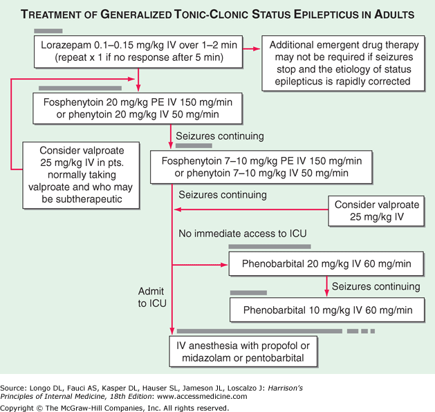
The duration of seizure activity sufficient to meet the definition of status epilepticus has traditionally been specified as 15–30 minutes. However, a more practical definition is to consider status epilepticus as a situation in which the duration of seizures prompts the acute use of anticonvulsant therapy. For GCSE, this is typically when seizures last beyond 5 minutes.

image

Status epilepticus is a medical emergency. Initial management includes maintenance of the airway and 50% dextrose (25–50 mL) intravenously in case hypoglycemia is responsible. If seizures continue, an intravenous bolus of lorazepam, 4 mg, is given and repeated once after 10 minutes if necessary; alternatively, 10 mg of diazepam is given intravenously over the course of 2 minutes, and the dose is repeated after 10 minutes if necessary. This is usually effective in halting seizures for a brief period but occasionally causes respiratory depression.

Regardless of the response to lorazepam or diazepam, phenytoin (18–20 mg/kg) is given intravenously at a rate of 50 mg/min; this provides initiation of long-term seizure control. The drug is best injected directly but can also be given in saline; it precipitates, however, if injected into glucose-containing solutions. Because arrhythmias may develop during rapid administration of phenytoin, electrocardiographic monitoring is prudent. Hypotension may complicate phenytoin administration, especially if diazepam has also been given. In many countries, injectable phenytoin has been replaced by fosphenytoin, which is rapidly and completely converted to phenytoin following intravenous administration. No dosing adjustments are necessary because fosphenytoin is expressed in terms of phenytoin equivalents (PE); fosphenytoin is less likely to cause reactions at the infusion site, can be given with all common intravenous solutions, and may be administered at a faster rate (150 mg PE/min). It is also more expensive.

If seizures continue, phenobarbital is then given in a loading dose of 10–20 mg/kg intravenously by slow or intermittent injection (50 mg/min). Respiratory depression and hypotension are common complications and should be anticipated; they may occur also with diazepam alone, although less commonly. If these measures fail, general anesthesia with ventilatory assistance and neuromuscular junction blockade may be required. Alternatively, intravenous midazolam may provide control of refractory status epilepticus; the suggested loading dose is 0.2 mg/kg, followed by 0.05–0.2 mg/kg/h.

After status epilepticus is controlled, an oral drug program for the long-term management of seizures is started, and investigations into the cause of the disorder are pursued.

Diazepam:-Sufficient cerebral levels are reached within one minute of a standard intravenous injection, and rectal administration produces peak levels at about 20 minutes. Diazepam is rapidly redistributed after acute administration, and thus has a relatively short duration of action. After repeated dosing, diazepam accumulates, resulting in higher peak levels, which persist. This can result in sudden and unexpected CNS depression and cardiorespiratory collapse. Diazepam is metabolised by hepatic microsomal enzymes. Respiratory depression, hypotension, and sedation are the principal side effects. Sudden apnoea can occur, especially after repeated injections or if the injection is administered at too fast a rate.

Bolus intravenous doses of diazepam should be given in an undiluted form at a rate not exceeding 25 mg/minute, using the Diazemuls formulation

Lorazepam:- has a lesser volume of distribution and is less lipid soluble than diazepam. Its pharmacokinetic characteristics result in a slower onset of action, but a longer duration of action. Lorazepam is indicated in the early stage of status epilepticus only, where its lack of accumulation in lipid stores, strong cerebral binding, and long duration of action due to its distribution half-life are very significant advantages over diazepam.

Initial injections of lorazepam are effective for about 12 hours (longer than with diazepam), but repeated doses are much less effective, and the drug has no place as long-term therapy.

Lorazepam is administered by intravenous bolus injection. As distribution is slow, the rate of injection is not critical. In adults, a bolus dose of 0.07 mg/kg (to a maximum of 4 mg) is given, and this can be repeated once after 20 minutes if no effect has been observed. In children under ten years, bolus doses of 0.1 mg/kg are recommended. Long-term infusion of lorazepam should not be used. It is usually available as a 1 ml ampoule containing 4 mg of lorazepam.

The initial infusion of phenytoin takes 2030 minutes in an adult, and the onset of action is slow. It is therefore often administered in conjunction with a short-acting drug with a rapid onset of action, such as diazepam

PHENYTOIN:-It is a highly effective anticonvulsant, with the particular advantage of a long duration of action.The usual phenytoin solutions have a pH of 12 and, if added to bags containing large volumes of fluid at lower than physiological pH (for example, 5% glucose), precipitation may occur in the bag or tubing; use in a solution of 0.9% sodium chloride (normal saline) (5-20 mg/ml) is safer. There is also a serious risk of precipitation if other drugs are added to the infusion solution.

The rate of infusion of phenytoin solution should not exceed 50 mg/minute, and it is prudent to reduce this to 2030 mg/minute in the elderly. The adult dose is 1520 mg/kg; this usually amounts to about 1000 mg and therefore takes at least 20 minutes to administer. Regrettably, a common and potentially serious mistake is to give a lower dose which results in suboptimal cerebral levels.

PHENOBARBITONE:-Phenobarbitone is a drug of choice for the treatment of established status epilepticus. It is highly effective, has a rapid onset of action, and prolonged anticonvulsant effects. It has stable and non-reactive physical properties, as well as convenient pharmacokinetics. Wide experience has been gained of its use in adults and in children, and few drugs are as well tried in the newborn. It has stronger anticonvulsant properties than most other barbiturates, and may be preferentially concentrated in metabolically active epileptic foci. As well as excellent anticonvulsant properties, it may also have cerebral-protective action. Acute tolerance to the antiepileptic effect is unusual, in contrast to the benzodiazepines and, once controlled, seizures do not tend to recur. Indeed, there is evidence to suggest that given with barbiturate anaesthesia, it can reduce the relapse rate with anaesthetic withdrawal.
The main disadvantages of phenobarbitone are its potential to cause sedation, respiratory depression, and hypotension; although in practice these effects seem slight except at high levels or with rapidly rising levels, its safety at even high doses is well established. The well-known chronic side effects of phenobarbitone in long-term therapy are of little relevance in the emergency situation of status epilepticus. The drug is eliminated slowly and, although this is of no importance on initial phenobarbitone loading, on prolonged therapy there is a danger of accumulation and blood level monitoring is essential. In the newborn period dosing is more difficult than in adults, as the pharmacokinetics change rapidly during the first weeks and months of life. The drug has a strong tendency to autoinduction. Phenobarbitone is a stable preparation, which does not easily decompose, and the drug is not absorbed by plastic. It should not be used in a solution containing other drugs (for example, phenytoin), as this may result in precipitation.
The usual recommended adult intravenous loading dose of phenobarbitone is 10 mg/kg (doses of up to 20 mg/kg have been used and recommended), given at a rate of 100 mg/minute (i.e. a total of about 700 mg in seven minutes). This should be followed by daily maintenance doses of 1-4 mg/kg. In neonates, initial phenobarbitone loading doses of between 12 and 20 mg/kg have been recommended to produce therapeutic levels, with subsequent supplementation of 3-4 mg/kg per day, to a maximum dose of 40 mg/kg. In older children, loading doses of between 5 and 20 mg/kg are recommended and maintenance doses of 1-4 mg/kg, although much higher doses have been safely given. After loading, maintenance doses can be given by the oral, intravenous, or intramuscular route. Phenobarbitone is usually presented in 1 ml ampoules containing 200 mg of phenobarbitone sodium.


Which of the following is not an adverse effect of
phenytoin – (AI 90)
a) Gum hyperplasia
b) Osteomalacia
c) Gynecomastia
d) Megaloblastic anaemia

In epilepsy, EEG is – (Kerala 90)
a) Diagnostic
b) Useless in diagnosis
c) Complimentary in diagnosis
d) may or may not be useful

Investigation of choice to diagnose epilepsy is –
a) MRI b) EEG (Kerala 94)
c) CT scan d) Angiogram

EEG with spike and dome pattern is characteristic
of…. Epilepsy – (AIIMS 85)
a) Jacksonian b) Grandmal
c) Petitmal d) Temporal lobe

Drug of choice for psychomotor epilepsy is

a) Valproic acid b) Carbamazepine (AIIMS 87) c) Ethosuximide d) Barbiturate
e) Phenytoin

Temporal lobe epilepsy was first recognized in 1881 by John Hughlings Jackson, who described "uncinate fits" seizures arising from the uncal part of temporal lobe and the "dreamy state."

In the 1940s, Gibbs et al introduced the term psychomotor epilepsy.The international classification of epileptic seizures (1981) replaced the term psychomotor seizures with complex partial seizures. Complex means that there is some alteration of consciouness, versus a simple partial seizure, which means there is no alteration of consciouness. The ILAE classification of the epilepsies uses the term temporal lobe epilepsy and divides the etiologies into cryptogenic (presumed unidentified etiology), idiopathic (genetic), and symptomatic (cause known, eg, tumor).

Carbamazepine (or a related drug, oxcarbazepine), lamotrigine and phenytoin are currently the drugs of choice approved for the initial treatment of focal seizures, including those that evolve into generalized seizures. Overall they have very similar efficacy, but differences in pharmacokinetics and toxicity are the main determinants for use in a given patient. For example, an advantage of carbamazepine (which is also available in an extended-release form) is that its metabolism follows first-order pharmacokinetics, and the relationship between drug dose, serum levels, and toxicity is linear. Carbamazepine can cause leukopenia, aplastic anemia, or hepatotoxicity and would therefore be contraindicated in patients with predispositions to these problems. Oxcarbazepine has the advantage of being metabolized in a way that avoids an intermediate metabolite associated with some of the side effects of carbamazepine. Oxcarbazepine also has fewer drug interactions than carbamazepine. Lamotrigine tends to be well tolerated in terms of side effects. However, patients need to be particularly vigilant about the possibility of a skin rash during the initiation of therapy. This can be extremely severe and lead to Stevens-Johnson syndrome if unrecognized and if the medication is not discontinued immediately. This risk can be reduced by slow introduction and titration.

Psychomotor epilepsy is Temporal lobe epilepsy

True about juvenile mycoclonic epilepsy – (PGI 03)
a) Focal seizure
b) Generalised seizure
c) Myoclonus
d) Responses to Sodium Valproate
e) Spike and waves in EEG

All the following are indications for brain imaging
in epilepsy, except – (J & k 05)
a) Epilepsy starts after the age of 5 years
b) Seizures have focal features clinical
c) EEG shows a focal seizure source
d) Control of seizures is difficult

Gustatory hallucinations are most commonly
associated with – (Corned 08)
a) Temporal lobe epilepsy
b) Grand mal epilepsy
c) Anxiety disorders
d) Tobacco dependence

Cause of death which is common in case of coma –
a) Tongue falling back and obstructing airway
b) Secretions obstructing airway (TN 03)
c) Cardiac arrest
d) Epilepsy

1501. Which vitamin deficiency is most commonly seen in a pregnant mother who is on phenytion
therapy for Epilepsy? All India 2006
a) Vitamin B6 b) Vitamin B12 c) Vitamin A d) Folic Acid

In Petit Mal epilepsy all findings are seen except JIPMER ’98
a) Transient loss of conciousness b) Non specific ECG pattern
c) No tonic-clonic phase d) Ethosuccamide is Drug of choice

A 15 year old boy with epilepsy on treatment with
combination of valproate and phenytoin has good
control of seizures. Levels of both drugs are in the
therapeutic range. All of the following adverse effects
can be attributed to valproate except :
A. Weight gain of 5 kg
B. Serum alanine aminotransaminase 150 IU/L
C. Rise in serum ammonia level by 201.tg/dL
D. Lymphadenopathy

Which of the following statements is incorrect in
relation to pregnant women with epilepsy?
A. The rate of congenital malformation is increased
in the offspring of women with epilepsy.
B. Seizure frequency increases in approximately
70% of women.
C. Breast feeding is safe with most anticonvulsants.
D. Folic acid supplementation may reduce the risk of
neural tube defect

An elderly male on ventilator has received atracurium
infusion for 3 days. He now develops epileptic fits.
Probable cause for his epilepsy is:
A. Allergy to drug
B. Accumulation of Atracurium
C. Accumulation of Laudanosine
D. Ventilator failure

Palatal Myoclonus is seen in – (SGPGI 05)
a) Epilepsy
b) Multiple sclerosis
c) Cerebellar infarction
d) Guillain Barre syndrome

Deja Vu is seen in – (Kerala 94)
a) Normal persons b) Temporal lobe epilepsy
c) Psychosis d) All of the above

Arun, diagnosed to have epilepsy recently and put
on phenytoin. He was previously on
antidepressents. He has developed fatigue and
anorexia. His Hb=8.0; TC=7500 ; ESR=30 in first
hour; SGOT=35; SGPT=35 and B-=0.6. the test
for diagnosis would be – (AIDE 99)
a) Chest x-ray b) Urine culture
•c) MCV d) Blood culture

All are used in treatment of epilepsy excepta)
Ethosuximide b) Reserpine (Kerala 94)
c) Acetazolamide d) Vigabatrine

Carbamazepine is useful in the following
conditions EXCEPT- (Kann 95)
a) Grandmal epilepsy b) Petitmal epilepsy
c) Psychomotor epilepsy d) Trigeminal neuralgia

Which is true regarding febrile seizures- (AI 93)
a) 50% recurrance
b) Long term phenytoin required
c) Interictal EEG normal
d) Status epilepsy is common

Hypsarrythmia in a child is due to

a) Grandmal epilepsy (AIIMS 91, Delhi 93)
b) Petitmal epilepsy
c) Myoclonic epilepsy
d) Reflex epilepsy

All of the following are true of febrile seizures except

a) Most commonly seen between 9 months and 5 years (AP 97)
b) Does not last more than 10 minutes
c) Almost invariably develop into epilepsy
d) Prognosis is good

WhIch of the following has the worst
prognosis – (JIPMER 2001)
a) Rolandic epilepsy b) Versive epilepsy
c) Absence epilepsy d) Infantile spasm

Which one of the following in the characteristic
feature of juvenile myoclonic epilepsy ? (AIIMS 06)
a) Myoclonic seizures frequently occur in morning
b) Complete remission is common
c) Response to anticonvulsants is poor
d) Associated absence seizures are present in
majority of patients

A 18-month old baby presents with recurrent
episodes of excessive crying followed by cyanosis,
uncon sciousness and occassional seizures since 9
months of age. The most likely diagnosis
is – (UPSC 98)
a) Epilepsy b) Anoxic spells
c) Breath holding spells d) Vasovagal attack

Posted in Medical | 10 Comments »

Central Nervous System I MCQs

Posted by Dr KAMAL DEEP on June 4, 2012

Action potentials normally are generated by the opening of sodium channels and the inward movement of sodium ions down the intracellular concentration gradient. Depolarization of the neuronal membrane opens potassium channels, resulting in outward movement of potassium ions, repolarization, closure of the sodium channel, and hyperpolarization. Sodium or potassium channel subunit genes have long been considered candidate disease genes in inherited epilepsy syndromes, and recently such mutations were identified. These mutations appear to alter the normal gating function of these channels, increasing the inherent excitability of neuronal membranes in regions where the abnormal channels are expressed.

All of the following are neurologic channelopathies
except – (AI 05)
a) Hypokalemic periodic paralysis
b) Episodic ataxia type 1
c) Familial hemiplegic migraine
d) Spinocerebellar ataxia 1

Ans is ‘d’ i.e., Sphinocerebellar Ataxia -1 [Ref Harrison 16 1"/e p. 2339 & 15’h/e p. 2321]

All of the following are calcium channelopathies,
except – (AIIMS May 05)
a) Episodic ataxia -1
b) Spinocerebellar ataxia-6
c) Familial hemiplegic migraine
d) Hypokalemic periodic paralysis

Episode weakness is due to – (PG1 87)
a) Hypokalaemia b) Dermatomyositis
c) Paramyotonia congenita d) Myasthenia gravis
e) Hypophosphatemia

Cataract is associated with PGI 88
a) Pseudo muscular hypertrophy b) myotonia congenita c) myotonic dystrophy d) SLE

In a patient, muscle cramps on exercise, +ve
myoglobulinemia, the disorder is – (PGI 98)
a) Pompe’s disease b) Myotonia congenita
c) Myotonic dystrophy d) Mc ardle’s disease

Table 366-1 Examples of Neurologic Channelopathies


Category Disorder Channel Type Mutated Gene Chap. Ref.
Genetic
Ataxias

Episodicataxia-1

Episodicataxia-2

Spinocerebellar ataxia-6

K

Ca

Ca

KCNA1

CACNL1A

CACNL1A

373
Migraine

Familial hemiplegic migraine 1

Familial hemiplegic migraine 3

Ca

Na

CACNL1A

SCN1A

14
Epilepsy

Benign neonatal familial convulsions

Generalized epilepsy with febrile convulsions plus

K

Na

KCNQ2, KCNQ3 SCN1B 369
Periodic paralysis

Hyperkalemic periodic paralysis

Hypokalemic periodic paralysis

Na

Ca

SCN4A

CACNL1A3

387
Myotonia

Myotonia congenita

Paramyotonia congenita

Cl

Na

CLCN1

SCN4A

387
Deafness Jervell and Lange-Nielsen syndrome (deafness, prolonged QT interval, and arrhythmia) K KCNQ1, KCNE1 30
  Autosomal dominant progressive deafness K KCNQ4  
Autoimmune
Paraneoplastic

Limbic encephalitis

Acquired neuromyotonia

Cerebellar ataxia

Lambert-Eaton syndrome

Kv1

Kv1

Ca (P/Q type)

Ca (P/Q type)

101

101

101

101


Whereas the specific clinical manifestations of channelopathies are quite variable, one common feature is that manifestations tend to be intermittent or paroxysmal, as occurs in epilepsy, migraine, ataxia, myotonia, or periodic paralysis. Exceptions are clinically progressive channel disorders such as autosomal dominant hearing impairment.

Posted in Medical | 1 Comment »

Dermatology (Skin) MCQs

Posted by Dr KAMAL DEEP on December 26, 2011

Capture

Skin is the largest organ in the body. In a 70-kg individual, the skin weighs over 5 kg and covers a surface area approaching 2 m2. Human skin consists of a stratifi ed, cellular epidermis and an underlying dermis of connective tissue.Beneath the dermis is a layer of subcutaneous fat, which is separated from the rest of the body by a vestigial layer of striated muscle. The epidermis is mainly composed of keratinocytes and is typically 0.05–0.1 mm in thickness. It is formed by division of cells in the basal layer which give rise to the spinous layer. This layer contains cells that move outwards and progressively differentiate, forming the granular layer and the stratum corneum. The cellular progression from the basal layer to the skin surface takes about 30 days but is accelerated in diseases such as psoriasis. The ‘bricklike’ shape of keratinocytes is provided by a cytoskeleton made of keratin intermediate fi laments. As the epidermis differentiates, the keratinocytes become fl attened. This process involves the fi lament aggregating protein, filaggrin, a protein component of keratohyalin granules. Indeed, keratin and fi laggrin comprise 80–90% of the mass of the epidermis. The outermost layer of the epidermis is the stratum corneum, where cells (now called corneocytes) have lost nuclei and cytoplasmic organelles. The corneocyte has a highly insoluble, cornified envelope within the plasma membrane, formed by cross-linking of soluble protein precursors, including involucrin and loricrin, the latter contributing 70–85% to the mass of the cornifi ed cell envelope; it also contains several lipids (fatty acids, sterols and ceramides) released from lamellar bodies within the upper, living epidermis. Other cells in the epidermis are the melanocytes, Langerhans’ cells and Merkel cells. Melanocytes are dendritic cells that distribute packages of melanin pigment in melanosomes to surrounding keratinocytes to give skin its colour. The number of melanocytes does not differ much between white and black skin. Rather it is the nature of the melanin and the size of the melanosomes that account for the different appearances. The Langerhans’ cells are also dendritic in nature, although these are of mesenchymal origin and originate from bone marrow. Langerhans’ cells are antigen-presenting cells and process antigens encountered by the skin to local lymph nodes and thus have a key role in adaptive immune responses in the skin. Merkel cells are probably derived from keratinocytes. They have a role as mechanosensory receptors in response to touch. Human skin contains pilosebaceous follicles and sweat glands. The hair follicles comprise pockets of epithelium that are continuous with the superficial epidermis but which also envelop a small papilla of dermis at their base. A bundle of smooth muscle, the arrector pili, extends at an angle between the surface of the dermis and a point in the follicle wall. Above the insertion, there are holocrine sebaceous glands which open into the pilary canal. In some sites, such as the axillae, the follicles may be associated with apocrine glands. Also derived from the epidermis and opening directly to the skin surface are the eccrine sweat glands. The epidermis is attached to the dermis via a complex network of proteins and glycoproteins that extend from inside basal keratinocytes into the superficial dermis. Besides adhesion, the dermal–epidermal junction components also contribute to cell migration (for example during wound healing) as well as epithelial– mesenchymal signalling events. Over 30 different macromolecules (collagens, laminins, integrins) interact within a basement membrane zone that is less than 200 μm across. The dermis is a supporting matrix or ground substance in which polysaccharides and proteins are linked to produce macromolecules that have a remarkable capacity for retaining water. The thickness of the dermis varies from less than 0.5 mm to more than 5 mm. There are two principal types of protein fibre: collagen and elastic tissue. Collagen is the major extracellular matrix protein comprising 80–85% of the dry weight of the dermis. Twenty-nine different collagens have been identified in vertebrate tissue (depicted by Roman numerals in the order of their discovery, from I to XXIX), of which at least 12 are expressed in skin. The main interstitial dermal collagens are types I and III whereas the principal basement membrane collagen (at the dermal–epidermal junction and around dermal blood vessels, nerves and appendages) is type IV collagen. Triple-helical collagen monomers polymerize into fibrils and fibres, which then become stabilized by the complex formation of both intra- and intermolecular crosslinks. Collagen fibres are extremely tough and provide skin with its tensile strength. Elastic fibres account for no more than 2–4% of the extracellular matrix in the dermis and consist of two components, elastin and elastin-associated microfibrils, which together give skin its elasticity and resilience. Elastic microfibrils are composed of several proteins, including fibrillin, which surround the elastin and which can extend throughout the dermis in a web-like configuration to the junction between the dermis and the epidermis. The dermis also contains a number of non-collagenous glycoproteins including fibronectins, fibulins and integrins. These extracellular matrix components facilitate cell adhesion and cell motility. Between the dermal collagen and elastic tissue is the ground substance made up of glycosaminoglycan/proteoglycan macromolecules. These contribute only 0.1–0.3% of the total dry weight of the dermis but provide a vital role by maintaining hydration, mostly due to the high water-binding capacity of hyaluronic acid. About 60% of the total weight of the dermis is water. The dermis has a very rich blood supply, although no vessels pass through the dermal–epidermal junction. There is a superficial and a deep vascular plexus. The motor innervation of the skin is autonomic, and includes a cholinergic component to the eccrine sweat glands and adrenergic components to both the eccrine and apocrine glands, to the smooth muscle and the arterioles and to the arrector pili muscle. The sensory nerve endings are of several kinds; some are free, some terminate in hair follicles and others have expanded tips.

Human sweat glands are generally divided into two types: apocrine and eccrine.

The eccrine gland is the primary gland responsible for thermoregulatory sweating in humans. Eccrine sweat glands are distributed over nearly the entire body surface.Acetylcholine is the primary neurotransmitter released from cholinergic sudomotor nerves and binds to muscarinic receptors on the eccrine sweat gland, although sweating can also occur via exogenous administration of α- or β-adrenergic agonists.

Mental or emotional sweating usually appears on the palms and soles.

Apart from eccrine glands, the skin also contains apocrine sweat glands. Eccrine glands do not show cytological changes during secretion whereas apocrine glands are characterized by decapitation secretion, in which part of the cell is pinched off and released into the lumen. Apocrine glands are located only in genital,axillary and mammary areas, where they are always connected to a hair follicle. Apocrine glands have a low secretory output, and hence no significant role in thermoregulation.

A flat discolouration on skin  1 cm is called
a) Macule b) Plaque c) Boil d) Papule e) Wheal

Table 51-1 Description of Primary Skin Lesions


Macule: A flat, colored lesion, <2 cm in diameter, not raised above the surface of the surrounding skin. A "freckle," or ephelid, is a prototype pigmented macule.
Patch: A large (>2 cm) flat lesion with a color different from the surrounding skin. This differs from a macule only in size.
Papule: A small, solid lesion, <0.5 cm in diameter, raised above the surface of the surrounding skin and, hence, palpable (e.g., a closed comedone, or whitehead, in acne).
Nodule: A larger (0.5–5.0 cm), firm lesion raised above the surface of the surrounding skin. This differs from a papule only in size (e.g., a dermal nevomelanocytic nevus).
Tumor: A solid, raised growth >5 cm in diameter.
Plaque: A large (>1 cm), flat-topped, raised lesion; edges may either be distinct (e.g., in psoriasis) or gradually blend with surrounding skin (e.g., in eczematous dermatitis).
Vesicle: A small, fluid-filled lesion, <0.5 cm in diameter, raised above the plane of surrounding skin. Fluid is often visible, and the lesions are translucent [e.g., vesicles in allergic contact dermatitis caused by Toxicodendron (poison ivy)].
Pustule: A vesicle filled with leukocytes. Note: The presence of pustules does not necessarily signify the existence of an infection.
Bulla: A fluid-filled, raised, often translucent lesion >0.5 cm in diameter.
Wheal: A raised, erythematous, edematous papule or plaque, usually representing short-lived vasodilatation and vasopermeability.
Telangiectasia: A dilated, superficial blood vessel.

Fluid filled in epidermis and dermis is called
a) Wheal b) Macule c) Papule d) Vesicle

A vesicle is a circumscribed, elevated lesion that contains fluid . Often the vesicle walls are so thin that they are translucent and the serum, lymph, blood, or extracellular fluid is visible. A vesicle with a diameter greater than 0.5 cm is a bulla. Vesicles and bullae arise from cleavage at various levels of the skin; the cleavage may be within the epidermis (i.e., intraepidermal vesication), or at or below the dermal-epidermal interface (i.e., subepidermal). Cleavage just beneath the stratum corneum produces a subcorneal vesicle or bulla , as in impetigo. Intraepidermal vesication may result from intercellular edema (spongiosis), as characteristically seen in delayed hypersensitivity reactions of the epidermis (e.g., in contact eczematous dermatitis) and in pompholyx. Loss of intercellular bridges, or desmosomes, is known as acantholysis, and this type of intraepidermal vesication  is seen in pemphigus vulgaris, where the cleavage is usually just above the basal layer. In pemphigus foliaceus, the cleavage occurs just below the subcorneal layer. Viruses cause a curious “ballooning degeneration” of epidermal cells , as in herpes zoster, herpes simplex, variola, and varicella. Viral bullae often have a depressed (“umbilicated”) center. Pathologic changes at the dermal-epidermal junction may lead to subepidermal vesicles and bullae , as are seen in pemphigoid, bullous erythema multiforme, porphyria cutanea tarda, dermatitis herpetiformis, and some forms of epidermolysis bullosa. The thickness of the wall of a bulla may be estimated by its translucency and flaccidity. The amount of pressure required to collapse the lesion may help predict whether the bulla is intraepidermal or subepidermal. It has been said that a relatively large, tense bulla suggests pemphigoid, whereas a flaccid bulla suggests pemphigus. There is, however, no reliable means of distinguishing these two diseases except by histologic examination of the lesion and immunofluorescence.

A 7yr. old child presenting with recurrent dry scaly macules (white, small) on face has
a) Indeterminate Leprosy b) Post dermal leishmaniasis
c) Pitryasis alba d) Early vitiligo

Capture

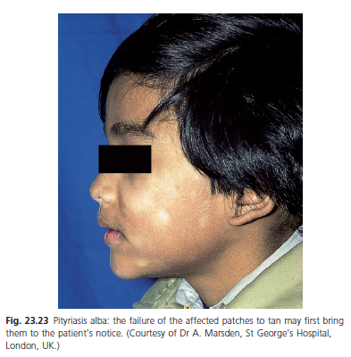
                                                                                                                        Pityriasis alba

Pityriasis alba is a common finding (5% of children) that is probably more usual in patients with the atopic diathesis .The condition appears in most instances before puberty. The face, neck, and arms are the most common sites. The lesions begin as a nonspecific erythema and gradually become scaly and hypopigmented. The hypopigmentation is transient and caused by mild dermal inflammation and the UV screening effect of the scaly skin. The condition gradually improves after puberty. Treatment consists of lubrication. Mild inflammation responds to group V topical steroids, but the degree of pigmentation is not affected by any treatment.
The condition is often confused with vitiligo and tinea versicolor. Vitiligo does not scale. The potassium hydroxide preparation is positive in tinea versicolor.
The age incidence, the fine scaling and the distribution of the lesions usually suggest the diagnosis. Conspicuous hypopigmentation may lead to a misdiagnosis of vitiligo. Naevus de pigmentosus most commonly presents at birth or before 3 years of age, and most often causes single, well-marginated lesions on the trunk .However, this condition may be difficult to distinguish from pityriasis alba when it occurs on the face and in cases of later onset. Discoid eczema in an atopic child is intensely pruritic, and the lesions are larger and more oedematous. In older children and adults, the lesions on the trunk, during their early erythematous phase, may be mistaken for psoriasis but the distribution and the relatively mild scaling should exclude this diagnosis. Mycosis fungoides, although relatively rare, may present with lesions clinically resembling pityriasis alba .This condition may also be difficult to distinguish histologically, so follow-up and repeat biopsy are sometimes required.

The test likely to help in diagnosis of a patient who presents with an itchy annular plaque on the face is:
A.Gram’s stain
B.Potassium hydroxide mount
C Tissue smear
D. Wood’s lamp examination

Woods lamp has a frequency of TN 04
a) 250 nm b) 300 nm c) 320 nm d) 360

The UV spectrum reaching the earth represents <10% of total incident solar energy and is arbitrarily divided into two major segments, UV-B and UV-A, constituting the wavelengths from 290–400 nm. UV-B consists of wavelengths between 290 and 320 nm. This portion of the photobiologic action spectrum is the most efficient in producing redness or erythema in human skin and hence sometimes is known as the "sunburn spectrum." UV-A includes wavelengths between 320 and 400 nm and is ~1000-fold less efficient in producing skin redness than is UV-B.

The wavelengths between 400 and 700 nm are visible to the human eye. The photon energy in the visible spectrum is not capable of damaging human skin in the absence of a photosensitizing chemical.

Uses of woods light includes
a) Urine examination in phorphyria b) examination of hair in T. capitis c) Sclerema d) all

A Wood’s lamp generates 360-nm ultraviolet (or "black") light that can be used to aid the evaluation of certain skin disorders. For example,

1. A Wood’s lamp will cause erythrasma (a superficial, intertriginous infection caused by Corynebacterium minutissimum) to show a characteristic coral pink color,

2. Wounds colonized by Pseudomonas to appear pale blue.

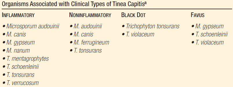
3. Tinea capitis caused by certain dermatophytes such as Microsporum canis or M. audouini exhibits a yellow fluorescence. Pigmented lesions of the epidermis such as freckles are accentuated, while dermal pigment such as postinflammatory hyperpigmentation fades under a Wood’s light.

4.Vitiligo appears totally white under a Wood’s lamp, and previously unsuspected areas of involvement often become apparent.

5.A Wood’s lamp may also aid in the demonstration of tinea versicolor and in recognition of ash leaf spots in patients with tuberous sclerosis.

A pinkish red fluorescence of urine with wood’s Lamp is seen in
a) Lead poisoning b) Porphyria cutanea tarda c) Erythromelagia d) Acrocyanosis

The wood’s lamp filter is made of AIIMS 79, PGI 80
a) Tin and chromium oxide b) Nickel oxide and silica c) Copper oxide Barium
oxide d) Zinc oxide

Coral red-fluorescence wood’s Lamp seen in

a) Porphyria cutanea tarda b) Erythrasma Livedo-reticularis
d) Hypomelanosis

Wood’s lamp (longwave ultraviolet light, “black” light) is essential for the clinical diagnosis of certain skin and hair diseases and of porphyria. Longwave ultraviolet radiation is obtained by fitting a high-pressure mercury lamp with a specially compounded filter made of nickel oxide and silica (Wood’s filter); this filter is opaque to all light except for a band between 320 and 400 nm. When using the Wood’s lamp, it is essential for the examiner to become dark-adapted in order to see the contrasts clearly. When the ultraviolet waves emitted by Wood’s lamp impinge on the skin, a visible fluorescence occurs. Wood’s lamp is particularly useful in the detection of the fluorescence of dermatophytosis ( Microsporum) in the hair shaft (green) and of erythrasma (coral red) on the skin. Wood’s lamp also helps to estimate the variation in the “whiteness” of lesions in relation to the normal skin color, in dark-skinned and especially in fair-skinned persons; for example, the lesions seen in hypomelanotic macules in tuberous sclerosis and in tinea versicolor are not as white as the macules present in vitiligo, which are typically amelanotic. Circumscribed hypermelanosis, such as ephelides and melasma, is much more evident under Wood’s lamp, and in lentigo maligna melanoma and acrolentiginous melanoma the Wood’s lamp can be used to detect the total extent of the lesion as a guide to total excision. Melanin in the dermis, as in a Mongolian sacral spot, does not become accentuated under Wood’s lamp. Therefore, it is possible to localize the site of melanin (epidermal or dermal) by use of the Wood’s lamp; this phenomenon is not evident in patients with brown or black skin. The technique is as follows: a grading (minimal, moderate, marked) of the degree of pigmentation is made with visible light and compared with a grading of the degree of color change when examined with Wood’s lamp. In epidermal melanin pigmentation the pigment grade increases from minimal to marked, but dermal melanin has the same degree of pigment in both visible light and Wood’s lamp illumination.

Patients with PCT excrete increased amounts of porphyrins in the urine, which rarely may exhibit characteristic pink-red fluorescence when examined with a Wood’s lamp.

Commonest site of Atopic dermatitis is
a) Scalp b) Elbow c) Trunk d) Ante cubital fossa

Atopic dermatitis can be best diagnosed by
a) Clinical evaluation b) Patch test c) IgE levei d)Skin Biopsy

‘Itch is the disease’ is true for

a) Atopic dermatitis b) Insect bites c) Seborrheic dermatitis d)Tinea cruris

Kaposis variceliform eruption PGI 2004
a) Atopic dermatitis b) Dermatitis herpetiformis
c) Lymphoma d) All


Kaposi varicelliform eruption (KVE) is the name given to a distinct cutaneous eruption caused by herpes simplex virus (HSV) type 1, HSV-2, coxsackievirus A16, or vaccinia virus that infects a preexisting dermatosis. Most commonly, it is caused by a disseminated HSV infection in patients with atopic dermatitis (AD) and, for this reason, is often referred to as eczema herpeticum (EH).

Kaposi varicelliform eruption (KVE) is caused primarily by HSV-1, but can also be caused by HSV-2, coxsackievirus A16, or vaccinia virus infecting a preexisting dermatosis. Most commonly, it is caused by a disseminated HSV infection in patients with AD. For this reason, it is also referred to as EH.

KVE has also been associated with the following:

  • Pityriasis rubra pilaris

  • Neurodermatitis

  • Irritant contact dermatitis

  • Congenital ichthyosiform erythroderma

  • Ichthyosis vulgaris

  • Pemphigus foliaceus

  • Benign familial pemphigus (Hailey-Hailey disease)

  • Darier disease

  • Wiskott-Aldrich syndrome

  • Sézary syndrome

  • Seborrheic dermatitis

  • Skin grafts

  • Burns

  • Cowpox

  • Cutaneous T-cell lymphomas

  • Rosacea

    A 25 — year old presents with recurrent episodes of flexural eczema, contact urticaria, recurrent skin infections and severe abdominal cramps and diarrhea upon taking sea foods. He is suffering from All India 04
    a) Seborrheic dermatitis
    b) Atopic dermatitis
    c) Airborne contact dermatitis d) Nummular dermatitis

    Personal history of food allergy makes diagnosis of atopy more than contact dermatitis.


    Air-borne contact dermatitis can be diagnosed by:
    a) Skin biopsy. b) Patch test. c) Prick test. d) Estimation of serum IgE levels

    Contact dermatitis is a type of Hypersensitivity
    a) Type I b) Type II c) Type III d) Type IV

    Berloque dermatitis is due to contact with: a) Metals b) Cosmetics c) Food d) Plants

    Commonest cause of air borne contact dematitis is
    a) Crysanthemum b) Parthenium c) Calotropis d) Bourgainvilia

    Type I hypersensitivity is characterized by an allergic reaction occurring immediately following an individual’s second contact with the responsible antigen (the allergen). Upon initial exposure to a soluble allergen, B cells are stimulated to differentiate into plasma cells and produce specific IgE with the help of T cells (figure 33.2). This IgE is sometimes called a reagin, and the individual has a hereditary predisposition for its production. Once synthesized, IgE binds to the Fc receptors of mast cells (basophils and eosinophils can also be activated) and sensitizes these cells, making the individual allergic to the allergen. When a second exposure to the allergen occurs, the allergen attaches to the surfacebound IgE on the sensitized mast cells causing degranulation. Degranulation releases physiological mediators such as histamine, leukotrienes, heparin, prostaglandins, PAF (platelet-activation factor), ECF-A (eosinophil chemotactic factor of anaphylaxis), and proteolytic enzymes. These mediators trigger smooth muscle contractions, vasodilation, increased vascular permeability, and mucous secretion. The inclusive term for these responses is anaphylaxis [Greek ana, up, back, again; and phylaxis, protection]. Anaphylaxis can be divided into systemic and localized reactions. Systemic anaphylaxis is a generalized response that occurs when an individual sensitized to an allergen receives a subsequent exposure to it. The reaction is immediate due to the large amount of mast cell mediators released over a short period. Usually there is respiratory impairment caused by smooth muscle constriction in the bronchioles. The arterioles dilate, which greatly reduces arterial blood pressure and increases capillary permeability with rapid loss of fluid into the tissue spaces. Because of these reactions the individual can die within a few minutes from reduced venous return, asphyxiation, reduced blood pressure, and circulatory shock. Common examples of allergens that can produce systemic anaphylaxis include drugs (penicillin), passively administered antisera, peanuts, and insect venom from the stings or bites of wasps, hornets, or bees (figure 33.3). Localized anaphylaxis is called an atopic (“out of place”) allergy. The symptoms that develop depend primarily on the route by which the allergen enters the body. Hay fever (allergic rhinitis) is a good example of an atopic allergy involving the upper respiratory tract. Initial exposure involves airborne allergens—such as plant pollen, fungal spores, animal dander, and house dust mites—that sensitize mast cells located within the mucous membranes. Reexposure to the allergen causes the typical localized anaphylactic response.

    Once the responsible allergen has been identified, the individual should avoid contact with it. At times this is not possible, and desensitization is warranted. This procedure consists of a series of allergen doses injected beneath the skin to stimulate the production of IgG antibodies rather than IgE antibodies. The circulating IgG antibodies can then act as blocking antibodies to intercept and neutralize allergens before they have time to react with mast cell-bound IgE. Recent evidence suggests that suppressor T-cell activity also may cause a decrease in IgE synthesis. Desensitizations are about 65 to 75% effective in individuals whose allergies are caused by inhaled allergens

    Type II hypersensitivity is generally called a cytolytic or cytotoxic reaction because it results in the destruction of host cells either by lysis or toxic mediators. In type II hypersensitivity, IgG or IgM antibodies are directed against cell surface or tissue-associated antigens. They usually stimulate the complement pathway and a variety of effector cells (figure 33.5). The antibodies interact with complement (Clq) and the effector cells through their Fc regions. The damage mechanisms are a reflection of the normal physiological processes involved in interaction of the immune system with pathogens. A classic example of type II hypersensitivity is that resulting when a person receives a transfusion with blood from a donor with a different blood group.


    Type III hypersensitivity involves the formation of immune complexes (figure 33.6a). Normally these complexes are removed effectively by the fixed monocytes and macrophages of the monocyte-macrophage system. In the presence of excess amounts of some soluble antigens, the antigen-antibody complexes may not be efficiently removed. Their accumulation can lead to a hypersensitivity reaction from complement that triggers a variety of inflammatory processes. This inflammation causes damage, especially of blood vessels (vasculitis; figure 33.6b), kidney glomerular basement membranes (glomerulonephritis), joints (arthritis), and skin. Diseases resulting from type III reactions can be placed into three groups. First, a persistent viral, bacterial, or protozoan infection, together with a weak antibody response, leads to chronic immune complex formation and eventual deposition of the complex in host tissues. Second, the continued production of autoantibody to self-antigen during an autoimmune disease can lead to prolonged immune complex formation. This overloads the monocyte-macrophage system, and tissue deposition of the complexes occurs (e.g., in the disease systemic lupus erythematosus). Third, immune complexes can form at body surfaces (such as the lungs), following repeated inhalation of allergens from molds, plants, or animals. For example, in Farmer’s lung disease, an individual has circulating antibodies to fungi after being exposed repeatedly to moldy hay. These antibodies are primarily IgG. When the allergens (fungal spores) enter the alveoli of the lungs, local immune complexes form, leading to inflammation. Some group A streptococcal infections can produce an immunologically mediated acute glomerulonephritis. Although the mechanism is not completely understood, it is believed that complexes of antibody and streptococcal antigen are deposited within the kidney glomeruli and generate a type III hypersensitivity.

    Type IV hypersensitivity involves delayed T-cell-mediated immune reactions. A major factor in the type IV reaction is the time required for a special subset of TH1 cells (often called delayedtype hypersensitivity [TDTH] cells) to migrate to and accumulate near the antigens. This usually takes a day or more. Type IV reactions occur when antigens, especially those binding to tissue cells, are phagocytosed by macrophages and then presented to receptors on the TH1 cell surface in the context of class I MHC. Contact between the antigen and TH1 cell causes the cell to proliferate and release cytokines. Cytokines attract lymphocytes, macrophages, and basophils to the affected tissue. Extensive tissue damage may result. Examples of type IV hypersensitivities include tuberculin hypersensitivity (the TB skin test), allergic contact dermatitis, some autoimmune diseases, transplantation rejection, and killing of cancer cells. In tuberculin hypersensitivity a partially purified protein called tuberculin, which is obtained from the bacillus that causes tuberculosis (Mycobacterium tuberculosis; see section 39.1), is injected into the skin of the forearm (figure 33.7a). The response in a tuberculin-positive individual begins in about 8 hours, and a reddened area surrounding the injection site becomes indurated (firm and hard) within 12 to 24 hours. The TH1 cells that migrated into the injection site are responsible for the induration. The reaction reaches its peak in 48 hours and then subsides. The size of the induration is directly related to the amount of antigen that was introduced and to the degree of hypersensitivity of the tested individual. Other microbial products used in type IV skin testing are histoplasmin for histoplasmosis, coccidioidin for coccidioidomycosis, lepromin for leprosy, and brucellergen for brucellosis. Allergic contact dermatitis is caused by haptens (see figure 32.4) that combine with proteins in the skin to form the allergen that elicits the immune response. The haptens are the antigenic determinants, and the skin proteins are the carrier molecules for the haptens. Examples of these haptens include cosmetics, plant materials (catechol molecules from poison ivy and poison oak; figure 33.7b), topical chemotherapeutic agents, metals, and jewelry (especially jewelry containing nickel). Several important chronic diseases involve cell and tissue destruction by type IV hypersensitivity reactions. These diseases are caused by viruses, mycobacteria, protozoa, and fungi that produce chronic infections in which the macrophages and T cells are continually stimulated. Examples are leprosy, tuberculosis, leishmaniasis, candidiasis, and herpes simplex lesions.

  • The name sandworm eruption refers to Kerala 04
    a) Cercarial dermatitis
    b) Creeping eruption
    c) Migratory myiasis d) Guinea worm disease

  • Cutaneous larva migrans (abbreviated CLM) is a skin disease in humans, caused by the larvae of various nematode parasites of the hookworm family (Ancylostomatidae). The most common species causing this disease in the Americas is Ancylostoma braziliense.

    Colloquially called creeping eruption due to the way it looks, the disease is also somewhat ambiguously known as "ground itch" or (in some parts of the Southern USA) "sandworms", as the larvae like to live in sandy soil. Another vernacular name is plumber’s itch. The medical term CLM literally means "wandering larvae in the skin.


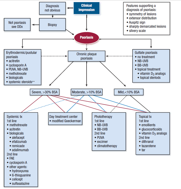
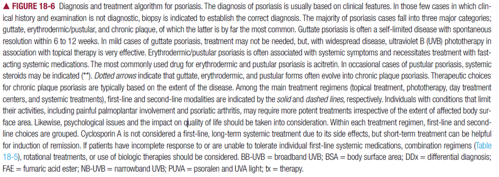
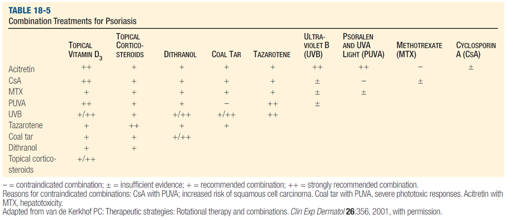
    PSORIASIS

  • Formation of spongiform microabscesses (Munro microabscesses) filled with granulocytes is a hallmark of psoriasis. The presence of these cells in psoriatic lesions is variable and becomes more pronounced with disease activity, e.g., in acute guttate or pustular psoriasis. Ultramicroscopically, degranulation is absent in these cells; instead, they appear to remain intact while migrating in response to chemotactic stimuli [CTCL:- The classic findings are characterized by finding lymphocytes with hyperchromatic and hyperconvoluted nuclei that cluster in the epidermis in microabscesses (Pautrier type)]

  • The treatment of choice for erythrodermic psoriasis is : SGPG I 04
    a) Corticosteroids b) Methotrexate c) Coaltar topically d) Topical corticosteroids

    Capture1

    Capture


    Capture


    Koebners phenomenon is seen in a) Lichen planus b) Psoriasis c) Icthyosis d) Pitriasis rubra e) Phemphigus

    Which of the following is pruritic
    a) Lichen planus b) Psoriasis c) Icthyosis d) secondary syphilis

    Photochemotherapy is used in A189
    a) Psoriasis b) Phemphigus c) Tinea capitis d) Tinea cruris

    All are common of psoriasis except a) Nail changes b) Extensor distribution c) Arthritis d) Squamo erythematous lesions

    Psoriasis is characterised by all except a) Definite pink plaque with clear margin c) Always associated with nail infection b) In children disappears in 2 weeks to reappear again d) Involves knees and elbows

    Non-cicatrical alopecia is present in TN 90
    Scieroderma b) Lichen planus c) Psoriasis d) parvo virus

    Psoralen with UV-A is used for treating a) Porphyria b) Psoriasis c) Pemphigus d) tinea versicolor

    Psoriasis is exacerbated by
    a) Lithium b) B-Blockers c) Antimalarials d) All of the above

    The important feature of psoriasis is a) Crusting b) Scaling c) Oozing d) Erythema

    Vitamin D analogue calcitriol is useful in the treatment of
    a) Lichen planus b) Psoriasis c) Pemphigus d) Leprosy

    Kobner’s phenomenon is seen in all the following PGI -04
    a. Lichen planus b. Herpes simplex c. Human papilloma virus infection
    d. Psoriasis e. All the above

    The Koebner (isomorphic) phenomenon refers to the fact that in persons with certain skin diseases, especially psoriasis, trauma is followed by new lesions in the traumatized but otherwise normal skin, and these new lesions are identical to those in the diseased skin.The Koebner reaction occurs in traumatized normal skin in vitiligo, psoriasis, and lichen planus, among other conditions. The Koebner phenomenon may occur in recent scars or at pressure points.

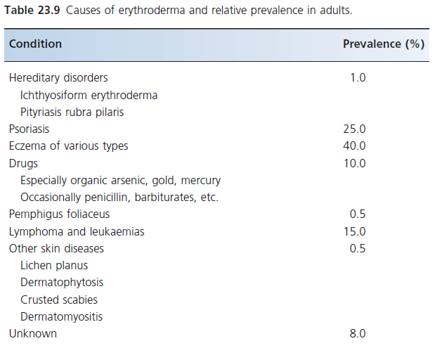
    Which of the following are causes for Erythroderma? PGI-05
    a. Psoriasis b. Lichen planus c. Eczema
    d. Pityriasis rubra pilaris e. Pityriasis versicoior

    Erythroderma is the term applied to any infl ammatory skin disease that affects more than 90% of the body surface. The term exfoliative dermatitis is used synonymously, although the degree of exfoliation is sometimes quite mild.

    image

    A wide range of drugs can cause erythroderma. Among the more commonly implicated are pyrazalone derivatives such as phenylbutazone, hydantoin derivatives, carbamazepine, cimetidine, gold salts and lithium.

    Pityriasis rubra pilaris (PRP) refers to a group of chronic disorders characterized by reddish orange, scaling plaques and keratotic follicular papules

    Treatment of Pustular psoriasis is: MAHA-05
    a. Thalidomide b. Retinoids c. Hydroxyurea d. Metholtrexate

    Koebners phenomenon is seen in a) Lichen planus b) Psoriasis c) Icthyosis d) Pitriasis rubra e) Phemphigus

    Causes of Cicatricial alopecia are PGI-04
    a. Disseminated lupus erythramatosis b. Alopecia areata c. Psoriasis
    d. Lichen planus e. Androgenic alopecia

    Bulkeley membrane is seen in; DNB 89, PGI 89
    a) Psoriasis b) Pemphigus c) Tinea d) Pityriasis rubra

    IN PSORIASIS WHEN THE SCALES ARE COMPLETELY SCRAPED OFF ,THE BASEMENT MEMBRANE IS EXPOSED & SEEN AS A MOIST RED SURFACE (BULKELEY’S MEMBRANE )THROUGH WHICH DILATED CAPILLARIES CAN BE SEEN AS RED SPOTS WORONOFF’S RING – PERILESIONAL HALO IN PSORIATIC PLAQUES USUALLY AFTER TREATMENT WITH UV LIGHT.DUE TO DEFICIENCY OF PG E2. GRATTAGE TEST -POSITIVE IN PSORIASIS,ALSO CANDLE WAX SIGN PRESENT.

    Antibody Brand name Approval date Type Target Indication
    (What it’s approved to treat)
    Example FDA approved therapeutic monoclonal antibodies[1]
    Abciximab ReoPro 1994 chimeric inhibition of glycoprotein IIb/IIIa Cardiovascular disease
    Adalimumab Humira 2002 human inhibition of TNF-α signaling Several auto-immune disorders
    Alemtuzumab Campath 2001 humanized CD52 Chronic lymphocytic leukemia
    Basiliximab Simulect 1998 chimeric IL-2Rα receptor (CD25) Transplant rejection
    Belimumab Benlysta 2011 human inihibition of B- cell activating factor SLE[disambiguation needed ]
    Bevacizumab Avastin 2004 humanized Vascular endothelial growth factor (VEGF) Colorectal cancer, Age related macular degeneration (off-label)
    Brentuximab vedotin Adcetris 2011 Chimeric CD30 Anaplastic large cell lymphoma (ALCL) and Hodgkin lymphoma
    Canakinumab Ilaris 2009 Human IL-1β Cryopyrin-associated periodic syndromes (CAPS)
    Cetuximab Erbitux 2004 chimeric epidermal growth factor receptor Colorectal cancer, Head and neck cancer
    Certolizumab pegol[19] Cimzia 2008 humanized inhibition of TNF-α signaling Crohn’s disease
    Daclizumab Zenapax 1997 humanized IL-2Rα receptor (CD25) Transplant rejection
    Denosumab Prolia , Xgeva 2010 Human RANK Ligand inhibitor Postmenopausal osteoporosis , Solid tumor`s bony metasteses
    Eculizumab Soliris 2007 humanized Complement system protein C5 Paroxysmal nocturnal hemoglobinuria
    Efalizumab Raptiva 2002 humanized CD11a Psoriasis
    Gemtuzumab Mylotarg 2000 humanized CD33 Acute myelogenous leukemia (with calicheamicin)
    Golimumab Simponi 2009 Human TNF-alpha inihibitor Rheumatoid arthritis, Psoriatic arthritis, and Ankylosing spondylitis
    Ibritumomab tiuxetan Zevalin 2002 murine CD20 Non-Hodgkin lymphoma (with yttrium-90 or indium-111)
    Infliximab Remicade 1998 chimeric inhibition of TNF-α signaling Several autoimmune disorders
    Ipilimumab ( MDX-101 ) Yervoy 2011 Human blocks CTLA-4 Melanoma
    Muromonab-CD3 Orthoclone OKT3 1986 murine T cell CD3 Receptor Transplant rejection
    Natalizumab Tysabri 2006 humanized alpha-4 (α4) integrin, Multiple sclerosis and Crohn’s disease
    Ofatumumab Arzerra 2009 Human CD20 Chronic lymphocytic leukemia
    Omalizumab Xolair 2004 humanized immunoglobulin E (IgE) mainly allergy-related asthma
    Palivizumab Synagis 1998 humanized an epitope of the RSV F protein Respiratory Syncytial Virus
    Panitumumab Vectibix 2006 human epidermal growth factor receptor Colorectal cancer
    Ranibizumab Lucentis 2006 humanized Vascular endothelial growth factor A (VEGF-A) Macular degeneration
    Rituximab Rituxan, Mabthera 1997 chimeric CD20 Non-Hodgkin lymphoma
    Tocilizumab ( or Atlizumab ) Actemra and RoActemra 2010 Humanised Anti- IL-6R Rheumatoid arthritis
    Tositumomab Bexxar 2003 murine CD20 Non-Hodgkin lymphoma
    Trastuzumab Herceptin 1998 humanized ErbB2 Breast cancer


    In addition to chimeric and humanized antibodies, there are other pharmaceutical purposes for the creation of chimeric constructs

    Etanercept:-. Etanercept, for example, is a TNFα blocker created through the combination of a tumor necrosis factor receptor (TNFR) with the immunoglobulin G1 Fc segment. TNFR provides specificity for the drug target and the antibody Fc segment is believed to add stability and deliverability of the drug.

    Alefacept:- Alefacept is a fusion protein: it combines part of an antibody with a protein that blocks the growth of some types of T cells.The exact mode of action is very complicated and involves dual mechanisms, one of which inhibits the activation of CD4+ and CD8+ T cells by interfering with CD2 on the T cell membrane thereby blocking the costimulatory molecule LFA-3/CD2 interaction. The second mechanism is inducing apoptosis of memory-effector T lymphocytes. If the T cells were to become activated they would stimulate proliferation of keratinocytes resulting in the typical psoriatic symptoms. Therefore, alefacept leads to clinical improvement of moderate to severe psoriasis by blunting these reactions.Combinations of therapeutic modalities have been utilized to meet the challenge of difficult to treat psoriasis.

    Pitting of nails is seen in PGI88
    a) Lichen planus b) Psoriasis c) Phemphigus d) Arsenic poisoning a) Leprosy

    Nail Involvement is not a feature of (UNSOLVED)?
    a) Dermatophytosis b) lichen planus c) DLE d) psoriasis

    Pterygium of nail is characteristically seen in: All India 2006
    a) Lichen planus. b) Psoriasis. c) Tinea unguium, d) Alopecia areata

    Tinea ungium effects
    A.Nail fold
    B.Nail plate
    C.Joints
    D.Inter digital space

    ONYCHOMYCOSIS
    Onychomycosis denotes any infection of the nail caused by dermatophyte fungi, nondermatophyte fungi, or yeasts. Tinea unguium, however, refers strictly to dermatophyte infection of the nail plate. The four clinical types of onychomycosis are: (1) distal subungual onychomycosis (DSO), (2) proximal subungual onychomycosis (PSO), (3) white superficial onychomycosis (WSO), and (4) candidal onychomycosis

    Least common site involvement in psoriasis is
    (A)Scalp
    (B)Nail involvement
    (C)CNS involvement
    (D)Arthritis

    Which of the following is wrong statements :
    (A)Koilonychia in Vit B12 deficiency
    (B)Oncholysis in Psoriasis
    (C)Mees lines in Arsenic poisoning
    (D)Pterygium of nails in Lichen Planus

    Griesofulvin is given for the treatment of fungal infection in finger nail dermatophytosis for :
    A.4 weeks
    B.6 weeks
    C.2 month
    D.3 month

  • Griseofulvin :-The duration of anti-fungal therapy may be 2 weeks for uncomplicated tinea corporis, 8–12 weeks for tinea capitis, or as long as 6–18 months for nail infections. Due to high relapse rates, griseofulvin is seldom used for nail infections. Common side effects of griseofulvin include gastrointestinal distress, headache, and urticaria.

  • Oral itraconazole and terbinafine are approved for onychomycosis. Itraconazole is given as either continuous daily therapy (200 mg/d) or pulses (200 mg bid for 1 week per month) administered with food. Fingernails require 2 months of continuous therapy or two pulses. Toenails require 3 months of continuous therapy or three pulses. Itraconazole has the potential for serious interactions with other drugs requiring the P450 enzyme system for metabolism. Terbinafine (250 mg/d) is also effective for onychomycosis. Therapy with terbinafine is continued for 6 weeks for fingernail infections and 12 weeks for toenail infections. Terbinafine has fewer drug-drug interactions, but caution should be used with patients who are on multiple medications.

    A middle aged man presents with paraesthesia of hands and feet. Examination reveals presence of `Mees’ lines in the nails and rain drop pigmentation in the hands. The most likely causative toxin for the above mentioned symptoms is :
    A.Lead
    B.Arsenic
    C.Thallium
    D.Mercury

  • NAIL INVOLVEMENT

    General Features of the Hair and Nails
    The distribution of the body hair, its texture, and amount should be noted as a part of the initial overall survey of the patient’s skin, as should examination of the nails. The nails (see Chap. 72) can provide evidence of latent skin disease (psoriasis, lichen planus, alopecia areata, congenital ectodermal defect), as well as suggest the presence of renal or liver disease. Beau’s lines (transverse indentations of the nails) and other variations on the theme of transverse white lines across the nails may be associated with a recent febrile or systemic illness, especially a renal or hepatic one. Telangiectasia in the periungual skin is a frequent and important diagnostic finding in systemic lupus erythematosus and dermatomyositis

    PSORIASIS:- Onychodystrophy, nail pitting, subungual keratosis, Beau’s lines (ridging), leukonychia, crumbling nail plate, and discoloration are more strongly associated with arthritis than with psoriasis alone, being found in 85 percent of those with arthritis versus 31 percent of those with the isolated cutaneous form of the disease. 47 The most prevalent findings are pitting and onycholysis. Onychodystrophy is closely related to involvement of the distal interphalangeal joint of the same digit, suggesting a broader acral dystrophic state. The appearance of onychodystrophy is closely related in time to the appearance of the arthritis.Nail pitting results from the presence of easily detachable parakeratotic cells in the superficial layers of the nail plate

    Nails in patients with alopecia areata may show fine pitting or, less commonly, mottled lunula, trachyonychia, or onychomadesis.

    Contact dermatitis of the proximal nail fold is responsible for chronic paronychia and nail plate surface abnormalities, such as irregular pitting and Beau’s lines. These are caused by mild involvement of the adjacent proximal nail matrix. Contact dermatitis of the hyponychium produces severe subungual hyperkeratosis and nail bed splinter hemorrhages. The differential diagnosis includes onychomycosis, psoriasis, parakeratosis pustulosa, pityriasis rubra pilaris, and keratoderma.


    Nail changes, including pitting, horizontal and longitudinal ridging, leukonychia, onycholysis, dyschromia, nail bed atrophy, and telangiectasia of the cuticle nail fold, have been observed in patients with SLE.

    DLE activity can localize to the nail unit. The nail can be impacted by other forms of cutaneous LE as well as SLE, producing nail fold erythema and telangiectasia, red lunulae, clubbing, paronychia, pitting, leukonychia striata, and onycholysis.


    In secondary syphilis, nail changes may result from involvement of either the nail matrix or the nail folds. In the nail plate, brittleness, splitting, onycholysis, pitting, lunular elkonyxis (pits) with fissuring, and dystrophy have been described.

    ECTRODACTYLY–ECTODERMAL DYSPLASIA–CLEFT LIP/PALATE (EEC) SYNDROME; SPLIT HAND–SPLIT FOOT–ECTODERMAL DYSPLASIA–CLEFT LIP/PALATE SYNDROME :-The nails are dystrophic in about four-fifths of individuals with transverse ridging, pitting, and slow growth

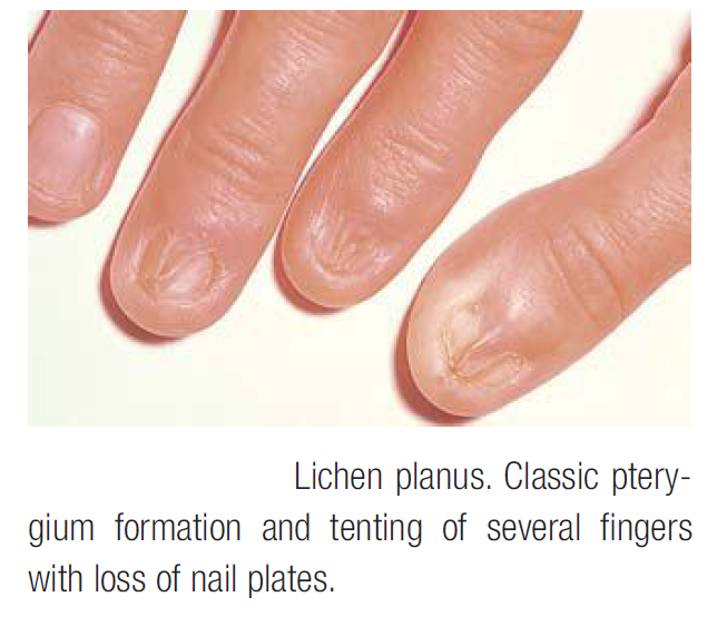
    LICHEN PLANUS OF THE NAILS Nail involvement occurs in 10 to 15 percent of patients. 23 Lichen planus limited to the nails is uncommon, and the initial involvement is followed, in many cases, by the development of typical lesions elsewhere. Lichen planus of nails is infrequent in children. Thinning, longitudinal ridging, and distal splitting of the nail plate (onychoschizia) are the most common findings. Onycholysis, longitudinal striation (onychorrhexis), subungual hyperkeratosis, or even absence (anonychia) of the nail plate can also be seen. Twenty-nail dystrophy (trachyonychia) may represent an isolated nail finding of lichen planus. Psoriasis and alopecia areata can also lead to these distinctive nail changes. Nail loss may result from ulcerative lichen planus involving the nail unit. Pterygium or forward growth of the eponychium with adherence to the proximal nail plate is a classic finding of lichen planus of the nail ( Fig. 49-12). An atrophic cicatrizing form of lichen planus with random and progressive nail loss in Asians and blacks has also been reported. The “tenting” or “pup-tent” sign is observed as a result of nail bed involvement that elevates the nail plate and may cause longitudinal splitting.

    Degeneration of basal cells occur in
    a) Lichen planus b) pemphigus c) psoriasis

    Compy’s sign (white patches due to degenerated squamous epithelium occurring on
    buccal Mucosae and gums) is seen in: JIPMER 80, AIIMS 85
    a) Moniliasis b) Pemphigus c) Lichen planus d) Measles

    30 year old male presents with pruritic flat-topped polygonal, shiny violaceous papules with flexural distribution. The most likely diagnosis is UPSC
    a) Psoriasis b) Pityriasis rosea c) lichen planus d) Lichenoid dermatitis

    Civatte bodies are found in
    a) Lichen planus b) Psoriasis c) Dermatophytosis d) Vitiligo

    Which of the following not photosensitive APPGE 04
    a) Porphyria b) DLE c) SLE d) Lichen Planus

    Causes of Cicatricial alopecia are PGI-04
    a. Disseminated lupus erythramatosis b. Alopecia areata c. Psoriasis
    d. Lichen planus e. Androgenic alopecia

    Kobner’s phenomenon is seen in all the following PGI -04
    a. Lichen planus b. Herpes simplex c. Human papilloma virus infection
    d. Psoriasis e. All the above

    Max Joseph’s space is a histopathological feature of:
    a) Psoriasis vulgaris. b) Lichen planus.
    c) Pityriasis rosea. d) Parapsoriasis.

    Wickham’s striae are seen in
    a) Lichen nitidus b)Lichen scrufosum c) Lichen planus d) DLE

    Basal cell degeneration is characteristic of
    a) Pemphigus b) Lichen planus c) Pemphigold
    d) Psoriasis

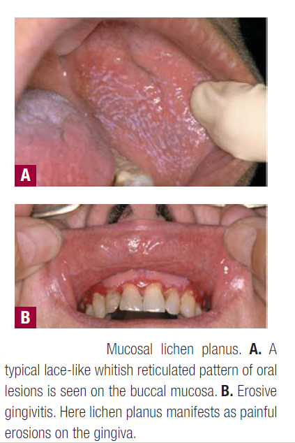
    The two major pathologic findings in lichen planus are basal epidermal keratinocyte damage and lichenoid-interface lymphocytic reaction. 1 The epidermal changes include hyperkeratosis, wedge-shaped areas of hypergranulosis, and elongation of rete ridges that resemble a sawtooth pattern .Multiple apoptotic cells or colloid-hyaline (Civatte) bodies are seen at the dermal–epidermal junction. Eosinophilic colloid bodies are present in the papillary dermis. They are PAS-positive and measure about 20 µm in diameter. A bandlike lymphocytic infiltrate is seen in the papillary dermis that abuts the epidermis. Many histiocytes and few plasma cells are seen. Plasma cells are more prominent in mucous membrane specimens. Few eosinophils are seen in drug-induced lichen planus or lichenoid drug eruptions. Melanin pigmentation is invariably present and is more pronounced in older, waning lesions and in dark-skinned individuals. Separation of the epidermis in small clefts (Max Joseph cleft formation) is occasionally seen .

    The classic cutaneous lesion of lichen planus is a faintly erythematous to violaceous, flat-topped, polygonal papule, sometimes showing a small central umbilication ( Fig. 49-1). A thin, transparent, and adherent scale may be discerned atop the lesion. Fine, whitish puncta or reticulated networks referred to as Wickham striae, named after the dermatologist who described the finding, are present over the surface of many well-developed papules. These are considered to be highly characteristic and are more easily observed after applying oil, xylene, or water and visualizing the lesions with a magnifying lens or a handheld dermatoscope. The surface alteration may result from localized thickening of the keratohyalin-containing cell layers of the stratum granulosum, although a focal increase in the activity of lichen planus may account for the morphologic alteration of Wickham striae.
    CaptureCapture

    Pterygium or forward growth of the eponychium with adherence to the proximal nail plate is a classic finding of lichen planus of the nail

    Pityriasis rosea

    Arvinder, 24 year old woman, presents with erythematous annular lesions with collarette scales predominantly arranged over trunk.
    Most likely diagnosis is
    a) Pityriasis rosea b) Pityriasis rubra pilaris c) Tenia versicolor
    d) Psoriasis

    Psoralen – A is used in the treatment of
    a) pemphigus b) Vitiligo c) pityriasis alba d) lcthyosis

    ‘Fir-tree’ type of distribution is seen in PGI 80, JIPMER 81
    a) Pityriasis Rosea b) Psoriasis c) Measles d) Secondary syhilis

    CaptureCapture2Capture3Capture4Capture5

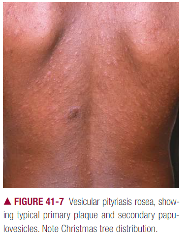
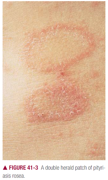
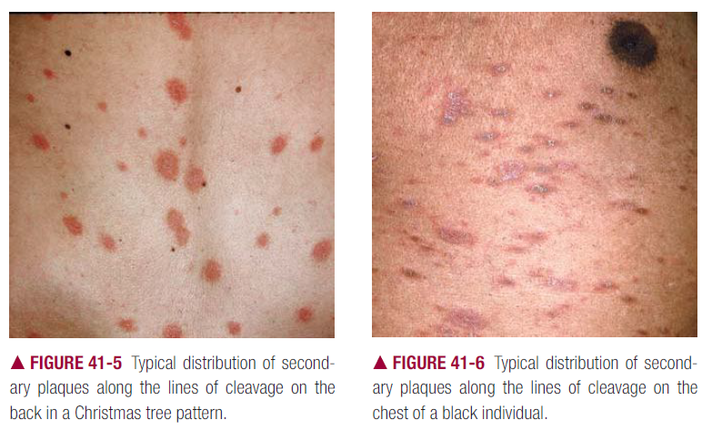
    The primary plaque (plaque primitive, herald patch, primary medallion, mother patch;)oval or round with a central, wrinkled, salmon-colored area and a darker red peripheral zone, separated by a collarette of fine scaling . The primary plaque is usually situated on the trunk in areas covered by clothes.

    Classic secondary rash small plaques resembling primary plaques in miniature, with the two red zones separated by the scaling ring,distributed with their long axes following the lines of cleavage of the skin, forming a “Christmas tree” pattern.

  • Pityriasis rubra pilaris (PRP)

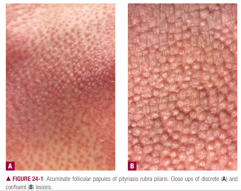
  • Pityriasis rubra pilaris (PRP) refers to a group of chronic disorders characterized by reddish orange, scaling plaques and keratotic follicular papules.

    The follicular papules so characteristic of the disease soon appear as keratotic follicular plugs surrounded by erythema .The follicular papules are not always present but are very prominent in black South Africans. They are seen most commonly on the dorsal aspects of the proximal phalanges , the elbows, and the wrists. They can occur on other areas of the extremities and even the trunk but appear rarely on the face. A more widespread eruption consisting of scaling orange-red plaques is observed on the trunk and extremities and occasionally on the face. The lesions have sharp borders, and islands of normal skin within them are characteristic .The lesions may expand and coalesce  and eventually cover the entire body .The scales vary from fine to thick, the latter being particularly common on the palms and soles, which also may fissure and have an intense yellow-orange hue, and may cover the scalp in thick sheets.

    Capture

    Capture

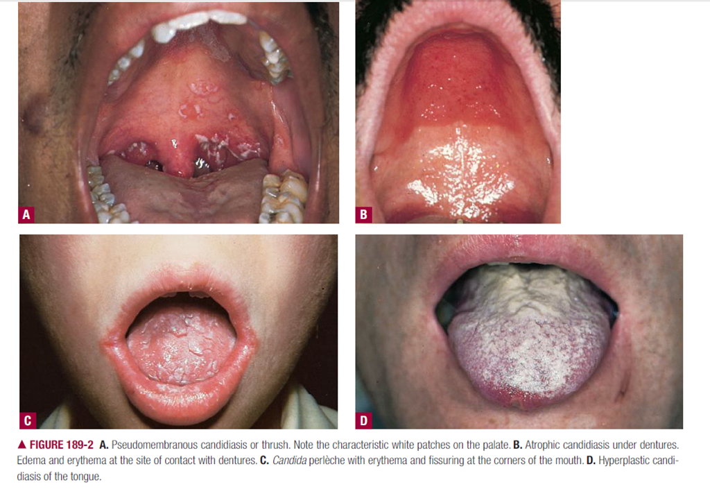
    Tricky Question:- A forty two-year-old engineer developed redness of the glans and radial fissuring of the prepuce 2 weeks ago. A potassium hydroxide preparation of scrapings from the glans showed pseudohyphae and buds. Which one of the following systemic illness should be screened for  a) Pulmonary tuberculosis b) Diabetes mellitus AIIMS 04
    c) Systemic candidiasis d)Chronic renal failure

    Drug of choice in systemic candidiasis is
    a) Amphotericin-B b) Griseofulvin c) Nystatin d) Ketoconazole

    Griseofulvin AND Terbinafine is not effective for candidiasis,deep fungal infections or pityriasis versicolor.

    DERMATOPHYTOSIS :-This commonly involves the inguinal area, i.e., tinea cruris, but rarely causes superficial infection of the scrotum or penis. Tinea cruris is always associated with tinea pedis, the dermatophyte being transferred by the hand from the feet to the crural area. Clinically, tinea cruris presents as an erythematous to tan plaque with well-demarcated, scalloped borders advancing inferiorly down the anterior thigh. Chronic scratching may induce an eczematous dermatitis or lichen simplex chronicus on the scrotum or, less commonly, on the penis. Chronic application of topical corticosteroids can mask the clinical presentation (tinea incognito).

    A washer man presents with thickness erosion & discoloration of web spaces of toes diagnosis is
    a) Psoriasis b) Tinea Unguum c) Both d) Candidiasis

    Tinea Capitis is caused by all except
    a) Epidermophyton b) Microsporum c) T. violaceum d) T. Schoenleinii

    Tinea capitis is caused by all except
    a) microsporum b) candida c) Epidermophyton d) Trichophyton

    Bulkeley membrane is seen in; DNB 89, PGI 89
    a) Psoriasis b) Pemphigus c) Tinea d) Pityriasis rubra

    Dhobi’s itch is:
    a) Tinea corporis b) Tinea cruris c) Tinea barbae d) Tinea capitis

    Boggy swelling on head with easily pluckable hair in a young child diagnosis a) Pyoderma capitis b) Tinea capitis c) Alopecia areata d) None

    Tinea incognito is due to inappropriate use of systemic & topical
    a) Antibiotics b) Antifungals c) Steroids d) Antiviral

    An eleven year old boy is having tinea capitis on his scalp. The most appropriate line of treatment is
    a) Oral griseofulvin therapy b) Topical griseofulvin therapy
    c) Shaving of the scalp d) Selenium sulphide shampoo

    All these fungal diseases causing tinea capitus are seen in India except PGI -04
    a. Tinea mentagrophytes b. Tinea ondoinii c. Tinea rubrum
    d. microsporum e. Epidermophyton

    Tinea versicolor is caused by PG186, AP 86, Kerala 87, AI-88, 77(90, Al 91
    a) Malaezzia furfur b) candida c) Trichophyton rubra d) Trichophyton, mentagrophyte

    Treatment of tinea versicolor
    a) Clotrimazole b) sod. sulphite c) selenium sulphide d) miconazole d) all of the above

    Girseofulvin is not useful in one of the following Karnat 89
    a) Tinea capitis b) Tinea cruris c) Tinea versicolor d) Tinea pedis

    Tinea cruris is common in a) Adult male b) Adult female c) female child d) male child


    While all dermatophyte species invade the stratum corneum of the skin;Epidermophyton floccosum never invades hair and only occasionally nail.T.rubrum rarely invades hair but frequently invades nail.

    Tinea Corporis:- Most commonly by T rubrum.All known dermatophytes can produce lesions of the glabrous skin.Some species have predilections for particular parts of the body; for example, M. audouinii, classically a cause of tinea capitis, and T. rubrum, which most commonly causes tinea pedis but these can, and on many occasions do, cause tinea corporis.

    Tinea capitis:-Most species of dermatophyte are capable of invading hair but some species (e.g. M. audouinii, T. schoenleinii and T. violaceum) have a distinct predilection for the hair shaft.The principal feature of tinea capitis in recent years has been the rise of M. canis as the dominant organism in infections in some parts of Europe , and the spread of T. tonsurans in urban communities in the USA . A similar rise in the prevalence of T. tonsurans has recently been recorded in urban areas of the UK and in some other European countries.T. tonsurans is now most common cause in USA.

    Imp:- Rook says M. audouinii is most common worldwide while fitzpatrick says M.canis is most common worldwide.

    Harrison says predominant organism is T. Tonsurans but it must be talking about USA though not mentioned specifically.

    E. fl occosum, T. concentricum and T. mentagrophytes var. interdigitale are exceptional in apparently never causing tinea capitis.

    image

    Tinea Barbae : The animal species T. verrucosum and T. mentagrophytes var. mentagrophytes are responsible for the great majority of cases.

    Tinea faciei:-T. mentagrophytes var. mentagrophytes and T. rubrum predominate but T. tonsurans, M. audouinii and M. canis are also common causes worldwide

    Tinea Pedis:- Three anthropophilic species, T. rubrum, T. mentagrophytes var. interdigitale and E. fl occosum, are together responsible for the vast majority of cases of foot ringworm throughout the world.

    Tinea cruris:-The causal species are those implicated in foot ringworm but in different proportions. T. rubrum is the main cause [1]; T. mentagrophytes var. interdigitale and E. fl occosum also account for some cases.

    Onychomycosis caused by dermatophytes:-The principal dermatophytes concerned are: (i) with associated foot and hand infections—T. rubrum, T. mentagrophytes var. interdigitale and E. fl occosum; (ii) with associated scalp infections—T. tonsurans, T. violaceum and T. soudanensei.

    image

    The treatment of dermatophyte infections usually involves the use of oral terbinafine, fl uconazole itraconazole, griseofulvin or one of several well-tried topical preparations .All those noted have been shown to be effective in a substantial majority of patients, provided they are used regularly.


    Favus. Infection with T. schoenleinii is now seen rarely and sporadically in a variety of countries, such as South Africa [32], Ethiopia, where it is still endemic, as well as the Middle East, Pakistan, USA, Canada, the UK and Australia. The classical picture of tinea capitis caused by this organism is characterized by the presence of yellowish cup-shaped crusts known as scutula.

    Tinea imbricata (Tokelau) resulting from T. concentricum, an anthropophilic dermatophyte found in southern Asia, the islands of the South Pacific and in Guatemala, southern Mexico and Brazil, causes a distinctive infection. It seems to affect mainly the native peoples of these areas, and although susceptibility may be inherited as an autosomal recessive character [13], it occurs in both sexes and at all ages. Occasional cases may be seen elsewhere in travellers from these regions [14]. The infection begins as a scaling ring; centrifugal spread follows, but within the area of central clearing a second wave of scaling soon arises. The process is repeated to give numerous concentric rings  and, as the natural history is normally extremely prolonged, the whole body may become affected. Pruritus is intense and may lead to lichenification. Hypopigmentation may accompany the lesions.

  • __________________________________________________________________________________________________________________________________________________________


    ALOPECIA


    Knowledge of the hair cycle is vital to understanding hair problems. The duration and rate of growth of the anagen (growth) phase normally vary at different body sites, in different individuals, and at various ages, and determine the ultimate length of hair in that area.

    1 Catagen is the transitional portion of the cycle between anagen and telogen and is short-lived (2 to 4 weeks) in duration. Telogen duration also varies greatly in different body sites, but interindividual variability appears more limited. In a normal scalp, telogen is assumed to last 3 to 4 months and anagen to last 3-plus years. With age, there is both a diminution in anagen duration and an increase in the time interval between two anagen cycles.

    2 At any given time, ˜90 percent of the scalp hair is in anagen and 10 percent in telogen; this is subject to some seasonal variability.

    3 At the end of anagen, each hair bulb moves from its location in the subcutaneous tissue or dermis (depth of location is determined by whether the follicle is terminal or vellus) to a more superficial location by means of shrinkage and remolding of that portion of the follicle below the “bulge” where the arrector pili muscle inserts. The concentric layers of the inner root sheath, which anchor the hair shaft in the follicle, are only present to the bottom of the isthmus of the follicle, the region to which the hair bulb ascends in telogen . Consequently, the hair shaft in telogen is no longer anchored securely in the tissue, as it was in anagen, and it may be dislodged with the gentle traction of shampooing, combing, brushing, etc. The recapitulation of the anagen follicle and initiation of growth of a new anagen hair leads to the shedding of any remaining telogen hair in the follicular canaL.


    image

    Non cicatrical alopecia is present in TN 90
    A) Scleroderma b) Lichen Planus c) Psoriasis d) Parvovirus

     click here. for D/D of alopecia.

    Scarring hair loss
    Primary
    Lymphocyte associated
    • Lichen planus
    • Lupus erythematosus
    • Pseudopelade of Brocq
    • Follicular mucinosis
    Neutrophil associated
    • Folliculitis decalvans
    • Diffecting folliculitis
    • Folliculitis keloidalis nuchae
    Secondary
    • Trauma
    • Radiotherapy
    • Some dermatophyte infections
    • Primary neoplasia
    • Secondary neoplasia (metastasis)


    Non-scarring hair loss
    On an abnormal scalp
    Psoriasis
    • Seborrhoeic dermatitis
    • Some dermatophyte infections
    • Traction alopecia
    On a normal scalp
    Diffuse
    • Telogen hair loss
    • Anagen hair loss
    Patchy
    Alopecia areata
    • Triangular alopecia
    • Trichotillomania
    Secondary syphilis
    With a distinct pattern
    • Male pattern baldness
    • Female pattern baldness
    Other causes of hair loss
    • Hair shaft abnormalities
    • Congenital hypotrichosis
    • Loose anagen syndrome
    Table

    Causes of Alopecia


    I. Nonscarring alopecia
    A. Primary cutaneous disorders
    1. Telogen effluvium
    2. Androgenetic alopecia
    3. Alopecia areata
    4. Tinea capitis
    5. Traumatic alopeciaa
    B. Drugs
    C. Systemic diseases
    1. Systemic lupus erythematosus
    2. Secondary syphilis
    3. Hypothyroidism
    4. Hyperthyroidism
    5. Hypopituitarism
    6. Deficiencies of protein, iron, biotin, and zinc
    II. Scarring alopecia
    A. Primary cutaneous disorders
    1. Cutaneous lupus (chronic discoid lesions)b
    2. Lichen planus
    3. Central centrifugal cicatricial alopecia
    4. Folliculitis decalvans
    5. Linear scleroderma (morphea)
    B. Systemic diseases
    1. Discoid lesions in the setting of systemic lupus erythematosusb
    2. Sarcoidosis
    3. Cutaneous metastases

    a Most patients with trichotillomania, pressure-induced alopecia, early stages of traction alopecia.

    b While the majority of patients with discoid lesions have only cutaneous disease, these lesions do represent one of the 11 American College of Rheumatology criteria (1982) for systemic lupus erythematosus.

    Alopecia areata is treated by
    a) minoxidil b) Tranquilizers
    c) whitfields oinment d) parenternal penicillin

    Treatment of alopecia areata is with either immunosuppressives (local or systemic) or with irritants/immunogens, and is generally tailored to the severity of the disease. For localized patchy alopecia areata, intralesional steroids given at 4- to 6-week intervals are usually effective, with the main side effect being local dermal/subcutaneous atrophy related to the depth and concentration of injected steroid. Topical glucocorticoids classes I to V are also effective but take several months for initiation of hair growth, rather than the weeks for intralesional steroids. Side effects of topical steroids are generally limited to acne/hypertrichosis on the face from inadvertent transfer from the scalp and local epidermal atrophy with the more potent steroids. Systemic steroids, particularly short courses (less than 8 weeks) of tapering doses, are often used either alone or in conjunction with topical agents. In this setting, acne and weight gain are commonly seen side effects. PUVA is another immunosuppressant treatment that may be effective in alopecia areata, particularly in patients with extensive scalp and body hair loss. Between 30 and 80 treatments may be necessary before hair induction occurs, and there is an increased risk of photodamage/photoaging and skin cancer with PUVA use. The immunostimulation of topical irritants, especially anthralin, or topical immunogens (diphencyprone, squaric acid dibutylester) can be very effective in alopecia areata, but their use runs the risk of intolerable irritation if the dose titration is inappropriate. The particular mechanism of action of contact dermatitis that makes it a treatment for alopecia areata is purely speculative at this time, but it may include enhanced clearance of the putative follicular antigens through recruitment of new T cells and antigenic competition and interference with the initial or continued production of proinflammatory cytokines by the follicular keratinocytes. Five percent topical minoxidil, as a nonspecific hair growth promoter, may be a useful drug as a single agent or as an adjuvant with topical anthralin. There is a low incidence of local dryness/irritation and facial hypertrichosis with topical minoxidil. Although rare, the potential for systemic effects of topical minoxidil, particularly in young children, must be considered and the total amount applied kept to the recommended = 2 mL/day. Patients with alopecia areata need psychological support and physical means of camouflaging their hair loss. The latter often requires the use of a wig, which should be considered an integral part of treatment in patients with extensive scalp hair loss.

    Alopecia areata is due to a) Estrogen Stimulation c) Lichenoid reaction D) Androgen stimulation
    d)auto immune

    The pathogenesis of alopecia areata is still obscure, although most authors tend to classify alopecia areata as an autoimmune disease. As opposed to normal hairs, strong major histocompatibility complex (MHC) class I and class II immunoreactivity are found in lesional alopecia areata follicles, which also display aberrant expression of adhesion molecules known to direct hematopoietic cell migration.

    Pseudo pelade is synonym of: AIIMS 81, PGI 84
    a) Alopecia steatoides b) Premature alopecia c) Traction d) Cicatricial

    image

    Pseudopelade of Brocq is a rare clinical syndrome of scarring alopecia and fibrosis, in which distinct pathologic features are absent. It is generally accepted that pseudopelade of Brocq is the end stage of follicular fibrosis caused by a primary inflammatory dermatosis such as lichen planus, lupus erythematosus, pustular-scarring forms of folliculitis, or fungal infections, including favus, scleroderma, and sarcoidosis.

    PSEUDOPELADE OF BROCQ In clinical terms, pseudopelade of Brocq implies flesh- to pink-colored, irregularly shaped alopecia that may begin in a moth-eaten pattern with eventual coalescence into larger patches of alopecia . There has been considerable debate as to the specificity of this diagnosis versus an assignation of the term to describe all noninflammatory scarring alopecias, including the end-stage of a variety of initially inflammatory conditions. Histologically, the lesions are characterized by a perifollicular and perivascular lymphocytic infiltrate primarily at the level of the follicular infundibulum, loss of sebaceous epithelium, and fibrotic streams into the subcutis without interface or follicular plugging changes. 103 Elastin stains may distinguish pseudopelade (persistent elastic fibers around the midshaft of the follicle) from lichen planopilaris and lupus erythematosus (loss of elastic fibers in this location). 104 Direct immunofluorescence is negative in the majority of cases. It is unclear what treatment specifically helps.


    Cradle cap is a special form of:
    a) Pityriasos capitis b) Cephalohematoma
    d) Down’s syndrome c) Alopecia steatoides

    Boggy swelling on head with easily pluckable hair in a young child diagnosis a) Pyoderma capitis b) Tinea capitis c) Alopecia areata d) None

    Exclamation mark hairs are seen in
    a) Syphilis b) Alopecia areata c) Psoriasis d) Dermatophytosis

    image

    These short broken hairs, whose distal ends are broader than the proximal ends, illustrate their inherent sequence of events: follicular damage in anagen and then a rapid transformation to telogen. White or graying hairs are frequently spared and probably account, in cases of fulminant alopecia areata, for the mysterious phenomenon of “going gray overnight.” There is an increased incidence of autoimmune diseases in patients with alopecia areata, particularly thyroid-related disease, and there is a higher prevalence of pigmentary defects in patients with alopecia areata. 53 Nails in patients with alopecia areata may show fine pitting or, less commonly, mottled lunula, trachyonychia, or onychomadesis.


    Exclamatory Point hairs are seen in TN 2003
    a) Alopecia areata b) lcthyosis Vulgaris c) Tinea capitis
    d) Dermatomyositis

    Alopecia aerata is SGPGI 04
    a) Cicatrical scar b) Non cicatrical c) Fungal infection d) None

    Causes of Cicatricial alopecia are PGI-04
    a. Disseminated lupus erythramatosis b. Alopecia areata c. Psoriasis
    d. Lichen planus e. Androgenic alopecia
    Ref: Harrison 15th edn. page 316

    Exclamation mark" hairs are seen in? Karnat-05
    a. Tinea capitis b. Alopecia areata c. Lichen planus pilaris d. Trichotillomania

    Which of the following conditions causes alopecia without scarring? J&K-05
    a. DLE b. Herpes Zoster c. Alopecia areata d. Lichen planus pilaris

    Piebaldism refers to KARNATAKA – PGMEE – 2006.
    a. Androgenetic alopecia b. Erythema nodosum leprosum
    c. Association of vitiligo with white forelock d. None of the above
    (Ref. Harrison’s 16th edition page 968, 971)

    Which of the following is contraindicated in Androgenic Alopecia :
    (A)Testosterone
    (B)Cyproterone
    (C)Acetate Finasteride
    (D)Minoxidil

    A man aged 30years presents with, alopecia, boggy scalp swelling and easily pluckable hair. Next step in establishing Diagnosis would be
    A.KOH smear
    B.Culture sensitivity
    C.Biopsy
    D.None of the above

  • Posted in Medical | 29 Comments »

    PICC (Peripherally Inserted Central Catheter) Insertion Technique

    Posted by Dr KAMAL DEEP on December 5, 2011

    Untitledimage

    image

    1. The insertion site (below or above the antecubital fossa), 2. The sternocleidomastoid triangle at the medial third of the clavicle, and 3. The 3rd right costosternal junction of the sternum.

    sum of the two linear distances measured between the three landmarks (with the inserted arm abducting laterally at 45-degree angle from the body) is the optimal PICC length.

    image

    PICC INSERTION TECHNIQUE


    1. Guidewire / SWG Insertion Techniques:

    Kits/Sets are available with a variety of Guidewires/SWGs. Guidewires are provided in different diameters, lengths, and tip configurations for specific insertion techniques. Become familiar with the guidewire(s) to be used with the specific technique chosen, before beginning the actual PICC insertion procedure.

    Image guidance may be used to gain initial venous access.

    Catheter Insertion with an 80 cm Guide wire:

    Use single 45 cm guidewire for venous access and 80 cm soft tip guidewire for catheter placement. Image guidance or fluoroscopy is used to gain initial venous access; catheter placement with 80 cm guidewire is done under fluoroscopy.

    • Gain venous access with 45 cm guidewire and peel-away sheath.

    • Load PICC onto 80 cm guidewire until soft tip of wire extends beyond tip of catheter.

    • While maintaining control of distal end of guidewire, advance soft tip/catheter tip as a unit through peel-away sheath to desired depth.

    • Once catheter is in desired location, remove guidewire.

    Catheter Insertion with an 130 cm Guidewire:

    Use single 45 cm guide wire for venous access and 130 cm soft tip guidewire for catheter placement. Image guidance or fluoroscopy is used to gain initial venous access; catheter placement with 130 cm guidewire is done under fluoroscopy.

    • Gain venous access with 45 cm guidewire.

    • Insert soft end of 130 cm guidewire through peel-away sheath to desired depth.

    • Thread catheter over guidewire and advance catheter over guidewire through sheath into vessel into correct position.

    • Once catheter is in desired location, remove guidewire.

    NOTE: Some clinicians will gain access with 130 cm guide wire and thread catheter over guide wire once wire has been correctly positioned in the SVC. This technique is done under fluoroscopy.

    Identify insertion vein:

    • Apply tourniquet above anticipated insertion vein.

    • Identify appropriate vein for insertion. Use direct visualization technologies, if available, and assess vein health.

    5. Release tourniquet and leave in place beneath the arm.

    6. Measure patient to assure placement of catheter in the SVC:

    • Extend arm laterally 45 to 90 degrees from trunk.

    • Measure distance from insertion site along presumed anatomical course of vessel to be catheterized.

    • Catheter tip should lie in distal one-third of SVC above right atrium and parallel to SVC wall.

    ◊ If a catheter stabilization device will be used, add ½ to 1 inch (1.2 to 2.5 cm) to catheter measurement (STATLOCK®); if another device is used, check manufacturer recommendations.

    ◊ If using upper arm circumference assessment; for consistency in measurement, measure from an anatomical point and record.

    7. Position patient as appropriate for insertion site:

    • Extend arm laterally 45 to 90 degrees from trunk.

    8. Prepare work area.

    2. Prep Puncture Site:

    1. Prep and drape peripheral puncture site.

    2. Perform skin wheal with a local anesthetic as needed.

    3. Gain Initial Venous Access:

    See specific guidewire instructions, Guidewire Insertion Techniques (page 7) under Accessory Component Instructions section.

    1. Insert soft tip of guidewire through introducer needle into vein. Advance guidewire to desired depth.

    Warning: Do not insert stiff end of soft tip guidewire into vessel as this may result in vessel damage.

    Warning: Do not cut guidewire to alter length.

    Warning: Do not withdraw guidewire against needle bevel to minimize the risk of possible severing or damaging of guidewire.

    2. Remove needle:

    Hold guidewire in place while removing introducer needle.

    Caution: Maintain firm grip on guidewire at all times.

    3. Enlarge puncture site, if necessary:

    Use scalpel positioned away from the guidewire to enlarge cutaneous puncture site. Do not cut guidewire. Retract scalpel to the protected position.

    4. Sheath Placement:

    1. Thread tapered tip of peel-away sheath/dilator assembly over guidewire. Grasping near skin advance assembly with slight twisting motion to a depth sufficient to enter vessel. Dilator may be partially withdrawn to further facilitate advancement of sheath into the vessel. A slight twisting motion of the peel-away might help sheath advancement.

    Caution: Do not withdraw tissue dilator until the sheath is well within the vessel to minimize the risk of damage to sheath tip. Sufficient guidewire length must remain exposed at hub end of sheath to maintain a firm grip on guidewire.

    2. Check sheath placement by holding sheath in place, withdraw guidewire and dilator sufficiently to allow venous blood flow. Holding sheath in place, remove guidewire and dilator as a unit.

    Warning: Do not leave the dilator in place as an indwelling catheter to minimize the risk of possible vessel wall perforation.

    Warning: Do not apply undue force on guidewire to minimize the risk of possible breakage.

    5. Catheter Advancement:

    Advance catheter according to the guidewire used. Review detailed instructions for 80 cm and 130 cm guidewire usage (page 7) under Accessory Component Instructions section.

    Warning: Do not apply excessive force in placing or removing catheter. Failure to do so can result in catheter breakage. If placement or withdrawal cannot be easily accomplished, an x-ray should be obtained and further consultation requested.

    1. Retract catheter guard.

    2. Insert catheter through peel-away sheath.

    • If resistance is met while advancing catheter, retract and/or gently flush while advancing.

    3. Stop advancing catheter 5 inches (13 cm) before reaching pre-established insertion length.

    4. Withdraw peel-away sheath over catheter until free from venipuncture site.

    5. Grasp tabs of peel-away sheath and pull apart, away from catheter, until sheath splits down entire length.

    6. Advance catheter to final indwelling position.

    Placement Wire (where provided):

    Caution: To minimize the risk of placement wire kinking, do not clamp extension line(s) when placement wire is in catheter.

    1. Complete catheter insertion.

    2. Remove placement wire.

    Warning: Remove placement wire and Luer-Lock sidearm assembly as a unit (see Figure 24). Failure to do so may result in wire breakage.

    6. Dressing:

    Replace dressing according to organizational policies, procedures, and practice guidelines. Change immediately if the integrity becomes compromised e.g. dressing becomes damp, soiled, loosened, or no longer occlusive.

    • Consult manufacturer’s recommendations for dressing specifics.

    • Transparent semipermeable membrane dressing should be changed every 7 days.

    • Gauze and tape should be changed every 48 hours.

    • Label dressing with type, size, and length of catheter; date and time; and initials of the clinician performing dressing change.

    7. Maintain Catheter Patency:

    Maintaining central venous catheter patency shall be done in accordance with organizational policies, procedures, and practice guidelines. All personnel who care for patients with central venous catheters must be knowledgeable about effective management to prolong catheter’s dwell time and prevent injury.

    Perform hand hygiene as required.

    1. Solution and frequency of flushing a venous access catheter should be established in hospital/agency policy.

    2. Catheter patency is established and maintained by:

    • flushing intermittently via syringe with heparinized saline or preservative-free 0.9% sodium chloride (USP)

    • continuous drip

    • positive pressure device

    3. The amount of heparin:

    • depends on physician preference,

    • hospital/agency protocol,

    • patient condition

    Caution: Assess patient for heparin sensitivity. Heparin-induced thrombocytopenia (HIT) has been reported with the use of heparin flush solutions.

    The volume of flush solution should be:

    • equal to at least twice the priming volume of the catheter and any add-on devices

    Catheter priming volume is printed on product packaging.

    5. When using any central venous catheter for intermittent infusion therapy, proper flushing (heparinization) using a positive-pressure flushing technique will help prevent occlusion. Neutral as well as positive displacement valve systems have also been shown to help prevent occlusion.

    6. All valves need to be properly cleansed with an appropriate antiseptic before being accessed.

    7. The SASH or SAS method of flushing will help eliminate occlusions due to incompatible solutions:

    • Saline • Administer drug • Saline • Heparin (if used)

    8.Catheter Removal Procedure

    1. PICC removal shall be performed:

    • following order of authorized prescriber

    • in accordance with organizational policies, procedures, and practice guidelines

    2. A PICC shall be removed immediately upon patient assessment for:

    • suspected contamination

    • unresolved complication

    • discontinuation of therapy

    3. As indicated, place patient in supine position to minimize the risk of potential air embolism.

    4. Remove dressing.

    Warning: Do not use scissors to remove dressing, to minimize the risk of cutting catheter.

    5. Open catheter stabilization device retainer wings and remove catheter from catheter stabilization device posts.

    6. Remove catheter by slowly pulling it parallel to skin. If resistance is met when removing the catheter, catheter should not be forcibly removed and the physician should be notified.

    Caution: Do not exert excessive force while removing the catheter; to minimize the risk of catheter breakage.

    7. Upon removal of catheter:

    • measure and inspect

    • ensure entire catheter length has been removed

    8. Direct pressure should be applied at site until hemostasis is achieved.

    9. Apply alcohol swab to catheter stabilization device adhesive and gently lift pad off of skin (if applicable).

    10. Dress insertion site. Sterile occlusive dressing should be applied and site assessed every 24 hours until site is epithelialized. Residual catheter track may remain an air entry point until completely sealed (usually 24 to 72 hrs); dependent upon amount of time catheter was indwelling.

    11. Document catheter removal procedure on patient’s chart per hospital/agency protocol.

    Include:

    • catheter condition

    • length of catheter removed

    • patient’s tolerance of the procedure

    • any nursing interventions needed for removal

    image

    image

    image

    Untitled

    image

    NOTE: These “standard” insertion sites were defined to correspond to the catheter lengths determined by the formulas. Any deviation from the defined site requires appropriate adjustment of catheter length. For example, a PICC inserted 2.5 cm above the antecubital fossa would be 5 cm shorter than the value from the chart, based on the patient’s height

    image

    These recommended lengths should always be critically appraised prior to use in the individual patients. For patients with unusual proportions, e.g. short or long neck, lengthy arm or upper body proportions, case-by-case decision-making is important.


    Catheter Trimmer:

    NOTE: There should be very limited resistance when cutting catheter with supplied trimmer. Any greater resistance is likely to be caused by the placement wire – which has not been sufficiently retracted. If so, do not use catheter.
    Catheter Trimmer is a one time use trimming device.
    • To trim catheter with Catheter Trimmer, retract placement wire 1-1/2 inches minimum (4 cm) behind where catheter is to be cut. The placement wire is to be withdrawn through septum (see Figure 4).


    image

    Kink proximal end of placement wire at connector with side-port (see Figure 5). This minimizes the risk of placement wire extending beyond distal tip of catheter during insertion. (Do not attempt to advance placement wire through septum.)

    image


    Peel back contamination guard exposing catheter portion to be trimmed. Using trimming device, cut catheter straight across (90° to catheter cross-section) to maintain a blunt tip. Warning: Do not cut placement wire when trimming catheter to minimize the risk of foreign embolism. Caution: Check that there is no wire in cut catheter segment, after trimming catheter. If there is any evidence that placement wire has been cut or damaged, catheter should not be used.

    Trim Catheter:
    If necessary, review detailed instructions for Catheter Trimmer device under Accessory Component Instructions section.
    1. Identify catheter type:
    • BFT (Blue FlexTip®)
    • Non-BFT
    2. Peel back contamination guard exposing catheter portion to be trimmed.
    3. Review catheter marking pattern below. The catheter is marked so clinician can easily identify desired amount of catheter to be trimmed; length of catheter that remains or as with BFT catheter – both.

    4. Using the trimming device, cut catheter straight across (90° to catheter cross-section) to maintain a blunt tip. NOTE: There should be very limited resistance when cutting catheter with supplied trimming device. If using a catheter with a placement wire, any greater resistance is likely to be caused by the placement wire which has not been sufficiently retracted. If so, do not use catheter. 5. Inspect cut surface for clean cut and no loose material. Warning: Do not cut placement wire when trimming catheter to minimize the risk of foreign embolism. Caution: Check that there is no wire in cut catheter segment, after trimming catheter. If there is any evidence that placement wire has been cut or damaged, catheter should not be used

         BFT double numbering pattern:

    image

    ◊ First number designates centimeters from tip of catheter.
    ◊ Second number designates centimeters from hub of catheter.
    ◊ This double numbering pattern permits clinician to easily identify centimeters of catheter to be trimmed and also identifies centimeters of catheter remaining.
    ◊ Record both numbers.

             Non-BFT numbering pattern:

    image


    Raulerson syringe

    Number designates centimeters of catheter to be trimmed and also gives amount of catheter remaining

    Reducing Risks: There are two key benefits to using the Raulerson syringe. First, it lessens the exposure to blood and can lower the risk of air embolism. Second, it enables you to place the catheter in fewer steps, with less risk of dislodging needle from vessel.

    image

    imageimage

    verify Catheter tip position
    Based on hospital policy, utilize
    one of the following methods for tip
    placement verification:
    • Radiographic imaging (Chest Film,
    Fluoroscopy)
    • Physiological Feedback such as EKG
    • Other approved methods.
    Appropriate tip placement for Central
    Venous Catheters other than Dialysis
    Catheters is in the lower one-third
    of the Superior Vena Cava or at the
    Cavoatrial Junction.
    If catheter tip is malpositioned,
    reposition and reverify tip position.

    image


    The following designations are examples for port usage and do not represent the
    only way the lumens can be used:

    Proximal: Blood Sampling
    Medications
    Blood Administration

    Medial: Total Parenteral Nutrition
    Medications (only if TPN use is not
    anticipated)

    Distal: CVP Monitoring
    Blood Administration
    High Volume or Viscous Fluids
    Colloids
    Medication
    4th Lumen: Infusion
    Medication

                                                                           ANATOMY OF ARM VEINS

    clip_image002

    clip_image004

    clip_image006

    clip_image008

    clip_image010

    clip_image012clip_image014

    A, Anatomy of vessels in the anticubital fossa, noting

    the brachial artery (BA), basilic vein (BAV), and deep brachial vein

    (DBV). B, Corresponding ultrasound image. The vein collapses with

    probe pressure to distinguish it from the artery.

    clip_image016

    Posted in Medical | Tagged: , , , | 10 Comments »

    Percutaneous dilatational tracheostomy (PDT)

    Posted by Dr KAMAL DEEP on September 12, 2011

    The author wish to acknowledge the excellent guidance of Dr Harjit Singh Mahay, Sr. Intensivist ; New Delhi in this topic.

    If the first two tracheal rings cannot be distinctly identified, the procedure is aborted and an open tracheostomy is performed.

    Equipment required at the bedside to perform the PDT pro-cedure includes the following:
    •PDT introducer set (Ciaglia Blue Rhino Percutaneous Tracheostomy Introducer Set #C-PTIS-100-HC)
    •Bronchoscope with video monitor display and bron-choscopy endotracheal tube adapter
    •Continuous electrocardiographic (ECG) monitor
    •Blood pressure monitoring device
    •Pulse oximeter
    •Free-flowing intravenous catheter
    •Mechanical ventilator
    •Suction
    •Resuscitation (“crash”) cart
    •Open tracheostomy instrument tray (unopened)

    Types of PCT
    Ciaglia technique
    Grigg’s technique
    Blue rhino / Rhino horn technique
    Fantony’s technique
    Perc twist technique

    Surgical Tracheostomy preferable to PCT in the
    following situations:
    Coagulation abnormality
    High level of ventilatory support (FiO2 >.7 & PEEP
    > 10 cm H2O)
    Unstable / Fragile cervical spine
    Neck injury
    Unfavorable neck anatomy (previous surgery, tumor
    etc.)
    Obesity

    PDT

    8

    1234

    567

    9

    Posted in Medical | Tagged: , , , , | Leave a Comment »

    CENTRAL VENOUS ACCESS

    Posted by Dr KAMAL DEEP on July 26, 2011

    Internal Jugular Approach

    The author wish to acknowledge the excellent guidance of Dr Harjit Singh Mahay , Sr. Intensivist in this topic.

    Percutaneous Cannulation of The Internal Jugular Vein (ALL APPROACHES)

    Jugular System
    The anatomy of the IJ vein is relatively constant, regardless of body habitus. The vein drains the cranium, beginning as the superior jugular bulb, which is separated from the floor of the middle ear by a delicate bony plate. The IJ vein emerges deep to the posterior belly of the digastric muscle. At its origin the IJ vein courses adjacent to the spinal accessory, vagus, and hypoglossal nerves, as well as the internal carotid artery. Several tributary veins enter the IJ vein at the level of the hyoid bone. The IJ vein, the internal (and, later, the common) carotid artery, and the vagus nerve course together in the carotid sheath, with the IJ vein occupying the anterior lateral position.The only structure that maintains a fixed anatomic relationship with the IJ vein is the carotid artery. The vein invariably lies lateral and slightly anterior to the carotid artery, and the course of the artery serves as a guide to venous cannulation. At the level of the thyroid cartilage, the IJ vein can be found just deep of the sternocleidomastoid muscle .

    The IJ vein emerges from under the apex of the triangle of the two heads of the sternocleidomastoid muscle and joins the subclavian vein behind the clavicle. As the vein approaches its supraclavicular junction with the subclavian vein, it assumes a more medial position in the triangle formed by the two heads of the sternocleidomastoid muscles, following the anterior border of the lateral head. In this lower cervical region, the common carotid artery assumes a deep paratracheal location. The brachial plexus is separated from the IJ vein by the scalenus anterior muscle. The phrenic nerve is anterior to the scalenus anterior muscle. Although quite deep, the stellate ganglion lies anterior to the lower brachial plexus.

    Unlike the subclavian vein, the IJ vein is quite distensible. The vessel diameter is increased with performance of a Valsalva maneuver and the assumption of the head-down (Trendelenburg) tilt position. Prolonged palpation of the carotid pulse will decrease the diameter of the IJ vein.Rotating the head 90 degrees toward the opposite side or extending the neck will not change the size of the IJ vessel significantly. Severe rotation of the head will bring the sterno-cleidomastoid muscle anterior or medial to the IJ vein and may make cannulation impossible without first traversing the carotid artery. The diameter of the IJ vessel is largest below the cricoid ring, where it may reach 2 to 2.5 cm.


    Untitled

    79926-79931-80298-1465749

    Neck anatomy showing the course of the internal jugular vein (IJV).

    ijv

    Untitled,


    image

    image


    image

    ijv3

    IJV2

    IJVAPP

    POSTERIOR APPROACH

    Diagram of external jugular line going up over the edge of the sternocleidomastoid muscle, the main landmark for the posterior approach. Identify the point of insertion for the introducer needle along the posterior edge of the sternocleidomastoid muscle at the level just superior to where the external jugular vein crosses the muscle. This is typically one third of the distance between the mastoid and the clavicle.

    • Typically, the vein is entered within 1-3 cm; if the vein is missed, draw the needle all the way back to the skin before redirecting it.
    • If the bevel of the introducer needle is swung back and forth in the deeper tissues, a vessel or nerve may be inadvertently lacerated. Redirecting the needle more lateral, toward the sternoclavicular joint as opposed to the notch, often allows the vein to be entered

    image

    Surface anatomy and various approaches to cannulation of the internal jugular vein. A: Surface anatomy. B: Anterior approach. C: Central approach. D: Posterior approach.

    MIDDLE

    ANTERIOR

    POST

    The anterior and posterior approaches are identical in technique, differing only in venipuncture site and plane of insertion. For the anterior approach (see Fig. )the important landmark is the midpoint of the sternal head of the SCM, approximately 5 cm from both the angle of the mandible and the sternum. At this point, the carotid artery can be palpated 1 cm inside the lateral border of the sternal head. The index and middle fingers of the left hand gently palpate the artery, and the needle is introduced 0.5 to 1 cm lateral to the pulsation. The needle should form a 45-degree angle with the frontal plane and be directed caudally parallel to the carotid artery toward the ipsilateral nipple. Venipuncture occurs within 2 to 4 cm, sometimes only while the needle is slowly withdrawn. If the initial attempt is unsuccessful, the next attempt should be at a 5-degree lateral angle, followed by a cautious attempt more medially, never crossing the plane of the carotid artery.

    The posterior approach (see Fig. )uses the EJV as a surface landmark. The needle is introduced 1 cm dorsally to the point where the EJV crosses the posterior border of the SCM or 5 cm cephalad from the clavicle along the clavicular head of the SCM. The needle is directed caudally and ventrally toward the suprasternal notch at an angle of 45 degrees to the sagittal plane, with a 15-degree upward angulation. Venipuncture occurs within 5 to 7 cm. If this attempt is unsuccessful, the needle should be aimed slightly more cephalad on the next attempt.

    DSC01597DSC01599DSC01600DSC01602DSC01603DSC01604DSC01605DSC01606_2DSC01607_2DSC01608DSC01609DSC01610DSC01611DSC01612DSC01613_2DSC01614DSC01615_2DSC01616_2DSC01618DSC01619DSC01620DSC01621_2DSC01632DSC01633DSC01634DSC01635DSC01636DSC01637DSC01638DSC01639DSC01640DSC01647DSC01649DSC01648DSC01650DSC01641DSC01642DSC01643DSC01644DSC01645DSC01646DSC01651_2DSC01652DSC01653DSC01654DSC01655DSC01656IJ line triangle

    Click To Enlarge

    catheter


    sleeve

     Sandwich Technique
    For multi-lumen catheters, bulky catheter hubs or difficult to dress areas
    1.
    Use two 3M™ Tegaderm™ Transparent Film Dressings. Fold one dressing across the
    width with the printed liner facing outward. The location of the fold may vary
    depending on the distance between the catheter exit site and the hub, but typically
    would be near the centre of the dressing.
    2. Remove the printed liner. Keeping the dressing folded, gently lift the catheter and slip
    the dressing underneath, positioning the fold at the exit site (or sutures). Secure one
    half to the skin; and lay the catheter on top of the exposed adhesive of the other half.
    (Figure 1) Smooth down the portion of the dressing on the skin. Do not remove the
    frame.
    3. Remove the printed liner from the second dressing and place it on top of the catheter.
    Smooth one half over the exposed adhesive of the first dressing, and the remaining half
    over the skin above the exit site. (Figure 2) Firmly smooth both dressings from the exit
    site outward to enhance adhesion.
    4. Pinch the dressings together around the catheter and remove the frame
    from both dressings, smoothing down the edges as the frames are removed. (Figure 3)

    sandwitch

    A sandwich, loop-line, or bridge technique was used to apply each of the dressings. Two mechanisms of displacement were tested: dressing adherence to skin and dressing adherence to line. Dressing to skin adherence was tested on a relatively hairless part of the upper arm. Weights were added sequentially until the dressing peeled off. Dressing to line adherence was tested by applying the dressing to a 7F Dual Lumen Bard Hickman line passing through a piece of foam (measuring 13 × 12 cm). Weights were attached to the line until the cuff was pulled through the foam.

    Dressing to skin adherence was poorest for the clear dressings, followed by Mefix and Sleek, and greatest for a combination of Tegaderm and Mefix. Dressing to line adherence was improved using a sandwich technique instead of a loop-line technique and most secure when a bridge technique was used to the thicker shaft of the line.

    The dressings used for securing Hickman lines are not all equally secure. The least effective is the IV 3000 loop-line dressing. Tegaderm-Mefix bridge and Tegaderm-Mefix-Sleek combination dressings are the most secure and cost effective.

    image

    Infraclavicular Sub

    IJV3

    image

    Patient positioning for subclavian cannulation. B: Cannulation technique for supraclavicular approach

    Capture

    needle

    2

    • Needle insertion site options include the following:
      • One centimeter inferior to the junctions of the middle and medial third of the clavicle
      • Inferior to the clavicle at the deltopectoral groove
      • Just lateral to the midclavicular line, with the needle perpendicular along the inferior lateral clavicle
      • One fingerbreadth lateral to the angle of the clavicle
    • Sternal notch: Direct the insertion needle toward this target in the coronal plane.

    The 18-gauge thin-wall needle is preferable for SV cannulation .The patient is placed in a 15- to 30-degree Trendelenburg position, with a small bedroll between the shoulder blades. The head is turned gently to the contralateral side and the arms are kept to the side. The pertinent landmarks are the clavicle, the two muscle bellies of the SCM, the suprasternal notch, the deltopectoral groove, and the manubriosternal junction. For the infraclavicular approach, the operator is positioned next to the patient’s shoulder on the side to be cannulated. For reasons cited earlier, the left SV should be chosen for pulmonary artery catheterization; otherwise, the success rate appears to be equivalent regardless of the side chosen. Skin puncture is 2 to 3 cm caudal to the clavicle at the deltopectoral groove, corresponding to the area where the clavicle turns from the shoulder to the manubrium. Skin puncture should be distant enough from the clavicle to avoid a downward angle of the needle in clearing the inferior surface of the clavicle, which also obviates any need to bend the needle. The path of the needle is toward the suprasternal notch. After skin infiltration and liberal injection of the clavicular periosteum with 1% lidocaine, the 18-gauge thin-wall needle is mounted on a 10-mL syringe. Skin puncture is accomplished with the needle bevel up, and the needle is advanced in the plane described above until the tip abuts the clavicle. The needle is then “walked” down the clavicle until the inferior edge is cleared. In order to avoid pneumothorax, it is imperative the needle stay parallel to the floor and not angle down toward the chest. This is accomplished by using the operator’s left thumb to provide downward displacement in the vertical plane during needle advancement, until the needle advances under the clavicle.

    As the needle is advanced further, the inferior surface of the clavicle should be felt hugging the needle. This ensures that the needle tip is as superior as possible to the pleura. The needle is advanced toward the suprasternal notch during breath holding or expiration, and venipuncture occurs when the needle tip lies beneath the medial end of the clavicle. This may require insertion of the needle to its hub. Blood return may not occur until slow withdrawal of the needle. If venipuncture is not accomplished on the initial effort, the next attempt should be directed slightly more cephalad. If venipuncture does not occur by the third or fourth attempt, another site should be chosen, as additional attempts are unlikely to be successful and may result in complications

    When blood return is established, the bevel of the needle is rotated 90 degrees toward the heart. The needle is anchored firmly with the left hand while the syringe is detached with the right. Blood return should not be pulsatile, and air embolism prophylaxis is necessary at all times. The guidewire is then advanced through the needle to 15 cm and the needle withdrawn. To increase the success rate of proper placement of the catheter, the J-wire tip should point inferiorly .The remainder of the procedure is as previously described. Triple-lumen catheters should be sutured at 15 to 16 cm on the right and 17 to 18 cm on the left to avoid intracardiac tip placement .

    For the supraclavicular approach  the important landmarks are the clavicular insertion of the SCM muscle and the sternoclavicular joint. The operator is positioned at the head of the patient on the side to be cannulated. The site of skin puncture is the claviculosterno-cleidomastoid angle, just above the clavicle and lateral to the insertion of the clavicular head of the SCM. The needle is advanced toward or just caudal to the contralateral nipple just under the clavicle. This corresponds to a 45-degree angle to the sagittal plane, bisecting a line between the sternoclavicular joint and clavicular insertion of the SCM .The depth of insertion is from just beneath the SCM clavicular head at a 10- to 15-degree angle below the coronal plane. The needle should enter the jugulosubclavian venous bulb after 1 to 4 cm, and the operator may then proceed with catheterization.

    Table 2. Common Errors in Technique

    Error Percent of Failures (n = 277)
    Inadequate landmark identification 14.7
    Improper insertion position 32.3
    Insertion of needle through periosteum 21.9
    Taking too shallow a trajectory with needle 16.1
    Aiming the needle too cephalad 7.5
    Failure to keep needle in place for wire passage 7.5

    Q:-Why is air embolism a complication of a lacerated wall
    of the internal jugular vein?

    Ans:- Air embolism is a serious complication of a lacerated
    wall of the internal jugular vein. Because the wall of
    this large vein contains very little smooth muscle, its injury
    is not followed by contraction and retraction (as
    occurs with arterial injuries). Moreover, the outer coat
    of the vein is attached to the fascia of the carotid sheath,
    which hinders the collapse of the vein.


    Q:-In subclavian vein catheterization using the infraclavicular
    approach, the following problems may occur, even
    when great care is exercised: (a) The needle may hit the
    clavicle; (b) the needle may hit the first rib; (c) the needle
    may hit the subclavian artery. How would you deal
    with these problems?

    Ans:- When performing a subclavian vein catheterization
    using the infraclavicular approach, the clavicle may
    be hit by the advancing needle. The needle may then
    be “walked” along the lower surface of the clavicle until
    its posterior edge is reached and then inserted into
    the subclavian vein. The needle may hit the first rib.
    This is due to the fact that the needle is pointing downward
    and not upward. The needle may hit the subclavian
    artery. This is recognized by feeling the pulsatile
    resistance to the advancing needle and the presence of
    bright red blood in the catheter. It indicates that the
    needle has passed too deeply posterior to the scalenus
    anterior muscle and perforated the wall of the subclavian
    artery .The needle should be
    partially withdrawn and the vein approached again.

    Posted in Medical | Tagged: , , , , , | 5 Comments »

    Preventive & Social Medicine

    Posted by Dr KAMAL DEEP on June 11, 2011

    Epidemic typhus is transmitted by a) Flea b) Mite c) Tick d) Louse

    Hard tick transmits
    a) oroyo fever b) Oriental sore c) Leishmaniasis d) Tick typhus

    Which one of the following is transmitted by Soft tick ? UPSC — 2006
    a) Tick typhus b) Tularaemia c) Relapsing fever d) Colorado tick fever

    The diseases transmitted by various insects are

    Ä     Anopheles mosquito

    o       Malaria

    o       Filaria (not in India)

    Ä     Culex mosquito

    o       Bancroftian Filariasis

    o       Japanese Encephalitis

    o       West Nile fever

    o       Viral Arthritis (epidemic / polyarthritis)

    Ä     Aedes mosquito

    o       Yellow fever (not in India)

    o       Dengue

    o       Dengue Haemorrhagic fever

    o       Chikungunya fever

    o       Chikungunya haemorrhagic fever

    o       Rift Valley fever

    o       Filaria (not in India)

    Ä     Mansonoides mosquito

    o       Malayan (Brugian) filariasis

    o       Chikungunya fever

    Ä     Housefly

    o       Typhoid and paratyphoid fever

    o       Diarrhoea

    o       Dysentry

    o       Cholera

    o       Gastro-enteritis

    o       Amoebiasis

    o       Helminthic infestations

    o       Poliomyelitis

    o       Conjucntivitis

    o       Trachoma

    o       Anthrax

    o       Yaws

    Ä     Sandfly

    o       Kala-azar

    o       Oriental Sore

    o       Sandfly fever

    o       Oraya Fever

    Ä     Tsetse Fly

    o       Sleeping Sickness

    Ä     Louse

    o       Epidemic Typhus

    o       Relapsing Fever

    o       Trench Fever

    o       Pediculosis

    Ä     Rat Flea

    o       Bubonic Plague

    o       Endemic Typhus

    o       Chiggerosis

    o       Hymenolepis diminuta

    Ä     Black Fly

    o       Onchocerciasis

    Ä     Reduvid Bug

    o       Chagas Disease

    Ä     Hard Tick

    o       Tick Typhus

    o       Viral Encephalitis

    o       Viral Fevers

    o       Viral Hemorrhagic fevers (eg KFD)

    o       Tularemia

    o       Tick Paralysis

    o       Human Babesiosis

    Ä     Soft Tick

    o       Q Fever

    o       Relapsing Fever

    Ä     Trombiculid Mite

    o       Scrub Typhus

    o       Rickettsial Pox

    Ä     Itch-mite

    o       Scabies

    Ä     Cyclops

    o       Guinea Worm

    o       Fish Tape worm

    Ä     Cockroaches

    o       Enteric Pathogens

    About Delphi method

    a. Formation of a team to undertake and monitor a Delphi on a given subject.

    b. Selection of one or more panels to participate in the exercise. Customarily, the panelists are experts in the area to be investigated.

    c. Development of the first round Delphi questionnaire

    d. All are true

    Answer

    d. All are true

    The Delphi method is a systematic interactive forecasting method for obtaining forecasts from a panel of independent experts. The carefully selected experts answer questionnaires in two or more rounds. After each round, a facilitator provides an anonymous summary of the experts’ forecasts from the previous round as well as the reasons they provided for their judgments. Thus, participants are encouraged to revise their earlier answers in light of the replies of other members of the group. It is believed that during this process the range of the answers will decrease and the group will converge towards the “correct” answer. Finally, the process is stopped after a pre-defined stop criterion (e.g. number of rounds, achievement of consensus, stability of results) and the mean or median scores of the final rounds determine the results.

    Delphi is based on well-researched principles and provides forecasts that are more accurate than those from unstructured groups. The technique can be adapted for use in face-to-face meetings, and is then called mini-Delphi or Estimate-Talk-Estimate (ETE). Delphi has been widely used for business forecasting and has certain advantages over another structured forecasting approach: prediction markets.

    Fowles (1978) describes the following ten steps for the Delphi method:

    Formation of a team to undertake and monitor a Delphi on a given subject.

    Selection of one or more panels to participate in the exercise. Customarily, the panelists are experts in the area to be investigated.

    Development of the first round Delphi questionnaire

    Testing the questionnaire for proper wording (e.g., ambiguities, vagueness)

    Transmission of the first questionnaires to the panelists

    Analysis of the first round responses

    Preparation of the second round questionnaires (and possible testing)

    Transmission of the second round questionnaires to the panelists

    Analysis of the second round responses (Steps 7 to 9 are reiterated as long as desired or necessary to achieve stability in the results.)

    Preparation of a report by the analysis team to present the conclusions of the exercise

    The most important issue in this process is the understanding of the aim of the Delphi exercise by all participants. Otherwise the panelists may answer inappropriately or become frustrated and lose interest. The respondents to the questionnaire should be well informed in the appropriate area but the literature suggest that a high degree of expertise is not necessary. The minimum number of participants to ensure a good group performance is somewhat dependent on the study design. Experiments by Brockhoff suggest that under ideal circumstances, groups as small as four can perform well.

    Posted in Medical | Tagged: , , , , , , | 3 Comments »

    Trauma MCQs & Notes

    Posted by Dr KAMAL DEEP on June 3, 2011

    BLUNT TRAUMA ABDOMEN

    image

    Blunt abdominal trauma initially is evaluated by FAST examination in most major trauma centers, and this has largely supplanted DPL .FAST is not 100% sensitive, however, so diagnostic peritoneal aspiration is still advocated in hemodynamically unstable patients without a defined source of blood loss to rule out abdominal hemorrhage FAST is used to identify free intraperitoneal fluid in Morison’s pouch, the left upper quadrant, and the pelvis. Although this method is exquisitely sensitive for detecting intraperitoneal fluid of >250 mL, it does not reliably determine the source of hemorrhage nor grade solid organ injuries.28,29 Patients with fluid on FAST examination, considered a “positive FAST,” who do not have immediate indications for laparotomy and are hemodynamically stable undergo CT scanning to quantify their injuries. Injury grading using the American Association for the Surgery of Trauma grading scale is a key component of nonoperative management of solid organ injuries. Additional findings that should be noted on CT scan in patients with solid organ injury include contrast extravasation (i.e., a “blush”), the amount of intra-abdominal hemorrhage, and presence of pseudoaneurysms . CT also is indicated for hemodynamically stable patients for whom the physical examination is unreliable. Despite the increasing diagnostic accuracy of multislice CT scanners, CT still has limited sensitivity for identification of intestinal injuries. Bowel injury is suggested by findings of thickened bowel wall, “streaking” in the mesentery, free fluid without associated solid organ injury, or free intraperitoneal air.30 Patients with free intra-abdominal fluid without solid organ injury are closely monitored for evolving signs of peritonitis; if patients have a significant closed head injury or cannot be serially examined, DPL should be performed to exclude bowel injury

    Blunt injuries to the abdomen – (JIPMER 82,80)
    a)May cause peritonitis
    b)Rarely need urgent laparotomy
    c)May cause intestinal obstruction
    d)May cause gastroduodenal ulceration

    Blunt trauma secondary to motor vehicle accidents, motorcycle accidents, falls, assaults, and striking of pedestrians remains the most frequent mechanism of abdominal injury. Penetrating abdominal wounds are usually caused by either gunshot or stab wounds and by a significantly smaller number of shotgun wounds.

    Based on the high frequency of intra-abdominal organ injury after gunshot wounds, mandatory abdominal exploration, with the rare exception of tangential and superficial wound trajectories restricted to the right upper quadrant, remains the standard form of management. Stab wounds to the abdomen, however, carry a significantly lower risk of intra-abdominal organ injury than do gunshot wounds, and several studies have recently favored a more selective approach, as opposed to mandatory exploratory laparotomy.

    The impetus for nonoperative management of solid organ injury in stable blunt trauma patients has expanded to penetrating trauma as well. With improved imaging, more stable patients sustaining a single solid organ injury after stab and gunshot wounds to the abdomen will be treated conservatively.

    The diagnostic approach differs for penetrating trauma and blunt abdominal trauma. As a rule, minimal evaluation is required before laparotomy for gunshot or shotgun wounds that penetrate the peritoneal cavity, because over 90% of patients have significant internal injuries.

    In contrast to gunshot wounds, stab wounds that penetrate the peritoneal cavity are less likely to injure intra-abdominal organs.

    Blunt abdominal trauma initially is evaluated by FAST examination in most major trauma centers, and this has largely supplanted DPL . FAST is not 100% sensitive, however, so diagnostic peritoneal aspiration is still advocated in hemodynamically unstable patients without a defined source of blood loss to rule out abdominal hemorrhage

    Hemodynamically stable patients sustaining blunt trauma are adequately evaluated by abdominal ultrasound or CT, unless other severe injuries take priority and the patient needs to go to the operating room before the objective abdominal evaluation. In such instances, DPL or focused abdominal sonography for trauma (FAST) is usually performed in the operating room to rule out intra-abdominal bleeding requiring immediate surgical exploration.

    Hemodynamically stable blunt trauma patients are evaluated by ultrasound in the resuscitation room, if available, or by DPL to rule out intra-abdominal injuries as the source of blood loss and hypotension.

    Hypotensive patients with isolated penetrating abdominal trauma who are hypotensive or in shock or have peritoneal signs should go to the operating room despite the mechanism of injury. Stab wound victims without peritoneal signs, evisceration, or hypotension benefit from wound exploration and DPL. Gunshot wound victims should generally undergo exploration

    MC abdominal organ injured in blunt trauma abdomen is – (PGI 99)
    a) Spleen b) Liver
    c) Pancreas d) Stomach

    The spleen is the intra-abdominal organ most frequently injured in blunt trauma. Suspicion of a splenic injury should be raised in any patient with blunt abdominal trauma. History of a blow, fall, or sports-related injury to the left side of the chest, flank, or left upper part of the abdomen is usually associated with splenic injury. The diagnosis is confirmed by abdominal CT in a hemodynamically stable patient or during exploratory laparotomy in an unstable patient with positive DPL findings.

    The small bowel is the most frequently injured organ after penetrating injuries.The colon is the second most frequently injured organ after gunshot wounds and the third after stab wounds to the abdomen.

    Suspicion of a splenic injury should be raised in any patient with blunt abdominal trauma.

    More than 70% of all stable patients are currently being treated by means of a nonoperative approach. The classic criteria for nonoperative treatment include hemodynamic stability, negative abdominal examination, absence of contrast extravasation on CT, absence of other clear indications for exploratory laparotomy or associated injuries requiring surgical intervention, absence of associated health conditions that carry an increased risk for bleeding (coagulopathy, hepatic failure, use of anticoagulants, specific coagulation factor deficiency), and injury grade I to III.

    Recent series have also indicated that nonoperative management should be performed in patients older than 55 years, those with a large hemoperitoneum, and patients with injury grades IV and V, which in the past have been relative contraindications.

    Splenectomy is indicated for hilar injuries, pulverized splenic parenchyma, or any injury of grade II or higher in a patient with coagulopathy or multiple injuries. The authors use autotransplantation of splenic implants to achieve partial immunocompetence in younger patients.Drains are not used. Partial splenectomy can be employed in patients in whom only the superior or inferior pole has been injured. Hemorrhage from the raw splenic edge is controlled with horizontal mattress sutures, with gentle compression of the parenchyma.As in repair of hepatic injuries, in splenorrhaphy hemostasis is achieved by topical methods (electrocautery; argon beam coagulation; application of thrombin-soaked gelatin foam sponges, fibrin glue, or BioGlue), envelopment of the injured spleen in absorbable mesh, and pledgeted suture repair.

    Postsplenectomy sepsis is caused by encapsulated bacteria, Streptococcus pneumoniae, Haemophilus influenzae, and Neisseria meningitidis, which are resistant to antimicrobial treatment. In patients undergoing splenectomy, prophylaxis against these bacteria is provided via vaccines administered optimally at 14 days.

    Table 20-11 Spleen Injury Scale (1994 Revision)
    GRADE[*] TYPE OF INJURY DESCRIPTION OF INJURY
    I Hematoma Subcapsular, <10% surface area
    Laceration Capsular tear, <1 cm in parenchymal depth
    II Hematoma Subcapsular, 10%-50% surface area; intraparenchymal, <5 cm in diameter
    Laceration Capsular tear, 1-3 cm in parenchymal depth and not involving a trabecular vessel
    III Hematoma Subcapsular, >50% surface area or expanding, ruptured subcapsular or parenchymal hematoma; intraparenchymal hematoma, ≥5 cm or expanding
    Laceration >3 cm in parenchymal depth or involving the trabecular vessels
    IV Laceration Laceration involving the segmental or hilar vessels and producing major devascularization (>25% of spleen)
    V Laceration Completely shattered spleen
    Vascular Hilar vascular injury that devascularizes the spleen
    From Moore EE, Cogbill TH, Jurkovich GJ, et al: Organ injury scaling: Spleen and liver (1994 revision). J Trauma 38:323-324, 1995
    * Advance one grade for multiple injuries up to grade III.

    Investigation of choice for blunt trauma abdomen in unstable patient – (PGI 2000)
    a) X-ray abdomen b) USG
    c) Diagnostic Peritoneal lavage d) MRI e) CT scan

    A 30 year old female comes with hypovolemic shock after blunt trauma of the abdomen. An emergency USG of abdomen shows splenic tear. Which of the following is to be done – (PGI 01)
    a)CECT of the abdomen
    b)Diagnostic lavage of peritoneal cavity before proceeding
    c)Monitor patient to assess for progression
    d)Immediate surgery
    e)Chest X-ray

    Commonly injured in blunt abdominal injury is – (PGI 01)
    a) Midileum b) Prox. jejunum
    c) Midjejnum d) Distal ileum e) Ileocecal junction

    The postulated mechanisms involved in blunt intestinal injury include the following:

    1.Crushing injury of the bowel between the vertebral bodies and the blunt object, such as a steering wheel or handlebars

    2Deceleration shearing of the small bowel at fixed points, such as the ligament of Treitz and the ileocecal valve and around the mesenteric artery

    3.Closed-loop rupture caused by a sudden increase in intra-abdominal pressure

    Preferred incision for abdominal exploration in Blunt injury abdomen is – (AI 07)
    a)Always Midline incision
    b)Depending upon the organ
    c)Transverse incision
    d)Paramedian

    Emergent Abdominal Exploration

    Abdominal exploration in adults is performed using a generous midline incision because of its versatility. For children under the age of 6, a transverse incision may be advantageous. Making the incision is faster with a scalpel than with an electrosurgical unit; incisional abdominal wall bleeding should be ignored until intra-abdominal sources of hemorrhage are controlled. Liquid and clotted blood is evacuated with multiple laparotomy pads and suction to identify the major source(s) of active bleeding. After blunt trauma the spleen and liver should be palpated and packed if fractured, and the infracolic mesentery inspected to exclude injury. In contrast, after a penetrating wound the search for bleeding should pursue the trajectory of the penetrating device. If the patient has an SBP of <70 mmHg when the abdomen is opened, digital pressure or a clamp should be placed on the aorta at the diaphragmatic hiatus. After the source of hemorrhage is localized, direct digital occlusion (vascular injury) or laparotomy pad packing (solid organ injury) is used to control bleeding .If the liver is the source in a hemodynamically unstable patient, additional control of bleeding is obtained by clamping the hepatic pedicle (Pringle maneuver) .Similarly, clamping the splenic hilum may more effectively control bleeding than packing alone. When the spleen is mobilized, it should be gently rotated medially to expose the lateral peritoneum; this peritoneum and endoabdominal fascia are incised, which allows blunt dissection of the spleen and pancreas as a composite from the retroperitoneum .

    image

    Pringle

    The four points of probe placement in Focused Abdominal Sonogram for trauma (FAST) in blunt thoraco-abdominal trauma are – (Karnataka 04)
    a) Epigastrium (R) hypochondrium, (L) Lower chest, hypogastrium
    b) Epigastrium, (R) and (L) Hypochondria, (R) Iliac fossa
    c) Epigastrium, (R) and (L) Lumbar regions, hypogastrium
    d) Hypogastrium (R) and (L) Lumbar regions, (R) lower chest

    True in Pancreas trauma – (PGI June 06)
    a)Solitary involvement common
    b)Blunt injury usual cause
    c)Always surgery needed
    d)Amylase increase
    in 90% cases
    e)HRCT is investigation of choice

    Pancreatic injury is rare and accounts for approximately 10% to 12% of all abdominal injuries. The great majority of such injuries are caused by penetrating mechanisms and are often associated with significant injuries involv-ing other intra-abdominal organs. Blunt trauma to the abdomen caused by a direct blow or seat belt injury may compress the pancreas over the vertebral column and result in pancreatic disruption. Major abdominal vascular injuries are present in more than 75% of cases of penetrating pancreatic trauma, and injuries to solid organs and hollow viscera are common after blunt trauma.

    Increased levels of serum and urinary amylase after a blunt injury are not diagnostic, but a persistent elevation suggests pancreatic injury. Contrast-enhanced duodenography may reveal widening of the C-loop. DPL is not sensitive enough for the diagnosis of retroperitoneal injuries, but this test may be positive because of the high frequency of associated injures and should prompt abdominal exploration. Abdominal CT is of potential value, but its role is still unclear. The diagnosis of a pancreatic injury with the use of newer-generation CT scanners has improved significantly; however, some injuries may be identified only during follow-up scans obtained because of changes in clinical status

    Isolated pancreatic injuries are rare.

    The most frequent complications after pancreatic trauma are pancreatic fistula and peripancreatic abscess. These complications occur in approximately 35% to 40% of patients sustaining pancreatic injuries. Pancreatic fistulas, if well drained, will close spontaneously in the majority of patient

    In Renal injury following blunt injury to abdomen which is not done – (AIIMS 92)
    a)Prophylactic nephrectomy
    b)Diagnostic peritoneal lavage
    c)IVP
    d)Exploratory laparotomy

    Forty eight hours after sustaining a blunt abdominal injury, a 15 year old boy presents with hematuria and pain in the left side of abdomen. On examination, he has a pulse rate of 96/minute with a BP of 110/70 mmHg. His Hb is 10-8 gm% with a PCV of 31%. The blood urea is 32 mg%. Abdominal examination revealed tenderness in left lumbar region but no palpable mass. The most appropriate investigation to diagnose and find the extent of renal injury would be –
    a)Sonographic evaluation of abdomen (UPSC 05)
    b)Intravenous pyelography
    c)Contrast enhanced computed tomography
    d)MR urography

    Cullen’s sign in seen in – (Kerala 94)
    a)Acute cholecystitis
    b)Acute pancreatitis
    c)acute haemorrhagic pancreatitis
    d)Blunt injury abdomen

    Best diagnostic aid in blunt trauma abdomen is –
    a)CT scan (AIIMS 87)
    b)4 quadrant aspiration
    c)Pertioneeal lavage
    d)Ultrasound

    Commonest cause of hemobilia is – (JIMPER 92)
    a) Gall stones b) Trauma
    c) Cholangitis d) Hepatoma

    Hemobilia is defined as bleeding into the biliary tree from an abnormal communication between a blood vessel and bile duct. It is a rare condition that is often difficult to distinguish from common causes of gastrointestinal bleeding. The most common causes of hemobilia in modern times are iatrogenic trauma, accidental trauma, gall-stones, tumors, inflammatory disorders, and vascular disorders.

    Portal venous bleeding into the biliary tree is rare, minor, and self-limited unless the portal pressure is elevated. Arterial hemobilia, the most common source, can be dramatic, however. Clinical sequelae of hemobilia are related to blood loss and the formation of potentially occlusive blood clots in the biliary tree. The classic triad of symptoms and signs of hemobilia are upper abdominal pain, upper gastrointestinal hemorrhage, and jaundice.

    When hemobilia is suspected, the first evaluation is upper gastrointestinal endoscopy, which rules out other sources of hemorrhage and may visualize bleeding from the ampulla of Vater. Upper endoscopy is only diagnostic of hemobilia in about 10% of cases, however. If upper endoscopy is diagnostic and conservative management is planned, no further studies are necessary. Ultrasound or CT may be helpful in demonstrating intrahepatic tumor or hematoma. Evidence of active bleeding into the biliary tree may be seen on contrast-enhanced CT in the form of pooling contrast, intraluminal clots, or biliary dilation. CT may also show risk factors associated with hemobilia, such as cavitating central lesions and aneurysms. Arterial angiography is now recognized as the test of choice when significant hemobilia is suspected and will reveal the source of bleeding in about 90% of cases. Cholangiography demonstrates blood clots in the biliary tree, which may appear as stringy defects or smaller spherical defects. The latter may be difficult to distinguish from stones.


    In India, Splenectomy is most commonly performed
    for – (AIIMS 92)
    a) Hydatid cyst b) Carcinoma thyroid
    c) Trauma d) Portal hypertension

    Which of the following statements related to gastric injury is not true? AI2007
    A.Mostly related to penetrating trauma
    B.Treatment is simple debridement and suturing
    C.Blood in stomach is always related to gastric injury
    D.Heals well and fast

    Sabiston:- Gastric injuries often result from penetrating trauma. Less than 1% of such wounds are due to blunt trauma secondary to motor vehicle accidents, falls, cardiopulmonary resuscitation, or interpersonal violence.

    Most penetrating wounds are treated by débridement of the wound edges and primary closure in layers. Injuries with major tissue loss may best be treated by gastric resection.

    During initial evaluation a nasogastric tube should be inserted, and if the aspirate is positive for blood, injury to the stomach should be suspected.

    Ten days after a splenectomy for blunt abdominal trauma, a 23-year-old man complains of upper abdominal and lower chest pain exacerbated by deep breathing. He is anorectic but ambulatory and otherwise making satisfactory progress. On physical examination , his temperature is 38.2°C(108°C)rectally, and he has decreased breath sounds at the left lung base. His abdominal wound appears to be healing well, bowel sound are active and there are no perito-neal signs. Rectal examination is negative. The W.B.C. count is 12,500 mm3with a shift to left. Chest X-rays show platelike atelectasis of the left lung field. Abdominal X-rays show a nonspecific gas pattern in the bowel and an air-fluid level in the left upper quadrant. Serum amylase is 150 Somogyi units dl (normal 60 to 80). The most likely diagnosis is AI 2002
    A.Subphrenic abscess
    B.Pancreatitis
    C.Pulmonary embolism
    D.Subfascial wound infection

    It has been noted that splenic absence provides a relative dead space in the left upper quadrant, which often becomes occupied with blood clot or serum, creating a potential for subphrenic abscess.

    A case of blunt trauma is brought to the emergency, in a state of shock; he is not responding to IV crystal-loids; next step in his management would be: AI 2001
    A.Immediate laparotomy
    B.Blood transfusion
    C.Albumin transfusion
    D.Abdominal compression

    Which of the following is true about renal trauma AI1995
    A.Urgent IVP is indicated
    B.Exploration of the kidney to be done in all cases
    C.Lumbar approach to kidney is preferred
    D.Renal artery aneurysm is common

    The workup of patients with suspected urinary tract injuries depends on hemodynamic status. Patients sustaining penetrating abdominal injuries requiring immediate exploratory laparotomy may undergo one-shot IVP. Victims of blunt trauma with blood at the urethral meatus should undergo urethrocystography to rule out the presence of a urethral injury before bladder catheterization.Once urethral injury has been ruled out, cystography is performed by injecting 250 to 300 mL of contrast medium through the Foley catheter to maximally distend the bladder. Films should be obtained after full distention and after emptying the bladder. This postvoid film is important to identify posterior extravasation of contrast that is not seen on AP films obtained when the bladder is maximally distended Patients with pelvic fractures involving the anterior arch are particularly likely to have an associated bladder injury

    A patient sustained Traumatic injury to major abdominal vessels. It has been planned to explore the
    Suprarenal Aorta, the Caeliac Axis, the Superior Mesentric Artery, and the Left Renal Artery. What maneuvre for exposure is recommended: AI2007
    A.Cranial visceral Rotation
    B.Caudal visceral Rotation
    C.Left Medial Visceral Rotation
    D.Right Medical Visceral Rotation

    image

    A left medial visceral rotation is used to expose the abdominal aorta.

    image

    A right medial visceral rotation is used to expose the infrahepatic vena cava.


    image

    FAST Examination

    Focused assessment of the sonographic examination of the trauma patient (FAST) is a rapid diagnostic examination to assess patients with potential thoracoabdominal injuries. The test sequentially surveys for the presence or absence of blood in the pericardial sac and dependent abdominal regions, including the right upper quadrant (RUQ), left upper quadrant (LUQ), and pelvis. Surgeons perform FAST during the American College of Surgeons’ advanced trauma life support (ATLS) secondary survey. Although minimal patient preparation is needed, a full urinary bladder is ideal to provide an acoustic window for visualization of blood in the pelvis.

    FAST is designed to assess fluid accumulation (presumed to be blood) in dependent areas of the pericardial sac and abdomen while the patient is in the supine position. It is important to note that FAST needs to be performed in a specific sequence. The pericardial area is visualized first so that blood within the heart can be used as a standard to set the gain. Most modern US machines have presets so that gain does not need to be reset each time that the machine is turned on. Periodically, however, especially if multiple types of examinations are performed with different transducers, gain needs to be checked to make sure that intracardiac blood appears anechoic. This maneuver ensures that hemoperitoneum will also appear anechoic and will therefore be readily detected on the US image. The abdominal part of FAST begins with a survey of the RUQ, which is the location within the peritoneal cavity where blood most often accumulates and is therefore readily detected with FAST. Investigators from four level I trauma centers examined true-positive US images of 275 patients who sustained either blunt (220 patients) or penetrating (55 patients) injuries. They found that regardless of the injured organ (with the exception of patients who had an isolated perforated viscus), blood was most often identified on the RUQ image of FAST.This can be a time-saving measure because when hemoperitoneum is identified on a FAST examination in a hemodynamically unstable patient, that image alone, in combination with the patient’s clinical picture, is sufficient to justify an immediate abdominal operation

    The technique of performing FAST is well documented. US transmission gel is applied to four areas of the thoracoabdomen, and the examination is conducted in the following sequence: pericardial area, RUQ, LUQ, and pelvis ( Fig. 13-3 ). A 3.5-MHz convex transducer is oriented for sagittal views and positioned in the subxiphoid region to identify the heart and examine for blood in the pericardial sac. Normal and abnormal views of the pericardial area are shown in Figure 13-4 . The subcostal image is not usually difficult to obtain, but a severe chest wall injury, a very narrow subcostal area, subcutaneous emphysema, or morbid obesity can prevent a satisfactory examination. Both of the latter conditions are associated with poor imaging because air and fat reflect the wave too strongly and prevent penetration into the target organ. If the subcostal pericardial image cannot be obtained or is suboptimal, a parasternal US view of the heart is performed.

    30 year old person met with a roadside accident.On admision his pulse rate was 120/minute, BP was 100/60 mmHg.Ultrasonagraphy examination revealed laceration of the lower pole of spleen and haemoperitoneum. He was resuscitated with blood and fluid. Two hours later, his pulse was 84/minute and BP was 120/70 mm Hg. The most appropriate course of management in this case would be-(ICS 98)
    a)Exploring the patient followed by splenectomy
    b)Exploring the patient followed by excusion of the lower pole of spleen
    c)Splenorrhaphy
    d)Continuation of conservatve treatment under close monitoring system and subsquent surgery if further inficated

    Ultrasound.

    It can be done expeditiously and is as accurate as DPL in detecting hemoperitoneum. It can also evaluate the liver and the spleen once free fluid is identified; however, this is not its main purpose. Portable machines can be used in the resuscitation area or in the emergency department in a hemodynamically unstable patient without delaying the resuscitation. Another advantage of ultrasound over DPL is its noninvasiveness. No further workup is necessary after a negative ultrasound in a stable patient. CT of the abdomen usually follows positive ultrasound findings in a stable patient. The advantages and disadvantages of abdominal ultrasound are listed in Box 20-3 . Its sensitivity ranges from 85% to 99% and its specificity from 97% to 100%.

    Advantages and Disadvantages of Ultrasound
    Advantages

    Noninvasive
    Does not require radiation
    Useful in the resuscitation room or emergency department
    Can be repeated
    Used during initial evaluation
    Low cost

    Disadvantages

    Examiner dependent
    Obesity
    Gas interposition
    Lower sensitivity for free fluid <500 mL
    False-negatives: Retroperitoneal and hollow viscus injuries

    Abdominal Computed Tomography

    CT is the most frequently used method to evaluate a stable blunt abdominal trauma patient. The retroperitoneum is best evaluated by CT. The indications and contraindications for abdominal CT are listed in Box 20-4 . The drawback of CT is the need to transport the patient to the radiology department.CT also evaluates solid organ injury, and in a stable patient with positive ultrasound findings, it is indicated to grade organ injury and to evaluate contrast extravasation. If contrast extravasation is seen, even with minor hepatic or splenic injuries, exploratory laparotomy or, more recently, angiography and embolization are indicated. Another indication for CT is in the evaluation of patients with solid organ injuries initially treated nonoperatively who have a falling hematocrit. The most important disadvantage of CT is its inability to reliably diagnose hollow viscus injury ( Box 20-5 ). Usually, the presence of free abdominal fluid on CT without solid organ injury should raise suspicion for mesenteric, intestinal, or bladder injury, and exploratory laparotomy is often warranted.

    Indications and Contraindications for Abdominal Computed Tomography
    Indications

    Blunt trauma
    Hemodynamic stability
    Normal or unreliable physical examination
    Mechanism: Duodenal and pancreatic trauma

    Contraindications

    Clear indication for exploratory laparotomy
    Hemodynamic instability
    Agitation
    Allergy to contrast media


    Advantages and Disadvantages of Abdominal Computed Tomography
    Advantages

    Adequate assessment of the retroperitoneum
    Nonoperative management of solid organ injuries
    Assessment of renal perfusion
    Noninvasive
    High specificity

    Disadvantages

    Specialized personnel
    Hardware
    Duration: Helical versus conventional
    Hollow viscus injuries
    Cost


    One of the most intriguing problems regarding the objective evaluation of blunt abdominal trauma by CT is what to do when free fluid without signs of solid organ or mesenteric injury is found

    Diagnostic Peritoneal Lavage

    DPL is a rapid and accurate test used to identify intra-abdominal injuries after blunt trauma in a hypotensive or unresponsive patient without obvious indication for abdominal exploration. Standard criteria for positive DPL findings in blunt trauma include aspiration of at least 10 mL of gross blood, a bloody lavage effluent, a red blood cell count greater than 100,000/mm3, a white blood cell count greater than 500/mm3, amylase level greater than 175 IU/dL, or detection of bile, bacteria, or food fibers. The indications and contraindications for DPL are listed in Box 20-2 . DPL is highly sensitive to the presence of intraperitoneal blood; however, its specificity is low, and because positive DPL findings prompt surgical exploration, a significant number of explorations will be nontherapeutic.

    Box 20-2

    Indications and Contraindications for Diagnostic Peritoneal Lavage
    Indications

    Equivocal physical examination
    Unexplained shock or hypotension
    Altered sensorium (closed head injury, drugs, etc.)
    General anesthesia for extra-abdominal procedures
    Cord injury

    Contraindications

    Clear indication for exploratory laparotomy
    Relative contraindications:

    Previous exploratory laparotomy
    Pregnancy
    Obesity

    Blunt hepatic injuries in hemodynamically stable patients without other indications for exploration are best served by a conservative, nonoperative approach. [49] [50] [51] These stable patients without peritoneal signs are better evaluated by ultrasound, and if abnormalities are found, a CT scan with contrast should be obtained  In the absence of contrast extravasation during the arterial phase of the CT scan, most injuries can potentially be treated nonoperatively. The classic criteria for nonoperative treatment of liver injuries include hemodynamic stability, normal mental status, absence of a clear indication for laparotomy such as peritoneal signs, low-grade liver injuries (grade I-III), and transfusion requirements of less than 2 units of blood. Recently, these criteria have been challenged and a broader indication for nonoperative management has been used. It has been demonstrated that most of these patients are monitored by serial hematocrit and vital signs rather than by serial abdominal examinations, which is the reason why intact mental status is not the sine qua non for nonoperative management. Furthermore, if the hematocrit drops, most patients will undergo a repeat CT scan to evaluate and quantify the hemoperitoneum. The overall reported success of nonoperative management of blunt hepatic injuries is greater than 90% in most series. Breaking it down by injury grade, the success rate of nonoperative treatment of injury grades I to III approaches 95%, whereas for injury grades IV and V the success rate decreases to 75% to 80%. With the use of angiography and superselective embolization in patients with persistent bleeding, the success rate may in fact be higher.

    DPL is a rapid and accurate test used to identify intra-abdominal injuries after blunt trauma in a hypotensive or unresponsive patient without obvious indication for abdominal exploration. Standard criteria for positive DPL findings in blunt trauma include aspiration of at least 10 mL of gross blood, a bloody lavage effluent, a red blood cell count greater than 100,000/mm3, a white blood cell count greater than 500/mm3, amylase level greater than 175 IU/dL, or detection of bile, bacteria, or food fiber

    For patients undergoing DPL evaluation, laboratory value cutoffs are different for those with thoracoabdominal stab wounds and for those with standard anterior abdominal stab wounds (see Table 7-6). An RBC count of >10,000/L is considered a positive finding and an indication for laparotomy; patients with a DPL RBC count between 1000/L and 10,000/L should undergo laparoscopy or thoracoscopy. Patients with stab wounds to the right upper quadrant can undergo CT scanning to determine trajectory and confinement to the liver for potential nonoperative care.Those with stab wounds to the flank and back should undergo triple-contrast CT to detect occult retroperitoneal injuries of the colon, duodenum, and urinary tract.

    Table 7-6 Criteria for “Positive” Finding on Diagnostic Peritoneal Lavage
    Anterior Abdominal Stab Wounds Thoracoabdominal Stab Wounds
    Red blood cell count >100,000/mL >10,000/mL
    White blood cell count >500/mL >500/mL
    Amylase level >19 IU/L >19 IU/L
    Alkaline phosphatase level >2 IU/L >2 IU/L
    Bilirubin level >0.01 mg/dL >0.01 mg/dL

    Treatment of choice for stab injury caecum –
    a)Caecostomy (Al 89)
    b)Ileo-transverse anastomosis
    c)Transverse colostomy
    d)Sigmoid colostomy

    Sabiston:-Penetrating colon injuries requiring resection (colostomy versus primary anastomosis) were recently evaluated in a prospective multicenter study. The type of colon management was not found by multivariate analysis to be a risk factor for abdominal complications. The authors concluded that once resection is necessary, the surgical method of colon management does not affect the incidence of abdominal complications, irrespective of associated risk factors, and that primary anastomosis should be considered in all patients.

    Haemostasis in scalp wound is best achived by –
    a)Direct presure over the wound (AIIMS 79,
    b)Catching and crushing the PGI 85) bleeders by haemostats
    c)Eversion of galea aponeurotica
    d)Coagulation of bleeders

    In penetrating injury of abdomen commonly affected is – (AIMS 94)
    a) Liver b) Large bowel
    c) Duodenum d) Small intestines

    Death in blunt trauma chest is due to -(AIIMS 98)
    a) Rupture oesophagus
    b) Tracheobronchial injury c)Pulmonary contusions
    d)Chylothorax

    A 12 hour old bullet injury to the left colon is ideally treated by – (AIIMS 99)
    a)Primary closure without drainage
    b)Primary closure with drainage
    c)Resection of affected segment with upper segment colostomy and lower segment as a mucus fistula
    d)Primary repair with loop colostomy

    Currently, three methods for treating colonic injuries are used: primary repair, end colostomy, and primary repair with diverting ileostomy. Primary repairs include lateral suture repair or resection of the damaged segment with reconstruction by ileocolostomy or colocolostomy. All suturing and anastomoses are performed using a running single-layer technique . The advantage of definitive treatment must be balanced against the possibility of anastomotic leakage if suture lines are created under suboptimal conditions. Alternatively, although use of an end colostomy requires a second operation, an unprotected suture line with the potential for breakdown is avoided. Numerous large retrospective and several prospective studies have now clearly demonstrated that primary repair is safe and effective in virtually all patients with penetrating wounds.Colostomy is still appropriate in a few patients, but the current dilemma is how to select which patients should undergo the procedure. Currently, the overall physiologic status of the patient, rather than local factors, directs decision making. Patients with devastating left colon injuries requiring damage control are clearly candidates for temporary colostomy. Ileostomy with colocolostomy, however, is used for most other high-risk patients

    Primary repair can be selected when known associated complicating factors have been excluded. General criteria for primary repair include early diagnosis (within 4-6 hours), absence of prolonged shock or hypotension, absence of gross contamination of the peritoneal cavity, absence of associated colonic vascular injury, less than 6 units of blood transfused, and no requirement for the use of mesh to permanently close the abdominal wall.Most patients with low-risk penetrating colonic injuries can be treated by primary closure or resection and primary anastomosis by following these guidelines. High-risk colon injuries or those associated with severe injuries will benefit from resection and colostomy. Exteriorization of the colonic repair has been performed infrequently because of extremely high rates of failure, repair breakdown, and infectious complications. Some surgeons use different approaches to treat injuries on the right side than on the left side of the colon; however, no prospective randomized data are available to compare primary repair performed on right-sided colonic injuries with end-colostomy for left-sided injuries

    Which one of the following veins should be avoided for intravenous infusion in the managment of abdominal trauma – (UPSC 2001)
    a) Cubital b) Cephalic
    c) Long saphenous d) External jugular

    Ans. is ‘d’ i.e., External jugular [Ref C.S.D.T. 8th/e p. 178]
    Percutoneously placed central venous cathater should not be used for initial Resusctitation, because lines are too long to permit rapid infusion and complications may occurs that can not be tolrated in emergency situation

    Schwartz:- A rule of thumb to consider is placement of femoral access for thoracic trauma and jugular or subclavian access for abdominal trauma. However, placement of jugular or subclavian central venous catheters provides a more reliable measurement of central venous pressure (CVP), which is helpful in determining the volume status of the patient and excluding cardiac tamponade. In hypovolemic patients under 6 years of age, an intraosseous needle can be placed in the proximal tibia (preferred) or distal femur of an unfractured extremity

    Which one of the following is not a part of the Revised Trauma score – (UPSC 2001)
    a)Glasgow coma scale
    b)Systolic blood pressure
    c)Pulse rate
    d)Respiratory rate

    Following trauma, which hormone is not released—
    a) Thyroxine b) Glucagon (AI 92)
    c) ADH d) GH

    Posted in Medical | Tagged: , , , , , , , , | 14 Comments »

    Surgery MCQs (Arterial,Venous Disorders & Lymphatic Disorders)

    Posted by Dr KAMAL DEEP on May 31, 2011

    Commonest cause of A-V fistulae is – (AI 88)
    a) Congenital b) Traumatic
    c) Surgical creation d) Tumour erosion

    Congenital A-V fistulas in the thigh will be associated with all except – (PGI 89)
    a)Increased cardiac output
    b)Increased skin tempreture
    c)Gigantism of limb
    d)Superficial venous engorgement

    Continuous murmur is not found in- (AIIMS 89)
    a)PDA
    b)Systemic A-V fistula
    c)Rupture of sinus of valsalva
    d)Double outlet right ventricle

    AV fistula leads to all except – (AIIMS 98)
    a)Sinus tachycardia
    b)Increased preload
    c)Cardiac arrythm ias
    d)Increased cardiac output

    AV fistula causes – (PGI 98)
    a) dec Diastolic b) inc Venous return
    c) dec Venous congestion d) inc Systolic fillin

    Nicoladoni branham sign is – (PG198)
    a)Compression cause bradycardia
    b)Compression cause tachycardia
    c)Hypotension
    d)Systolic filling

    True regarding AV fistula is – (PGI 02)
    a)Leads to cardiac failure
    b)Causes local gigantism
    c)Can cause ulcers
    d)Cause excess bleeding on injury

    e)Closes spontaneously

    Complications arising out of A – V fistula done for renal failure include the following EXCEPT –
    a)Infection (Jipmer 03)
    b)Thrombosis
    c)High output cardiac failure
    d)Necrosis of the distal part

    Ans. is ‘d’ i.e., Necrosis of the distal part [Ref Sabiston 161"/e p.1456; Love & Bailey 246/e p.950 ; 23"//e p.23]

    Pulsating varicose vein in a young adult is due to-
    a)Arteriovenous fistula (AIIMS 92)
    b)Sapheno femoral incompetence
    c)Deep vein thrombosis
    d)Abdominal tumour

    In Osler Weber Rendu syndrome A.V. fistulas occur.

    The clinical features depend on the location and size of the fistula. Frequently, a pulsatile mass is palpable, and a thrill and bruit lasting throughout systole and diastole are present over the fistula. With longstanding fistulas, clinical manifestations of chronic venous insufficiency, including peripheral edema; large, tortuous varicose veins; and stasis pigmentation become apparent because of the high venous pressure. Evidence of ischemia may occur in the distal portion of the extremity. Skin temperature is higher over the arteriovenous fistula. Large arteriovenous fistulas may result in an increased cardiac output with consequent cardiomegaly and high-output heart failure.

    The diagnosis is often evident from the physical examination. Compression of a large arteriovenous fistula may cause reflex slowing of the heart rate ( Nicoladoni -Branham sign).

    Physiological effect
    The combination of an uncontrolled leak from the high-pressure arterial system and an enhanced venous return and venous pressure results in an increase in pulse rate and cardiac output. The pulse pressure is high if there is a large and persistent shunt. Left ventricular enlargement and, later, cardiac failure occur. A congenital fistula in the young may cause overgrowth of a limb. In the leg, indolent ulcers may result from relative ischaemia below the short circuit.
    Clinical signs
    Clinically, a pulsatile swelling may be present if the lesion is relatively superficial. On palpation, a thrill is detected and auscultation reveals a buzzing continuous bruit. Dilated veins may be seen, in which there is a rapid blood flow. Pressure on the artery proximal to the fistula causes the   swelling to diminish in size, the thrill and bruit to cease, the pulse rate to fall [known variously as Nicoladoni’s (1875) or Branham’s (1890) sign] and the pulse pressure to return to normal.
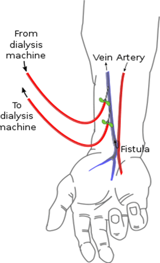
    220px-Radiocephalic_fistula_svg

    AV (arteriovenous) fistulas are recognized as the preferred access method. To create a fistula, a vascular surgeon joins an artery and a vein together through anastomosis. Since this bypasses the capillaries, blood flows rapidly through the fistula. One can feel this by placing one’s finger over a mature fistula. This is called feeling for "thrill" and produces a distinct ‘buzzing’ feeling over the fistula. One can also listen through a stethoscope for the sound of the blood "whooshing" through the fistula, a sound called bruit.

    Fistulas are usually created in the nondominant arm and may be situated on the hand (the ‘snuffbox‘ fistula’), the forearm (usually a radiocephalic fistula, or so-called Brescia-Cimino fistula, in which the radial artery is anastomosed to the cephalic vein), or the elbow (usually a brachiocephalic fistula, where the brachial artery is anastomosed to the cephalic vein). A fistula will take a number of weeks to mature, on average perhaps 4–6 weeks. During treatment, two needles are inserted into the fistula, one to draw blood and one to return it.

    The advantages of the AV fistula use are lower infection rates, because no foreign material is involved in their formation, higher blood flow rates (which translates to more effective dialysis), and a lower incidence of thrombosis. The complications are few, but if a fistula has a very high blood flow and the vasculature that supplies the rest of the limb is poor, a steal syndrome can occur, where blood entering the limb is drawn into the fistula and returned to the general circulation without entering the limb’s capillaries.

    Maximum tourniquet time for the upper limb is –
    a) 1/2 hour b) 1 hr (JIPMER 87)
    c) 1-1/2 hrs.
    d) 2 hrs
    e) 2-1/2 hrs

    Sabiston:-  Tourniquet Application:-       The tourniquet is used to provide a bloodless field so that clear visualization of all structures in the operative field is obtained. Penrose drains, rolled rubber glove fingers, or commercially available tourniquets can be used on digits. Great care must be taken in using any constrictive device on digits because narrow bands cause direct injury to underlying nerves and digital vessels. With the use of an arm tourniquet, the skin beneath the cuff must be protected with several wraps of cast padding. During skin preparation, this area must be kept dry to prevent blistering of the skin under an inflated cuff over moist padding. The cuff selected needs to be as wide as the diameter of the arm. Standard pressures used are 100 to 150 mm Hg greater than systolic blood pressure. The cuff is deflated every 2 hours for 15 to 20 minutes (5 minutes of reperfusion for every 30 minutes of tourniquet time) to revascularize distal tissues and to relieve pressure on nerves locally before reinflating the cuff for more extensive procedures.  Exsanguination of the extremities is performed by wrapping the extremity with a Martin’s bandage in all cases, except those involving infection or tumors. In these latter cases, because of the possibility of embolization by mechanical pressure, exsanguination by bandage wrapping needs to be avoided. Simple elevation of the extremity for a few minutes before tourniquet inflation suffices.

    Bailey:-  Tourniquet:-    A bloodless field is essential for accurate surgery. A well-padded tourniquet above the elbow, inflated to 75 mmHg pressure over the systolic blood pressure, is usually satisfac­tory. The time should not exceed 2 hours. An Esmarch bandage or a rubber-tube exsanguinator are effective, but should be avoided for tumour or infection cases lest the pathology is spread systemically. In the finger, a tourniquet can be made by placing a sterile glove on the patient, snipping off the tip and then rolling the glove down to the base of the finger.


    Thromboembolism after pelvic surgery is usually from the veins – (A189)
    a) iliac b) Calf
    c) Femoral d) Pelvic

    An obese patient develops acute oedematous lower limb following a Pelvic surgery. Deep vein thrombosis is suspected . The most useful investigation in this case would be – (UPSC 2002)
    a) Doppler imaging b) Fibrinogen uptake
    c) Venography d) Plethysmography

    Which of these is not a risk factor for thromboembolism – (TN 2001)
    a)Myocardial infarction
    b)Hypertension
    c)Estrogen therapy
    d)Superficial thrmbophlebitis

    Most common cause of death in patients with
    Burger’s disease is – (AIIMS 87)
    a) Gangrena b) Pulmonary embolism
    c) Myocardial infarction d) Carcinoma lung

    Which of the following best responds to sympathectomy – (JIPMER 86)
    a) Burger’s disease b) Hyperhydrosis
    c) Raynaud’s disease d) Acrocyanosis

    Intermittent claudication at the level of the hip indicates – (PGI 87)
    a)Popliteal artery occlusion
    b)Bilateral iliac artery occlusion
    c)Common femoral occlusion
    d)superficial femoral artery occlusion

    The artery commonly involved in cirsoid aneurysm is – (PGI 88)
    a) Occipital b) Superficial temporal
    c) Internal carotid d) External carotid

    Plusating tumours include all except – (PGI 88)
    a)Bone sarcoma
    b)Osteoclastoma
    c)Secondaries from hyper nephromas
    d)Secondary from prostate

    Preferred material for femoro popliteal bypass –
    a) Dacron b) PTFE (PGI 89)
    c) Saphenous vein d) Gortex

    Prosthetic materials For bypass of the aortoiliac segment the favoured material is Dacron (Fig. 15.21a). Prostheses come in two types: woven and knitted. Woven grafts tend to leak less when first exposed to blood flow during surgery, but newer knitted prostheses may be sealed with gelatin or collagen by the manufacturer and may leak even less than their woven counterparts. In the final analysis, there is probably little to choose between any of the styles of Dacron graft; all achieve satisfactory results. For bypass in the femoropopliteal region, if autogenous long saphenous vein (or other veins such as the short saphenous or arm vein) is not available, PTFE (Fig. 15.21b) or glutaraldehyde-tanned, Dacron-supported, human umbilical vein (Fig. 15.21c) may be employed. In general, any vein used requires a diameter of at least 3.5mm. For profundaplasty, a small piece of vein may be used or, alternatively, PTFE or Dacron. Suture materials for vascular surgery are usually monofilament in nature; polypropylene has been particularly popular. In the aorta it is usual to use 2/0 or 3/0 polypropylene. In the femoral artery at the groin it is usual to use 4/0 or 5/0 polypropylene. Finer sutures, up to 7/0, may be needed further down the limb. PTFE may (alternatively) be stitched using a suture of the same material. PTFE sutures tend to cause less bleeding through stitch holes in the graft substance.
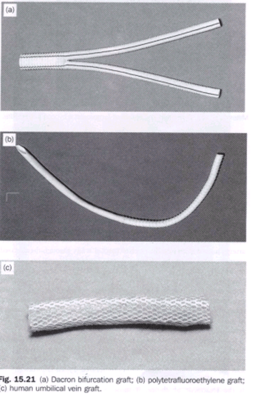
    image

    Most common cause of aneurysm of abdominal aorta is – (A196)
    a) Trauma b) Atherosclerosis
    c) Syphilis d) Cystic medial necrosis

    The most common complication of an aortic aneurysm size 8 cm is – (Delhi, PG 96)
    a) Rupture b) Intramural thrombosis
    c) Embolism d) Calcification

    Management of a cause of iliac artery embolism requires – (JIPMER 81, UPSC 86)
    a)Embolectomy
    b)Injection of vasodilators
    c)Hypotensive therapy
    d)Sympathectomy

    Intermittent claudicatin is caused by – (TN 89)
    a)Venous occlusion
    b)Arteria insufficiency
    c)Nerual compression
    d)Muscular dystrophy

    Burger’s disease is seen in – (PGI 88)
    a) Only male b) Age less than 40
    c) Age more than 40 d) Smoker

    In the abdomen, aneurysms of the …. commonly occur next only to the aorta …. – (PGI 88)
    a)Internal iliac artery
    b)External iliac artery
    c)Splenic artery
    d)Inferior mesentric artery

    In extraperitoneal approach, to left sympathectomy the following may be injured –
    a) Ureter b) Gonadal vessels
    c) A+B d) IVC

    Lumbar sympathectomy is indicated in – (TN 90)
    a)Intermittent claudication
    b)TAO with skin changes
    c)Burger’s disease
    d)Raynaud’s disease

    Commonest site of throboangitis obliterans is –
    a) Femoral artery b) Popiteal artery (A190)
    c) iliac artery d) Pelvic vessels


    Treatment of acute femoral embolus is- (AIIMS 91)
    a)Warfarin
    b)Heparin
    c)Immediate embolectomy
    d)Embolectomy after 5 days bed rest

    Ganglion which is spared in Lumbar sympathetomy is – (JIPMER 92)
    a) Ll b) L2
    c) L3 d) L4

    Surgical lumbar sympathectomy may be indicated in arterial disease. Surgical or chemical (phenol injection) sympathectomy may be used to treat rest pain or other troublesome sensory symptoms in arterial disease or in causalgia. The segment of the chain including the second and third lumbar ganglia is removed: preservation of the first lumbar ganglion is said to lessen the risk of ejaculatory problems 
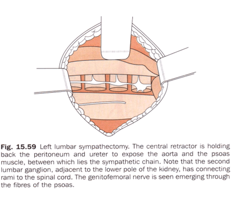
    image

    Vessels most commonly involved in thrombo angitis obliterans – (AIIMS 92)
    a)ilio-femoral
    b)Aorto-iliac
    c)Femora popliteal
    d)Arterior and Posterior tibial

    Following are used in treatment of Buergers disease except – (Al 93)
    a) Trental b) Anticoagulation
    c) Sympathectomy d) Antiplatelets

    Buerger’s disease affects all except – (PGI 01)
    a) Small arteries b) Small veins
    c) Medium -size arteries d) Multiparity
    e) First pregnancy after 30 years

    Buerger’s disease is associated with – (rGI 02)
    a) smoking b) Poor nutrition
    c) Alcohol d) Prolonged standing
    e) Superficial thrombophlebitis

    The most common cause of peripheral limb ischaemia in India is – (AIIMS NOV 05)
    a) Trauma b) Altherosclerosis
    c) Buerger’s disease d) Takayastu’s disease

    A 45-year-old male having a long history of cigarette smoking presented with gangrene of left foot. An amputation of the left foot was done. Representative sections from the specimen revealed presence of arterial thrombus with neutrophilic infiltrate in the arterial wall. The inflammation also extended into the neighbouring veins and nerves. The most probably diagnosis is – (AIIMS 06)
    a)Takayasu arteritis
    b)Giant cell arteritis
    c)Hypersensitivity angiitis
    d)Thromboangiitis obliterans

    Ans. is ‘d’ i.e., Thromboangiitis obliterans [Ref: Hariison 166/e p. 1487; Schwartz 86/e p. 792]

    THROMBOANGIITIS OBLITERANS :- Thromboangiitis obliterans (Buerger’s disease) is an inflammatory occlusive vascular disorder involving small and medium-sized arteries and veins in the distal upper and lower extremities. Cerebral, visceral, and coronary vessels may be affected rarely. This disorder develops most frequently in men <40 years. The prevalence is higher in Asians and individuals of eastern European descent. While the cause of thromboangiitis obliterans is not known, there is a definite relationship to cigarette smoking in patients with this disorder. In the initial stages of thromboangiitis obliterans, polymorphonuclear leukocytes infiltrate the walls of the small and medium-sized arteries and veins. The internal elastic lamina is preserved, and a cellular, inflammatory, thrombus develops in the vascular lumen. As the disease progresses, mononuclear cells, fibroblasts, and giant cells replace the neutrophils. Later stages are characterized by perivascular fibrosis, organized thrombus, and recanalization. The clinical features of thromboangiitis obliterans often include a triad of claudication of the affected extremity, Raynaud’s phenomenon, and migratory superficial vein thrombophlebitis. Claudication is usually confined to the calves and feet or the forearms and hands because this disorder primarily affects distal vessels. In the presence of severe digital ischemia, trophic nail changes, painful ulcerations, and gangrene may develop at the tips of the fingers or toes. The physical examination shows normal brachial and popliteal pulses but reduced or absent radial, ulnar, and/or tibial pulses. Arteriography is helpful in making the diagnosis. Smooth, tapering segmental lesions in the distal vessels are characteristic, as are collateral vessels at sites of vascular occlusion. Proximal atherosclerotic disease is usually absent. The diagnosis can be confirmed by excisional biopsy and pathologic examination of an involved vessel. There is no specific treatment except abstention from tobacco. The prognosis is worse in individuals who continue to smoke, but results are discouraging even in those who do stop smoking. Arterial bypass of the larger vessels may be used in selected instances, as well as local debridement, depending on the symptoms and severity of ischemia. Antibiotics may be useful; anticoagulants and glucocorticoids are not helpful. If these measures fail, amputation may be required.

    Lumbar sympathectomy:- Operative method. Using a transverse loin incision, an extraperitoneal approach is used in which the colon and peritoneum, to which the ureter clings, are stripped medially so as to expose the inner border of the psoas muscle .The sympathetic trunk lies on the sides of the bodies of the lumbar vertebrae; on the right side it is overlapped by the vena cava. Lumbar veins are apt to cross the trunk superficially. The sympathetic trunk is divided on the side of the body of the fourth lumbar vertebra. It is then traced upwards to be divided above the large second lumbar ganglion, which is easily recognised by the number of white rami which join it. Care should be taken not to mistake small lymph nodes, lymphatics, the genitofemoral nerve or the occasional tendinous strip of the psoas minor for the sympathetic chain. It is possible to perform the operation via an endoscope after the creation of a suitably expanded retroperitoneal tissue plane. Along with a decline in the recognised indications for sympathectomy there has been a move away from the operative approach in favour of the less hazardous chemical (phenol) sympathectomy.
    Chemical method. This is contraindicated in patients taking anticoagulants. Under radiographic fluoroscopic control, with the   patient in the lateral position, local anaesthetic is injected. A long spinal needle is then inserted  to seek the side of the vertebral body and to pass alongside it to reach the lumbar sympathetic chain. After confirming the needle position by injection of contrast agent, approximately 5 ml of phenol in water is injected. This is usually done at two sites: beside the bodies of the second and fourth lumbar vertebrae. Great care is needed to avoid penetrating the aorta, cava or ureter; the plunger of the syringe must always be drawn back before injection to exclude the presence of blood.

    In a lumbar sympathectomy the sympathetic chain in its usual position is likely to confused with the – (pal 81, AIIMS 80, 82)

    a) Ureter b) Psoas minor
    c) Genitofemoral nerve
    d) Ilioinguinal nerve e) Lymphatics

    The commonest cause of aneurysm formation
    is – (JIPMER 80, Delhi 89)
    a) Gun shot injury b) Syphilis
    c) Congenital factors
    d) Atherosclerosis

    Bullet wounds near major blood vessels should
    be explored only if- (PGI 81, AMC 85)
    a)The extremity is cold
    b)The fingers or toes are paralysed
    c)The pulse is weakened
    d)There in no pulse
    e)In all cases regardiess of physical findings

    A knitted Dacron artery graft (PGI 99, AIIMS 84)
    a)Is not porous
    b)Is eventually dissolived by tissue reaction
    c)Never gets infected
    d)Can be easily incised and the opening resutured

    The sequence of symptoms in pulmonary embolism is – (JIPMER 89, DNB 90)
    a)Fever, pain, dyspnoea
    b)Fever, dyspnoea
    c)Dysponea, pain, haemoptysis
    d)Dysponea, cough, purulent sputum

    A useful through temporary improvement in a patient’s ischaemic foot can be attained by giving intravenously – (PGI 79, Delhi 84)
    a) 10% Mannitol b) 10% Dextrose
    c) Dextran 40 d) Dextran 100

    Diabetic gangrene is due to – (Kerala 94)
    a)’schema
    b)Increased blood glucose
    c)Altered defence by host and neuropathy
    d)All of the above

    All are true about Embolic Arterial occlusion except-
    a)No previous history (JIPMER 95)
    b)Muscles are unaffected
    c)Pulse is absent
    d)Anaesthesia is present

    All are true about Raynauds phenomena except-
    a)Exposure to cold aggravate (Kerala 95)
    b)Spasm of vessels
    c)More common is females
    d)Atherosclerosis of vessels

    The commonest site of iodgement of a pulmonary embolus is in the territory at – (UPSC 95)
    a) Rt. lower lobe b) Rt. upper lobe
    c) Lt. lower lobe
    d) Lt. upper lobe

    Kaposi sarcoma is commonly seen in – (AMU 95)
    a) Upper limbs
    b) Lower limbs
    c) Head and Neck d) Trunk

    Which of the following causes meximum bleeding-
    a)Partial arterial severing (PGI 95)
    b)Complete arterial severing
    c)Artery caught between fractured ends of bones
    d)Intimal tear

    In a 40 years old male thrombus in the common femoral artery is because of – (AIIMS 97)
    a)Atheroma
    b)Thrombangits obliterans
    c)Reynauds disease
    d)Abdominal mass

    One of the following is not indicated for arterial
    leg ulcer – (PGI 96)
    a) Debridement b)
    Elevation of limb
    c) Head end of bed is raised d) Low dose aspirin

    Not used as graft material in peripheral vascular disease – (PGI 97)
    a) Dacron graft b) Vein
    c) PTFE d) PVC

    The commonest cause of arterio-venous fistula is-
    a)Penetrating injury (MP 97)
    b)Congenital
    c)Neoplasmic invasion of an artery and adjacent vein
    d)Aneurysm of the artery eroding a vein

    AV fistula leads to all except – (AIIMS 98)
    a)Sinus tachycardia
    b)Increased preload
    c)Cardiac arrythm ias
    d)Increased cardiac output

    Commonest peripheral aneurysm is – (SCTIMS 98) a) Popliteal b) Femoral
    c) Carotid d) iliac

    Popliteal aneurysm-All are true except-
    a)Presents as a swelling (SCTIMS 98) behind the knee
    b)Presents with symptoms due to complication
    c)Surgery is indicated in case of complication
    d)Uncommon among peripheral aneurysm

    Graft used in infra inguinal by pass is 4Jipmer 2K)
    a) PTFE b) Dacron
    c) Autologous vein d) Autologous artery

    True about Erythrocyanosis except — (A.P 96)
    a)Affects young girls
    b)Cold peripheries
    c)Palpable pulses
    d)Ulceration & gangrene of fingers

    Diabetic gangrene is due to AfE (TN 86)
    a)Vasospasm
    b)Atherosclerosis
    c)Peripheral neuritis
    d)Increased sugar in blood

    Pseudo aneurysms are most commonly due- (Jipmer a) Atherosclerosis b) Trauma 93)
    c) Congenital deficiency d) Infections

    Bilateral pulseless disease in upper limbs in caused
    by- (PGI 97)
    a) Aortoarteritis b) Coarctation of aorta
    c) Fibromuscular dysplsia d) Buerger’s disease

    Abdominal Aneurysm is characterized by all except – (PGI 2000)
    a)Elective surgery complication should be < 5%
    b)Emergency surgery complication < 10%
    c)Rarely asymptomatic before rupture

    d)Bigger the size it is more prone to rupture

    Dissection of which artery is seen in pregnancy –
    a) Carotid artery b) Aorta (PGI 2000)
    c) Coronary A d) Femoral artery

    Peripheral arterial occlusion (Sudden onset) is characterized all except – (PGI 2000)
    a) Paresthesia b) Rubor
    c) Pallor d) Pain

    In which one of the following conditions Dactylitis CANNOT be seen- (UPSC 02)
    a) Sickle – cell anemia b) Beta thalassemia
    c) Congenital syphilis d) Tuberculosis
    e) Sarcoidosis

    Not seen in sudden onset peripheral arterial occlusion – (Kerala 04)
    a) Pain b) Rubor
    c) Pallor d) Anesthesia

    Drug used for Burger’s disease – (MAHE 05)
    a) Xanthinol micotinate b) Propranolol
    c) GTN d) All the above

    Pseudoarterial aneurysm in drug abuser’s seen in -a) Radial b) Brachial (PGI June 05)
    c) Femoral d) Carotid
    e) Pedal

    Syndrome of internal iliac artery occlusion manifested by – (PGI June 05)
    a)Pain in calf
    b)Absent pulse at the dorsalis pedis artery
    c)Intermittent claudication

    d)Gangrene

    The most common cause of peripheral limb ischaemia in India is – (AIIMS NOV 05)
    a) Trauma b) Altherosclerosis
    c) Buerger’s disease d) Takayastu’s disease

    Lumbar sympathectomy is of value in the management of- (AI 05)
    a)Intermittent claudication
    b)Distal ischaemia affecting the skin of the toes
    c)Arteriovenous Fistula
    d)Back pain

    The most common cause of acquired arteriovenous fistuala is – (Al 06)
    a) Bacterial infection b) Fungal infection
    c) Blunt trauma d) Penetrating trauma

    Etiopathogenesis of diabetic foot include the following except- (UPSC 07)
    a)Myelopathy
    b)Osteoarthropathy
    c)Microangiopathy
    d)Infection

    Fogarty’s catheter is used for – (UPSC 07)
    a)Drainage of urinary bladder
    b)Parenteral hyperalimentation
    c)Removal of embolus from blood vessels
    d)Ureteric catheterisation

    Pseudoaneurysms in IV drug abusesrs seen commonly in – (PGI June 07)
    a)Brachial artery
    b)Radial artery
    c)Femoral artery

    Treatment of femoral artery naeurysm –
    a)Ultrasound guided compression of the neck of aneurysm (PGI June 07)
    b)Thrombin injection
    c)Bypass graft repair


    Venous Disorders:-

    Bailey & Love,Gray, Sabiston

    imageimageimageimage 

    Perforators are not present at – (AIIMS Nov 07)
    a) Ankle b) Medial calf
    c) Distal to calf d) Below inguinal ligament


    The superficial veins are in the subcutaneous connective tissue and are interconnected with and ultimately drain into the deep veins. The superficial veins form two major channels-the great saphenous vein and the small saphenous vein. Both veins originate from a dorsal venous arch in the foot:

    • the great saphenous vein originates from the medial side of the dorsal venous arch, and then ascends up the medial side of the leg, knee, and thigh to connect with the femoral vein just inferior to the inguinal ligament;
    • the small saphenous vein originates from the lateral side of the dorsal venous arch, ascends up the posterior surface of the leg, and then penetrates deep fascia to join the popliteal vein posterior to the knee; proximal to the knee, the popliteal vein becomes the femoral vein.

    The superficial and deep veins join at a number of points. The short saphenous vein terminates at the saphenopopliteal junction (SPJ) and the long saphenous vein at the saphenofemoral junction (SFJ) in the groin. Here the flow in the superficial veins joins that in the deep veins. There is, in addition, a number of places in the calf and thigh where flow in the superficial veins may also join that in the deep veins. These is the ankle, calf and thigh communicating or perforating veins (Fig. 16.3). The names of these veins come from their course from the superficial to the deep venous system in which they perforate the deep fascia of the leg. Near the ankle are the Cockett perforating veins, near the knee the Boyd perforators and in the thigh the Hunterian perforating vein. All veins in the upper and lower limbs contain valves every few centimetres which ensure that blood flows towards the heart.

    Multiple perforator veins traverse the deep fascia to connect the superficial and deep venous systems. Clinically important perforator veins are the Cockett and Boyd perforators. The Cockett perforator veins drain the medial lower leg and are relatively constant. They connect the posterior arch vein (a tributary to the GSV) and the posterior tibial vein. They may become varicose or incompetent in venous insufficiency states. The Boyd perforator veins connect the GSV to the deep veins approximately 10 cm below the knee and 1 to 2 cm medial to the tibia.

    Earliest sign of deep vein thrombosis is(AIIMS 87) a) Calf tenderness b) Rise in temperature c) Swelling of calf muscle d) Homan’s sign

     Injection sclerotherapy for varicose veins is by using – (PGI 88)
    a) Phenol b) Absolute alcohol
    c) 70% alcohol d) Ethanolamine oleate

    Sclerotherapy acts by destroying the venous endothelium. Sclerosing agents include hypertonic saline, sodium tetradecyl sulfate, and polidocanol. Concentrations of 11.7 to 23.4% hypertonic saline, 0.125 to 0.250% sodium tetradecyl sulfate, and 0.5% polidocanol are used for telangiectasias. Larger varicose veins require higher concentrations: 23.4% hypertonic saline, 0.50 to 1% sodium tetradecyl sulfate, and 0.75 to 1.0% polidocanol.83 Elastic bandages are wrapped around the leg after injection and worn continuously for 3 to 5 days to produce apposition of the inflamed vein walls and prevent thrombus formation. After the bandages are removed, elastic compression stockings should be worn for a minimum of 2 weeks. Complications from sclerotherapy include allergic reaction, pigmentation, thrombophlebitis, DVT, and possible skin necrosis.

    White leg is due to – (TN 90)
    a)Femoral vein thrombosis and lymphatic obstruction
    b)Deep femoral vein thrombosis
    c)Lymphatic obstruction only
    d)None of the above

    All of the following are seen in deep vein thrombosis except – (Al 90)
    a) Pain b) Discolouration
    c) Swelling d) Claudication

    The following is the commonest site for venous ulcer- (A IIMS 91)
    a)Instep of foot
    b)Lower 1/3 leg and ankle
    c)Lower 2/3 of leg
    d)middle 1/3 of leg

    The most important perforator of the Lower limb is between – (ALL INDIA 92 )
    a)Long saphenous and posterior tibial vein
    b)Short saphenous and posterior tibial vein
    c)Short saphenous and popliteal vein
    d)Long saphenous and femoral vein

    Best method for diagnosis of Deep vein thromvosis is – (JIPMER 92)
    a)Doppler examination
    b)Plethysmography
    c)Contrast phlebography
    d)1131 Fibrinogen studies

    Commonest complication varicose vein stripping is-
    a) Thrombo embolism b) Hemorrhage
    c) Ecchymosis d) Infection

    Investigation of choice for diagnosis of deep vein thrombosis – (AIIMS 92)
    a) Venogram
    b) Doppler
    c) Isotope scan d) Homans sign

    Pulsating varicose vein in ayoung adult is due to-
    a)Arteriovenous fistula (AIIMS 92)
    b)Sapheno femoral incompetence
    c)Deep vein thrombosis
    d)Abdominal tumour

    Which is not used in treatment of Superficial venous thrombosis – (AIIMS 92)
    a)Immediate anticoagulation
    b)Rest and elevation
    c)Analgesics
    d)Treat assosiated malignancy

    Most common complication of varicose vein stripping is – (JIPMER 78, AIMS 79,92)
    a) Infection b) Haemorrhage
    c) Ecchymosis d) Thrombo embolism

    An operated case of varicose veins has a recurrence rate of – (AIIMS 80, AP 89)
    a) About 10% b) About 25%
    c) About 50% d) Over 60%

    What is acceptable in the management of femoral vein thrombosis – (AIIMS81, PGI 86)
    a)Bed rest and spiral elastic bandages
    b)A venogram
    c)Thrombectomy
    d)Embolectomy
    e)A mobin udin umbrella inserted into the vein

    Operative Venous Thrombectomy

    In patients with acute iliofemoral DVT, surgical therapy is generally reserved for patients who worsen with anticoagulation therapy and those with phlegmasia cerulea dolens and impending venous gangrene. If the patient has phlegmasia cerulea dolens, a fasciotomy of the calf compartments is first performed. In iliofemoral DVT, a longitudinal venotomy is made in the common femoral vein and a venous balloon embolectomy catheter is passed through the thrombus into the IVC and pulled back several times until no further thrombus can be extracted. The distal thrombus in the leg is removed by manual pressure beginning in the foot


    Operations for varicose veins are best accomplished by – (PGI 81, AIIMS 84, 86)
    a)Stripping
    b)Multiple subcutaneos ligatures
    c)Subfascial ligatures
    d)Division and ligation at the superficial venous system

    A 60-years old male has been operated for carcinoma of caecum and right hemicolectomyhas been done. On the fourth post – oprative day, the patient develops fever and pain in the legs. The most important clinical entity one should lookfor is – (UPSC 96)
    a)Urinary tract infection
    b)Intravenous line infection
    c)Chest infection
    d)Deep vein thrombosis

    All of following may be predisposing factors for deep vein thrombosis except – (AIIMS 95)
    a) Oral contrceptives b) Nephrotic syndrome
    c) Sickle cell anemia d) Thrombocytosis

    The duration of heparin therapy in deep vein thrombosis is – (CUPGEE 96)
    a) 7 – 10 days b) 15-20 days
    c) 3-4 days d) 1 month

    Cocket & Dodd’s operation is for (AP 96)
    a)Saphenofemoral flush ligation
    b)Subfascial ligation
    c)Deep vein thrombosis
    d)Diabetic foot

    In obstruction of inferior vena cava there is -(A197)
    a)Prominent thoraco epigastric vein
    b)Caput medusa
    c)Hemorrhoids
    d)Esophageal varices

    Most accurate & non invasive method for diagnosing deep vein thrombosis – (JIPMER 98)
    a)Doppler duplex
    b)Plethesmography
    c)Radioactive labelled fibrinogen
    d)Angiography

    Most common site for venous thrombosis -(JIPMER a) Popliteal vein b) Soleal vein 98)
    c) Femoral vein d) Internal iliac vein

    Deep vein thrombosis is caused by all except –
    a)Lower limb trauma (AIIMS 98)
    b)Hip and pelvic surgery
    c)Subungual melanoma
    d)Cushing’s syndrome

    Which of the following test is used to detect perforator incompetence in varicose- (JIPMER 2K)
    a) Trendelenberg test b) Fegan’s test- (localise)
    c) Morissey’s test d) Homan’s test
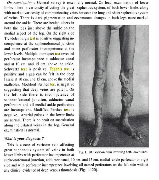
    image

    image

    image

    image

    image


    The deficiency of all of the following factors increases the incidence of thrombus formation except – (UPSC 2K)
    a) Lipoprotein A b) Protein – C
    c) Anti – thrombin III d) Protein – S

    The most common vein to get thrombosed is the – (AIIMS 99)
    a) Long saphenous b) Short saphenous
    c) Both d) Posterior tibial

    Brodie -Trendlenburg test demonstrates-
    a)Mid – thigh perforation (ORRISA 98)
    b)Deep vein thrombosis
    c)Sapheno — femoral incompetence
    d)Calf perforators

    An intern was doing saphenous cannulation for a burns patient. Then the patient developed sudden onset of pain along the medial border of the correponding foot. Which nerve must have been accidentally ligated – (AIIMS 2K)
    a) Sural nerve b) Deep peroneal nerve
    c) Saphenous nerve d) Genicular nerve

    In DVT all are seen except (CMC 2001)
    a)High fever
    b)Increased temperature at site
    c)Pain
    d)Tenderness

    Low Grade Pyrexia is seen not High.

    An obese patient develops acute oedematous lower limb following a Pelvic surgery. Deep vein thrombosis is suspected . The most useful investigation in this case would be – (UPSC 2002)
    a) Doppler imaging b) Fibrinogen uptake
    c) Venography d) Plethysmography

    In a patient on anticoagulant therapy, the INR is maintained at – (UPSC 2002)
    a)1.5 to 2.5 times the normal
    b)2.5 to 3.5 times the normal
    c)3.5 to 4.5 times the normal
    d)4.5 to 5.5 times the normal

    DVT, investigation of choice is – (PGI 97)
    a) Doppler b) Plethysmography
    c) Venography d) X-ray

    In diabetic ulcer, following site is involved-(PGI 97)
    a) Heel b) Head of metatarsal
    c) Webs d) Tips of toes

    For prophylaxis of deep vein thrombosis used is –
    a)Warfarin (PGI 97)
    b)Heparin
    c)Pneumatic shock garment
    d)Graded stocking

    Effective methods of VTE prophylaxis involve the use of one or more pharmacologic or mechanical modalities. Currently available pharmacologic agents include low-dose UFH, LMWH, synthetic pentasaccharides, and vitamin K antagonists. Mechanical methods include intermittent pneumatic compression (IPC) and graduated compression stockings. Aspirin therapy alone is notadequate for DVT prophylaxis. These prophylaxis methods vary with regard to their efficacy, and the 2008 ACCP Clinical Practice Guidelines stratify their uses according to the patient’s level of risk.

    Harrison:-
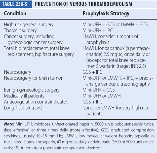
    image

    Schwartz:-

    Thromboembolism Risk and Recommended Thromboprophylaxis in Surgical Patients


    Level of Risk Approximate DVT Risk without Thromboprophylaxis (%) Suggested Thromboprophylaxis Options
    Low risk:- <10 No specific thromboprophylaxis
    Minor surgery in mobile patients   Early and "aggressive" ambulation
    Moderate risk:- 10–40 LMWH (at recommended doses), LDUH bid or tid, fondaparinux
    Most general, open gynecologic, or urologic surgery   Mechanical thromboprophylaxis
    Moderate VTE risk plus high bleeding risk    
    High risk:- 40–80 LMWH (at recommended doses), fondaparinux, oral vitamin K antagonist (INR 2–3)
    Hip or knee arthroplasty, hip fracture surgery    
    Major trauma, spinal cord injury   Mechanical thromboprophylaxis
    High VTE risk plus high bleeding risk    

    DVT = deep vein thrombosis; INR = International Normalized Ratio; LDUH = low-dose unfractionated heparin; LMWH = low molecular weight heparin; VTE = venous thromboembolism.


    Deep vein thrombosis is best diagnosed by-(PGI 97)
    a) Plethysmography b) Duplex ultrasound
    c) Radionuclide scan d) CT scan

    Which is true regarding Trendelenburg operation – (PGI 01)
    a)Sripping of the superficial varicose vein
    b)Flush ligation of the superficial varicose vein
    c)Ligation of the perforators
    d)Ligation of small tributaries at the distal end of superficial varicose vein
    e)Ligation of short saphenous vein

    Surgical treatment of varicose veins
    Surgical treatment of varicose veins is widely used and is effective in removing varicose veins of the main saphenous trunks, as well as their tributaries, down to a size of about 3 mm. Veins smaller than this are best treated by sclero­therapy. Surgical removal of varices is inappropriate where these form a major part of the venous drainage of the limb, for example where a deep vein thrombosis has destroyed the main axial limb veins and the patient relies on the superficial veins. This possibility may be suggested by the patient’s medical history and can be confirmed by duplex ultrasonography or venography.
    The main principles of surgical treatment are to ligate the source of the venous reflux (usually the SFJ or the SPJ) and to remove the incompetent saphenous trunks and the associated varices. Sapheno-femoral ligation alone, sometimes referred to as a ‘Trendelenburg procedure’, is associated with a high rate of recurrence of varices. Recent research has shown that it is necessary to remove the long saphenous vein to ensure that as much venous reflux as possible is eliminated. Similarly, communications between the many deep veins in the popliteal fossa and the short saphenous vein mean that some patients develop recurrences in the short saphenous vein due to the re-establishment of reflux from these veins. This problem may be eliminated by removing the short saphenous vein. Removal of the saphenous veins has the disadvantage that both veins are accompanied by a nerve that may be damaged in the vein stripping operation. To avoid nerve injury the long saphenous vein should not be removed below mid-calf level and great care should be exercised in removing the short saphenous vein.

    Sensory nerve injury is seen occasionally after removal of varicose veins. The saphenous nerve and its branches accompany the long saphenous vein in the calf, the sural nerve accompanies the short saphenous vein. Damage to the main part of these nerves occurs in about 1 per cent of operations, but small areas of anaesthesia may occur more frequently (in up to 10 per cent of patients). The adoption of inverting stripping techniques and avoidance of stripping the long saphenous vein below mid-calf level have reduced the risk of damage to these nerves. All patients should be warned before surgery that they may experience small areas of numbness and tingling after the operation. These changes are usually reversible but can be quite persistent.


    For which of the following venous abnormality is surgery indicated – (PGI 01)
    a)Deep vein incompetence with DVT
    b)Deep vein incompetence without DVT
    c)Varicosity > 3 cm
    b) Varicosity < 3 cm
    e) Saphenofemoral incompetence

    Imp:- Surgical treatment of varicose veins is widely used and is effective in removing varicose veins of the main saphenous trunks, as well as their tributaries, down to a size of about 3 mm. Veins smaller than this are best treated by sclero­therapy. Surgical removal of varices is inappropriate where these form a major part of the venous drainage of the limb, for example where a deep vein thrombosis has destroyed the main axial limb veins and the patient relies on the superficial veins. This possibility may be suggested by the patient’s medical history and can be confirmed by duplex ultrasonography or venography
    Saphenous vein ligation and stripping is still the more commonly performed procedure worldwide, and it may be the preferred therapy for patients with GSVs of very large diameter (>2 cm).

    Migratory thrombophlebitis is seen most commonly with- (PGI 02)
    a) Pancreatic ca b) Testicular ca
    c) Gastric ca d) Breast ca
    e) Liver ca

    Brodie-Tredenlenburg test is positive in – (PGI 02)
    a)Sapheno-Femoral incompetence
    b)Perforator competence above knee
    c)Deep vein incompetence
    d)Perforator competence below knee
    e)SVT

    Varicose veins are seen in – (PGI 02)
    a) DVT
    b) Superficial venous thrombosis

    c)AV fistula
    d)Prolonged standing
    e)Obesity

    Gold standard diagnostic test in varicose veins is
    a)Photoplethysmography (Jipmer 03)
    b)Duplex imaging
    c)Ultrasonography
    d)Radio – labeled fibrinogen study

    The initial therapy of documented deep venous thrombosis in a post operative case is –
    a)Subcutaneous heparin therapy (Karnataka 03)
    b)Intravenous heparin therapy
    c)Thropmbolytic therapy with urokinase
    d)Aspirin therapy

    Which one of the following reagents is not used as sclerosant in the treatment of bleeding varices –
    a) Ethyl alcohol .b) Ethanolamine oleate (ICS 05)
    c) Phenol d) Sodium morrhuate

    Site of diabetic for ulcer – (PGI June 05)
    a) Medial malleolus b) Lateral malleolus
    c) Heel d) Head of metatarsal
    e) Head of toes

    Harrison:-Approximately 15% of individuals with DM develop a foot ulcer (great toe or MTP areas are most common), and a significant subset will ultimately undergo amputation (14–24% risk with that ulcer or subsequent ulceration). Risk factors for foot ulcers or amputation include: male sex, diabetes >10 years’ duration, peripheral neuropathy, abnormal structure of foot (bony abnormalities, callus, thickened nails), peripheral arterial disease, smoking, history of previous ulcer or amputation, and poor glycemic control. Large callouses are often precursors to or overlie ulcerations.

    Which of the following statements is true regarding fat embolism – (AJIMS NOV 05)
    a)Most patients with major trauma involving long bones have urinary fat globules
    b)All patients with urinary fat globules develop fat embolism
    c)Peak incidence of respiratory insufficiency for pulmonary fat embolism is around day 7 after injury
    d)Heparin as an anticoagulant decreases mortality and morbidity in fat embolism syndrome

    Fat Embolism::- A certain degree of lung dysfunction occurs in all patients after long bone fractures, but clinically significant fat embolism syndrome as such develops in only 10 to 15 percent of these patients. Signs include hypoxia, tachycardia, mental status changes, and petechiae on the conjunctiva, axilla, or upper thorax. Fat globules in the urine are nondiagnostic, but lung infiltrates seen on chest radiograph confirm the presence of lung injury.
    The pathophysiology of fat embolism represents capillary endothelial breakdown causing pericapillary hemorrhagic exudates most apparent in the lungs and brain. Pulmonary edema and hypoxemia occur as a result of pulmonary exudates. Hypoxia and areas of cerebral edema may account for the variable neurologic abnormalities seen.
    The more severe cases of fat embolism involve fractures of the femur and tibia. Delays in fixation of bones and extensive reaming of the medullary canals contribute to perioperative morbidity and to the severity of fat embolism syndrome. Efforts to surgically correct fractures early and minimize trauma to the bone marrow lessen the degree of fat/bone marrow embolism. Patients with coexisting lung injury are at additional risk of fat embolism. Evidence suggests that fat may pass to the systemic circulation through a patent foramen ovale  or by transpulmonary passage.The chemical composition of the fat may even contribute to this process.  For this reason, it is preferable to minimize pulmonary artery hypertension to reduce transpulmonary passage of fat and limit pulmonary endothelial transudation of fluid.
    Treatment includes early recognition, oxygen administration, and judicious fluid management. Corticosteroids in large doses shortly after major trauma have been found to minimize the clinical presentation of fat embolism but are probably not necessary in most cases if oxygen therapy is administered. With appropriate fluid management, adequate ventilation, and the prevention of hypoxemia, outcome is usually excellent.


    Which of the following is true about varicocele except ? (Manipal 06)
    a)Incompetent valves of testicular vein are responsible for varicocele
    b)90% are on the left side
    c)Asymptomatic cases require surgery
    d)Femoral catheterization with spermatic vien ablation is done in recurrence

    Which one of the following is the investigation of choice for suspected deep vein thrombosis of the lower extremity ? (UPSC 07)
    a)Radioactive labelled fibrinogen uptake
    b)Ascending contrast phlebography
    c)D-dimer estimation
    d)Duplex ultrasonography

    With reference to varicocele, which one of the following is not true of it ? (UPSC 07)
    a)Varicosity of cremastric veins
    b)Left side is affected usually
    c)Feels like a bag of worms
    d)May lead to infertility


    All are done for a case of deep vein thrombosis except – (MAHE 07)
    a) Thrombolytic therapy b) Bandage
    c) Heparin d) Bed rest

    The practice of having a patient “out of bed into a chair” is one of the most thrombogenic positions that one could order a patient into. Sitting in a chair with the legs in a dependent position causes venous pooling, which in the postoperative milieu could easily be a predisposing factor in the development of thromboembolism

    Treatment regimens may include antithrombotic therapy, vena caval interruption, catheter-directed or systemic thrombolytic therapy, and operative thrombectomy

    studies, as well as the current ACCP guidelines, suggest that catheter-directed thrombolysis (with adjunctive angioplasty, venous stenting, and pharmacomechanical fragmentation and extraction) may be useful in selected patients with extensive iliofemoral DVT. Patients should have a recent onset of symptoms (<14 days), good functional status, decent life expectancy, and low bleeding risk.

    Table 24-2 Risk Factors for Venous Thromboembolism


    Acquired Inherited
    Advanced age Factor V Leiden
    Hospitalization/immobilization Prothrombin 20210A
    Hormone replacement therapy and oral contraceptive use Antithrombin deficiency
    Protein C deficiency
    Pregnancy and puerperium Protein S deficiency
    Prior venous thromboembolism Factor XI elevation
    Malignancy Dysfibrinogenemia
    Major surgery Mixed Etiology
    Obesity Homocysteinemia
    Nephrotic syndrome Factor VII, VIII, IX, XI elevation
    Trauma or spinal cord injury Hyperfibrinogenemia
    Long-haul travel (>6 h) Activated protein C resistance without factor V Leiden
    Varicose veins  
    Antiphospholipid antibody syndrome  
    Myeloproliferative disease  
    Polycythemia

    Early in the course of DVT development, venous thrombosis is thought to begin in an area of relative stasis, such as a soleal sinus vein or immediately downstream of the cusps of a venous valve in the axial calf veins. Isolated proximal DVT without tibial vein thrombosis is unusual. Early in the course of a DVT, there may be no or few clinical findings such as pain or swelling. Even extensive DVT may sometimes be present without signs or symptoms. History and physical examination are therefore unreliable in the diagnosis of DVT. In addition, symptoms and signs generally associated with DVT, such as extremity pain and/or swelling, are nonspecific. In large studies, DVT has been found by venography or DUS in ≤50% of patients in whom it was clinically suspected.Objective studies are therefore required to confirm a diagnosis of DVT or to exclude the presence of DVT.

    Investigation of Choice-Duplex USG

    With major advances in technology of imaging, magnetic resonance venography has come to the forefront of imaging for proximal venous disease. The cost and the issue of patient tolerance due to claustrophobia limit the widespread application, but this is changing. It is a useful test for imaging the iliac veins and the IVC, an area where duplex ultrasound is limited in its usefulness.

    Gold standard:-Injection of contrast material into the venous system is obviously and understandably the most accurate method of confirming DVT and the location.Not used usually.

    Clinical symptoms may worsen as DVT propagates and involves the major proximal deep veins. Massive DVT that obliterates the major deep venous channel of the extremity with relative sparing of collateral veins causes a condition called phlegmasia alba dolens or white leg .This condition is characterized by pain, pitting edema, and blanching. There is no associated cyanosis. When the thrombosis extends to the collateral veins, massive fluid sequestration and more significant edema ensues, resulting in a condition known as phlegmasia cerulea dolens.Phlegmasia cerulea dolens is preceded by phlegmasia alba dolens in 50 to 60% of patients. The affected extremity in phlegmasia cerulea dolens is extremely painful, edematous, and cyanotic, and arterial insufficiency or compartment syndrome may be present. If the condition is left untreated, venous gangrene can ensue, leading to amputation

    SVT

    Superficial vein thrombophlebitis (SVT) most commonly occurs in varicose veins but can occur in normal veins. When SVT recurs at variable sites in normal superficial veins, it may signify a hidden visceral malignancy or a systemic disease such as a blood dyscrasia and/or a collagen vascular disease. This condition is known as thrombophlebitis migrans. SVT also frequently occurs as a complication of indwelling catheters, with or without associated extravasation of injected material. Upper extremity vein thrombosis has been reported to occur in 38% of patients with peripherally inserted central catheters; 57% of these developed in the cephalic vein . Finally, suppurative SVT may occur in veins with indwelling catheters and may be associated with generalized sepsis.

    Rx of SVT by schwartz:- In patients with SVT not within 1 cm of the saphenofemoral junction, treatment consists of compression and administration of an anti-inflammatory medication such as indomethacin. In patients with suppurative SVT, removal of any existing indwelling catheters is mandatory, and excision of the vein may be necessary. If the SVT extends proximally to within 1 cm of the saphenofemoral junction, extension into the common femoral vein is more likely to occur. In these patients, anticoagulation therapy for 6 weeks and GSV ligation appear equally effective in preventing thrombus extension into the deep venous system.

    Rx of SVT by Harrisons:- Treatment is primarily supportive. Initially, patients can be placed at bed rest with leg elevation and application of warm compresses. Nonsteroidal anti-inflammatory drugs may provide analgesia but may also obscure clinical evidence of thrombus propagation. If a thrombosis of the greater saphenous vein develops in the thigh and extends toward the saphenofemoral vein junction, it is reasonable to consider anticoagulant therapy to prevent extension of the thrombus into the deep system and a possible pulmonary embolism.

    ____________________________________________________________________________________
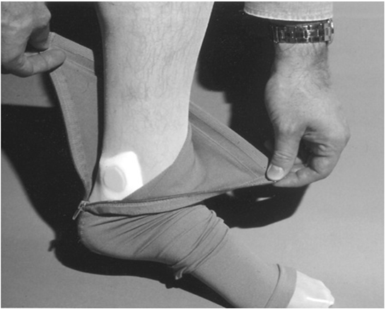
    image

    Elastic compression stocking with zippered side to facilitate treatment of chronic venous insufficiency

    Compression therapy is most commonly achieved with graduated elastic compression stockings. Graduated elastic compression stockings, initially developed by Conrad Jobst in the 1950s, were made to simulate the gradient of hydrostatic forces exerted by water in a swimming pool. Elastic compression stockings are available in various compositions, strengths, and lengths, and can be customized for a particular patient.

    To improve compliance, patients should be instructed to wear their stockings initially only as long as it is easily tolerated and then gradually to increase the amount of time the stockings are worn. Alternatively, patients can be fitted with lower-strength stockings initially followed by introduction of higher-strength stockings over a period of several weeks. Many commercially available devices, such as silk inner toe liners, stockings with zippered sides,and metal fitting aids ,are available to assist patients in applying elastic stockings.
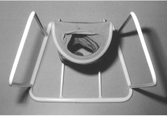
    image

    Metal fitting aid to assist in placement of elastic compression stockings

    Lymphatic Disorders

    Lymphedema is extremity swelling that results from a reduction in lymphatic transport, with resultant pooling of lymph within the interstitial space. It is caused by anatomic problems such as lymphatic hypoplasia, functional insufficiency, or absence of lymphatic valves.

    The original classification system, described by Allen, is based on the cause of the lymphedema.

    Primary lymphedema is further subdivided into

    1. Congenital lymphedema may involve a single lower extremity, multiple limbs, the genitalia, or the face. The edema typically develops before 2 years of age and may be associated with specific hereditary syndromes (Turner syndrome, Milroy syndrome, Klippel-Trénaunay-Weber syndrome).The familial version of congenital lymphedema is known as Milroy’s disease and is inherited as a dominant trait

    2.Lymphedema praecox is the most common form of primary lymphedema, accounting for 94% of cases. Lymphedema praecox is far more common in women, with the gender ratio favoring women 10:1. The onset is during childhood or the teenage years, and the swelling involves the foot and calf. The familial version of lymphedema praecox is known as Meige’s disease.

    3.Lymphedema tarda is uncommon, accounting for <10% of cases of primary lymphedema. The onset of edema is after 35 years of age.

    Secondary lymphedema is far more common than primary lymphedema. Secondary lymphedema develops as a result of lymphatic obstruction or disruption. Axillary node dissection leading to lymphedema of the arm is the most common cause of secondary lymphedema in the United States. Other causes of secondary lymphedema include radiation therapy, trauma, infection, and malignancy. Globally, filariasis (caused by Wuchereria bancrofti, Brugia malayi, and Brugia timori) is the most common cause of secondary lymphedema.In developed countries, the most common causes of secondary lymphedema involve resection or ablation of regional lymph nodes by surgery, radiation therapy, tumor invasion, direct trauma, or less commonly, an infectious process.

    Lymphoscintigraphy has emerged as the test of choice in patients with suspected lymphedema.It cannot differentiate between primary and secondary lymphedemas; however, it has a sensitivity of 70% to 90% and a specificity of nearly 100% in differentiating lymphedema from other causes of limb swelling.

    Email to Colleague Print Version

    Malignant cell in Hodgkin’s lymphoma is –

    a) Reed sternberg cell b) Lymphocytes 85)
    c) Histiocyte d) Reticulum cells

    Chronic lymphedema predisposes to all except – (PGI 89)
    a) Lymphangiosarcoma b) Marjolins ulcer
    c) Recurrent infections d) Thickening of skin
    In the lower extremity the swelling involves the dorsum of the foot, and the toes have a squared-off appearance. In advanced cases, hyperkeratosis of the skin develops, and fluid weeps from lymph-filled vesicles

    Recurrent cellulitis is a common complication of lymphedema. Repeated infection results in further lymphatic damage, worsening existing disease. The clinical presentation of cellulitis ranges from subtle erythema and worsening of edema to a rapidly progressive soft tissue infection with systemic toxicity

    Commonest cause of unilareral pedal edema in india is – (A190)
    a) Filariasis b) Post traumatic
    c) Post irradiation d) Milroy’s disease

    All are true about congenital lymphedema except-
    a)It is bilateral (AI 91)
    b)Involve lower limb
    c)Almost always manifests before puberty
    d)Acute lymphangitis may occur
    All are true?

    The commonest cause for lymphedema of upper limb is – (AI 91)
    a) Filariasis b) Congenital
    c) Neck surgery d) Post mastectomy irradiation

    Commonest cause of upper limb lymphedema is –
    a) Congenital b) Filariasis (AI 92)
    c) Post mastectomy d) Irradiation

    ???

    ANS given in all guides is Filariasis but it should be Post Mastectomy.

    Secondary lymphedema, more common than the primary form, usually develops following disruption or obstruction of lymphatic pathways associated with a disease process, or following surgery or radiotherapy. Worldwide, filariasis is the most common cause of secondary lymphedema usually affecting lower limbs.In the United States, the most common secondary lymphedema develops in the upper extremity following axillary lymph node dissection.The incidence varies considerably, depending on the definition of edema. Results of one series of published reports reveal a 14 percent rate of secondary lymphedema in postmastectomy patients who also had undergone irradiation therapy.

    Milroys disease is – (JIPMER 92)
    a)Edema due to filareasis
    b)Post cellulitic lymphedema
    c)Congenital lymphedema
    d)Lymphedema following surgery

    Investigation of choice in detecting small para-aortic lymph node is – (JIPMER 92)
    a) Ultra sound scan b) CT scan
    c) Lymphangiography d) Arteriography

    Not Sure

    Contrast lymphangiography. Although few centres now perform this technique, it remains the standard by which all other lymphatic imaging is judged and provides precise information about the anatomy of the lymphatic system. It is now generally reserved for preoperative evaluation of patients with megalymphatics who arc being considered for bypass or fistula ligation.

    CT scanning may be useful in identifying pathologic intra-abdominal lymph nodes and masses and in identifying the extent and localization of fluid. Lymphangiography and lymphoscintigraphy may help localize lymph leaks and obstruction; this information is particularly useful for surgical planning.

    Milroys disease is lymphedema which is-(AMU 85)
    a)Familial
    b)Follows filariasis
    c)Follows erysipelas
    d)A sequele to white leg

    Lymphangiography of the leg is performed by –
    a)An injection of sodium diatrizoate (Hypaque) subcutaneously between the toes
    b)Injecting sodium diatrizoate retrogradely under pressure into a small vein on the dorsum of the foot
    c)Dissecting lymphatics through an incision on the dorsum of the foot
    d)The use of an infusion pump

    Direct-contrast lymphangiography provides the finest details of the lymphatic anatomy. However, it is an invasive study that involves exposure and cannulation of lymphatics at the dorsum of the forefoot, followed by slow injection of contrast medium (ethiodized oil).

    Radiologic lymphology is performed by first visualizing the lymphatics by injecting colored dye into the hand or foot. The visualized lymphatic segment is exposed through a small incision and cannulated with a 27- to 30-gauge needle. An oil-based dye is then injected slowly into the lymphatics over several hours. The lymphatic channels and nodes are then visualized with traditional radiographs . Lymphangiography is reserved for patients with lymphangiectasia or lymphatic fistulas, and patients who are being considered for microvascular reconstruction

    Under local anaesthesia, a small transverse incision is made in the dorsum of the foot after I ml of isosulphan blue has been injected subcutaneously to identify the lymphatics. Lymphatics are dissected out under loupe magnification and a 30G needle used to infuse lipid-soluble contrast at a rate of I ml in 8 minutes to a maximum of 7 ml (taking about 1 hour) into each limb

    Finding the cause of unilateral lympoedema of the leg includes – (JIPMER 78,79, PGI 85)
    a)Taking a family history
    b)Looking for chronic infection in the foot
    c)Looking for early malignant disease of the testis…(Not early.It should have been late metastatic )
    d)Looking for filariasis
    e)Performing a casoni
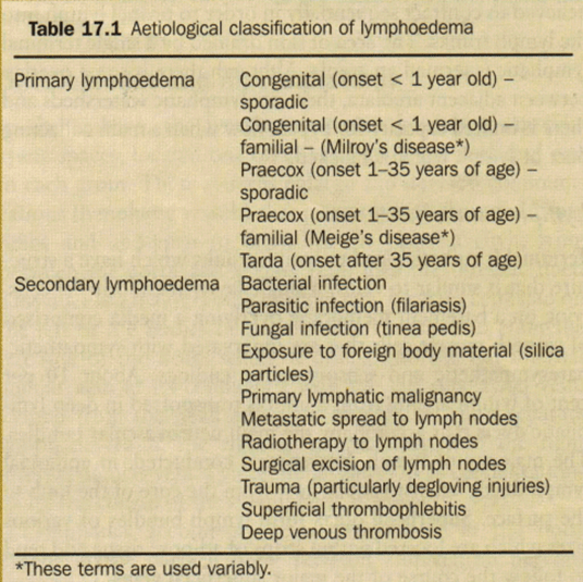
    image

    Treatment of Acute lymphangitis requires –
    a)Antibiotic and rest (JIPMER 81, AMC 84)
    b)Immediate lymphangiography
    c)Immediate multiple incisions
    d)No special treatment

    Acute inflammation of the Iymphatics
    Acute lymphangitis occurs when a deep or superficial infec­tion, often due to Streptococcus pyogenes or Staphylococcus aureus, spreads to the draining lymphatics and lymph nodes (lymphadenitis) where an abscess may form. Eventually this may progress to bacteraemia or septicaemia. The normal signs of infection (rubor, calor, dolor) are present and a red streak is seen in the skin along the line of the inflamed lymphatic. The part should be rested to reduce lymphatic drainage, elevated to reduce swelling and the patient treated with intravenous antibiotics based upon actual or suspected sensitivities. Failure to improve within 48 hours suggests inappropriate antibiotic therapy, the presence of undrained pus either in the lymph nodes or at the site of primary infection, or the presence of an underlying systemic disorder (malignancy, immunodeficiency). The lymphatic damage caused by acute lymphangitis may lead to recurrent attacks of infection and lymphoedema.
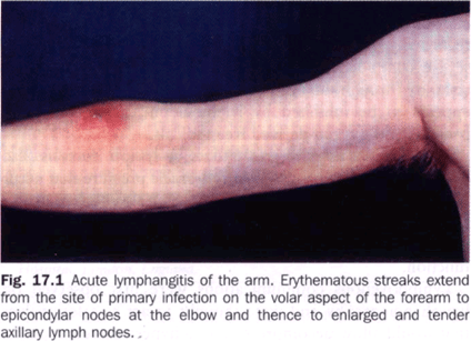
    image


    Total dose of radiation in Hodgkins dosease is – (JIPMER 95)
    a) 500 -1000 rad b) 1000 – 2000 rad
    c) 3000 – 5000 rad d) 5000 – 7000 rad

    The most important prognostic indicator of Hodgkin’s lymphoma is – (TN 95)
    a)Lymphocytic predominance histology
    b)Visceral involvement
    c)Hepatic involvement
    d)Involvement of spleen

    Diagnosis of Hodgkin’s disease is.confirmed by –
    a)CT scan (PGI 9 7)
    b)Bone marrow biopsy
    c)Lymph node biopsy
    d)Lymphangiography

    Grade I lymphedema means – (JIPMER 2K)
    a)Pitting edema upto the ankle
    b)Pitting edema upto the knee
    c)Non-pitting edema
    d)Edema disapearing after overnight rest
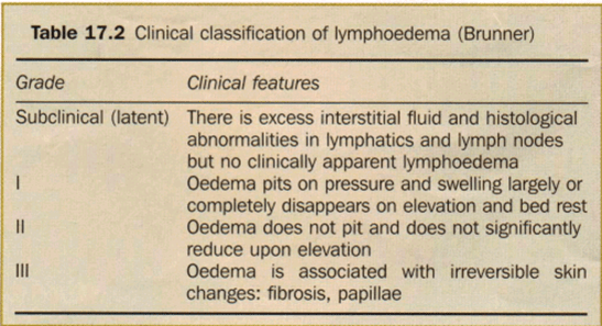
    image

    Lymphovenous anastomosis is done for – (PGI 97)
    a)Filarial lymphoedema
    b)Lymphoid cyst
    c)Cystic hygroma
    d)Malignant lymphoedema

    Surgery

    A variety of surgical procedures have been devised for the treatment of lymphedema. Surgical treatment involves either excision of extra tissue or anastomosis of a lymphatic vessel to another lymphatic or vein.125 In excisional procedures, part or all of the edematous tissue is removed. This does not improve lymphatic drainage but debulks redundant tissue. The microsurgical procedures involve the creation of a lymphaticolymphatic or lymphaticovenous anastomosis, which theoretically improves lymphatic drainage. No long-term follow-up data are available for these interventions, and therefore operative therapy for lymphedema is not well accepted worldwide. Furthermore, operative intervention can further obliterate lymphatic channels, worsening the edema.126

    Summary

    Lymphedema is a chronic condition caused by ineffective lymphatic transport, which results in edema and skin damage. Lymphedema is not curable, but the symptoms can be controlled with a combination of elastic compression stockings, limb elevation, pneumatic compression, and massage. Controlling the edema protects the skin and potentially prevents cellulitis

    True about lymphangioma – (PGI 03)
    a)It is a malignant tumour
    b)It is a congenital sequestration of lymphatic
    c)Cystic hygroma is a lymphangioma

    d)Laser excision is done
    e)Sclerotherapy is commonly done

    The most common site of lymphangiosarcoma is
    a)Liver (UPSC 04)
    b)Spleen
    c)Post mastectomy edema of arm
    d)Retroperitoenum

    Commonest cause of A/C Lymphadenitis in India –
    a)Barefoot walking (MAHE 05)
    b)TB
    c)Staphylococcal skin infection
    d)Lymphoma

    All of the following soft tissue sarcomas have a propensity for lymphatic spread except –
    a)Neurofibrosarcoma (AiimsNov 05)
    b)Synovial sarcoma
    c)Rhabdomyosarcoma
    d)Epitheloid sarcoma

    A 40 year old man presented with a flat 1 cm x 1 cm scaly, itchy black mole on the front of thigh. Examination did reveal any inguinal lymphadenopathy. The best course of management would be- (UPSC 07)
    a)FNAC of the lesion
    b)Incision biopsy
    c)Excisional biopsy
    d)Wide excision with inguinal lymphadenectomy

    In India, what is the most common cause of unilateral lymphoedema of lower limb ? (UPSC 07)
    a)Lymphoedema tarda
    b)Carcinoma of penis with metastatic nodes
    c)Filariasis
    d)Tubercular lymphadenopathy

    Posted in Medical | Tagged: , , , , , , | 18 Comments »

    Surgery MCQs (Burns)

    Posted by Dr KAMAL DEEP on May 30, 2011

    Exposure treatment is done for burns of the –
    a)Upper limb (JIPMER 87)
    b)Lower limbs
    c)Thorax
    d)Abdomen
    e)Head & neck

    Circumferential (encircling) burns of the trunk are generally unsuited for exposure because all of the burned surfaces cannot be adequately exposed to the air at one time.

    Granulating surfaces should never be exposed.


    Rule of nine to estimate surface area of a burnt patient was introduced by – (AIIMS 84)
    a) Mortiz kaposi b) Alexander wallace
    c) Joseph lister d) Thomas barclay

    Deep skin burns is treated with – (AIIMS 91)
    a)Split thickness graft
    b)Full thickness graft
    c)Amniotic membrane
    d)Synthetic skin derivatives

    In burns heat loss is by/ due to – (PGI 80,
    a)Dilatation of veins DELHI 80,92)
    b)Shock
    c)Exposed area by evaporation
    d)None of the above

    The cold water treatment of burns has the disadvantage that it increase the chances of –
    a) Pain b) Exudation (PGI 81, AIIMS 83)
    c) Infection d) None of the above

    Pus in burns form in – (PGI 79, DELHI 89)
    a) 2-3 Days b) 3-5 days
    c) 2-3 weeks d) 4

    One of folowing is not seen in severe burns -(Al 96)
    a) Hypovolemia b) Sepsis
    c) Duodenal ulcer d) Hyperthemia

    Undue restlessness in a patient during the immediate post burn period is often a manifestitation of-(Karp
    a) Hypoxia b) Hypovolemia 95)
    c) Hyperkalemia d) Anxiety


    image

    Sabiston:- First-degree burns are, by definition, injuries confined to the epidermis. These burns are painful and erythematous, blanch to the touch, and have an intact epidermal barrier. Examples include sunburn or a minor scald from a kitchen accident. First-degree burns do not result in scarring, and treatment is aimed at comfort with the use of topical soothing salves, with or without aloe, and oral nonsteroidal anti-inflammatory agents.

    Second-degree burns are divided into two types: superficial and deep. All second-degree burns have some degree of dermal damage, and the distinction is based on the depth of injury into this structure. Superficial dermal burns are erythematous and painful, blanch to touch, and often blister. Examples include scald injuries from overheated bathtub water and flash flame burns from open carburetors. These wounds spontaneously re-epithelialize from retained epidermal structures in the rete ridges, hair follicles, and sweat glands in 7 to 14 days. After healing, these burns may result in some slight skin discoloration over the long term. Deep dermal burns into the reticular dermis appear more pale and mottled, do not blanch to touch, but remain painful to pinprick. These burns heal in 14 to 35 days by re-epithelialization from hair follicles and sweat gland keratinocytes, often with severe scarring as a result of the loss of dermis

    Third-degree burns are full thickness through the epidermis and dermis and are characterized by a hard, leathery eschar that is painless and black, white, or cherry red. No epidermal or dermal appendages remain; thus, these wounds must heal by re-epithelialization from the wound edges. Deep dermal and full-thickness burns require excision with skin grafting to heal the wounds in timely fashion.

    Fourth-degree burns involve other organs beneath the skin, such as muscle, bone, and brain.

    Schwartz:- Burn wounds are commonly classified as superficial (first degree), partial thickness (second degree), full thickness (third degree), and fourth-degree burns, which affect underlying soft tissue. Partial-thickness burns are then classified as either superficial or deep partial thickness burns by depth of involved dermis. Clinically, first-degree burns are painful but do not blister, second-degree burns have dermal involvement and are extremely painful with weeping and blisters, and third-degree burns are hard, painless, and nonblanching

    Because full-thickness grafts are impractical for most burn wounds, split-thickness sheet autografts harvested with a power dermatome make the most durable wound coverings and have a decent cosmetic appearance.

    Schwartz:- Thighs make convenient anatomic donor sites, which are easily harvested and relatively hidden from an aesthetic standpoint. The thicker skin of the back is useful in older patients, who have thinner skin elsewhere and may have difficulty healing donor sites. The buttocks are an excellent donor site in infants and toddlers.

    The scalp is also an excellent donor site; the skin is thick and there are many hair follicles so it heals quickly. It has the added advantage of being completely hidden once hair regrows.

    Areas of cosmetic importance such as the face, neck, and hands should be grafted with nonmeshed sheet grafts to ensure optimal appearance.


    All requires hospitalization except – (Al 91)
    a)5% Burns in children
    b)10% Scalds in children
    c)Electrocution
    d)15% Deep burns in adults

    Indications for specialist referral in burns –
    a)> 20% superficial burn in adult (PGI 04)
    b)Only palms
    c)Scalds on head and face
    d)10% burns in infants
    e)10% deep burns in adult

    A burn patient is referred when – (PGI 04)
    a)10% superficial burn in child
    b)Scald in face

    c)25% superficial burn in adult
    d)25% deep burn in adult
    e)Burn in palm

    Improvements in burn care originated in specialized units specifically dedicated to the care of burned patients. These units consist of experienced personnel with resources to maximize outcome from these devastating injuries ( Box 22-1 ). Because of these specialized resources, burned patients are best treated in such places. Patients with the following criteria are referred to a designated burn center:

    1. Partial-thickness burns greater than 10% TBSA
    2. Burns involving the face, hands, feet, genitalia, perineum, or major joints
    3. Any full-thickness burn
    4. Electrical burns, including lightning injury
    5. Chemical burns
    6. Inhalation injury
    7. Burns in patients with preexisting medical disorders that could complicate management, prolong recovery, or affect outcome
    8. Any patient with burns and concomitant trauma (e.g., fractures) in which the burn injury poses the greater immediate risk for morbidity and mortality. In such cases, if the trauma poses the greater immediate risk, the patient may be initially stabilized in a trauma center before being transferred to a burn unit. Physician judgment is necessary in such situations, and decisions must be made in concert with the regional medical control plan and triage protocols
    9. Burned children in hospitals without qualified personnel or equipment to care for children
    10. Burns in patients who will require special social, emotional, or long-term rehabilitative intervention


    In 3’rd degree burns, all are seen except – (PGI 99)
    a)Vesicles are absent
    b)Painful
    c)Leathery skin
    d)Reddish due to Hb Infiltration

    Late deaths in burns is due to – (PGI 99)
    a) Sepsis b) Hypovolemia
    c) Contractures d) Neurogenic

    Burns with vesiculation, destruction of the epidermis and upper dermis is – (PGI 99)
    a) 1″ degree b) 2nd degree
    c) 3rd degree d) 4th degree

    True about burns – (PGI 2000)
    a)Hyperglycemia is seen in early burns
    b)Child with burns should have damp dressing
    c)Chemical powder burns should be kept dry
    d)3rd degree burns are painfull

    Metabolic derangements in severe burns are all except- (PGI 2000)
    a)cortico steroid secretion
    b)Hyperglycaemia
    c)secretion of HCl
    d)Neutrophil dysfunction

    Superficial burns; true is/are – (PGI 01)
    a)Always requires skin grafting
    b)Dry & inelastic
    c)Blister formation
    d)Painless
    e)Can be healed within 7 to 10 days

    Schwartz:- Clinically, first-degree burns are painful but do not blister, second-degree burns have dermal involvement and are extremely painful with weeping and blisters, and third-degree burns are hard, painless, and nonblanching

    Love and Bailey:-Superficial burns These have the ability to heal themselves by epithelialisation alone. Epidermal burns look red, are painful, blisters are not present, and they heal rapidly without sequelae. Superficial dermal burns are blistered and painful; they should heal by epithelialisation within 14 days without scarring, but some­times leave long-term pigmentation changes.burns heal in 14 to 35 days

    Hence superficial burns include first degree as well as second degree superficial ones with superficial dermis involvement.


    True statement about burn resuscitation -(PGI 03)
    a)Colloid preferred in initial 24 hrs
    b)Colloid preferred if burnt area is > 15 % of total BSA
    c)Half of the calculated fluid given in initial 8 hrs.
    d)Diuretics should be given to all pt of electric burn.


    The ideal temperature of water to cool the burnt surface is – (UPSC 02)
    a) 15° b) 10°
    c) 8° d) 6°

    The best guide to adequate tissue perfusion in the fluid management of a patient with burns, is to ensure a minimum hourly urine output of- (Karn a) 10-30 ml b) 30-50 ml 04)
    c) 50-70 ml d) 70-100 ml

    Bailey:- Fluid resuscitation:- It is important at an early stage to secure large-bore intra­venous lines. Samples are taken for haemoglobin, urea and electrolytes, and blood cross-matching. Blood gases and blood analysis for carbon monoxide or cyanide poisoning are required in the unconscious patient. Having estimated the percentage burned surface area and measured the body weight, initial fluid resuscitation can be planned. The simplest formula (for adults) is: 3—4 ml/kg body weight/% burn/in the first 24 hours.Half of this volume is given in the first 8 hours and the rest in the next 16 hours. Timings begin from the time of the burn, not the start of resuscitation. Hartmann solution is preferred, but other isotonic fluids may be used. Metabolic fluid requirements are also needed. Formulae are only a guide and the adequacy of fluid resuscitation is monitored by regular clinical assessment. A urinary catheter is essential. Urine output is the best guide to adequate tissue perfusion; in an adult one should aim for 30—50 ml/hour.

    Schwartz:- As in any critically ill patient, the target MAP is 60 mmHg to ensure optimal end-organ perfusion. Goals for urine output should be 30 mL/h in adults and 1 to 1.5 mL/kg per hour in pediatric patients. Because blood pressure and urine output may not correlate perfectly with true tissue perfusion, the search continues for other adjunctive parameters that may more accurately reflect adequate resuscitation. Some centers have found serum lactate to be a better predictor of mortality in severe burns; others have found that base deficit may be a better predictor of eventual organ dysfunction and mortality. Burned patients with normal blood pressures and serum lactate levels may still have compromised gastric mucosal blood flow. However, continuous measurement of gastric mucosal pH is logistically difficult and has not been widely implemented.

    A third degree cirumferential burn in the arm and forearm region, which of the following is most important for monitoring – (U.P.P.GM.E.E. 04)
    a)Blood gases
    b)Carboxy-oxygen level
    c)Macroglobiunria cryoglobinuria
    d)Peripheral pulse and circulation

    In 3″ degree burns, all are seen except –
    a)Vesicles are absent (UPPGMEE 04)
    b)Painful
    c)Leathery skin
    d)Reddish due to Hb infiltration

    IV rules for burns – (MAHE 05)
    a)% body surface area X weight in pounds X 4 = Volume in ml
    b)% body surface area X weight in Kgs X 4 = Volume in Lts
    c)% body surface area X weight in Kgs X 5 = Volume in ml
    d)% body surface area X weight in Kgs X 4 = Volume in ml

    Ans. is ‘d’ i.e., % body surface area X weight in Kgs [Ref : Bailey & Love 24th/e p. 272] X 4 = Volume in ml

    A myriad of formulas exist for calculating fluid needs during burn resuscitation, suggesting that no one formula benefits all patients. The most commonly used formula, the Parkland or Baxter formula, consists of 3 to 4 mL/kg per percent burned of lactated Ringer’s, of which half is given during the first 8 hours postburn, and the remaining half over the subsequent 16 hours. The concept behind the continuous fluid needs are simple. The burn (and/or inhalation injury) drives an inflammatory response that leads to capillary leak; as the plasma leaks into the extravascular space, crystalloid administration maintains the intravascular volume. Therefore, if a patient receives a large fluid bolus in a prehospital setting or emergency department, that fluid has likely leaked into the interstitium and the patient will still require ongoing burn resuscitation, according to the estimates.

    In a patient with the burn wound extending into the superficial epidermis without involving the dermis would present with all of the following EXCEPT –
    a)Healing of the wound (SGPGI 05) spontaneously without scar formation
    b)Anaesthesia at the site of burns
    c)Blister formation
    d)Painful

    What is the most important aspect of management of burn injury in the first 24 hours ? (UPSC 07)
    a) Fluid resuscitation b) Dressing
    c) Escharotomy d) Antibiotics

    The initial colonization of a burn is by which micro organisms – (JIPMER 80, UPSC 87)
    a) Proteus b) Pseudomonas
    c) Staphylococcus d) E. coli

    Cardiac arrest, ECG changes occurs in – (UP 07)
    a) Thermal burn b) Electrical burn
    c) Cold burn d) Ionising radiation injury

    Stress ulcers seen in burns are – (PGI 2000)
    a) Curling’s ulcer b) Cushing’s ulcer
    c) Meleney’s ulcer d) Rodent ulcer

    ‘Sterile needle test’ helps in differentiating –
    a)Healing proces (JIPMER 81, AIIMS 86 )
    b)Depth of burns
    c)Degenerative proces
    d)Infection

    Which of the following is not true of Curling’s ulcer – (KA RNAT 96)
    a)Seen in burned patients
    b)Are solitary penetrating ulcer
    c)Are shallow multiple erosions
    d)Has also been described in children after head injury or craniotomy

    An intern was doing saphenous cannulation for a burns pareint. Then the jpatient developed sudden onset of pain along the medial border of the correponding foot. Which nerve must have been accidentally ligated – (AIIMS 2K)
    a) Sural nerve b) Deep peroneal nerve
    c) Saphenous nerve d) Genicular nerve

    Head & neck involvement in burns in infant is –
    a) 9% b) 18% (PGI 2000)
    c) 27% d) 32%

    its 21%

    An adult whose both lower limbs are charred along with genitalia has – – – -burns -(PGI 80, AIIMS 84)
    a) 18% b) 19%
    c) 36% d) 37%

    Calculate the percentage of burns on the head, neck and face in a child of one year – (Al.. 88)
    a) 10% b) 16%
    c) 13% d) 15%…..NONE

    Children have a relatively larger proportion of body surface area in their head and neck, which is compensated for by a relatively smaller surface area in the lower extremities. Infants have 21% of TBSA in the head and neck and 13% in each leg, which incrementally approaches the adult proportions with increasing age. The Berkow formula is used to accurately determine burn size in children

    Berkow Diagram to Estimate Burn Size (%) Based on Area of Burn in an Isolated Body Part[*]
    BODY PART 0-1 yr 1-4 yr 5-9 yr 10-14 yr 15-18 yr ADULT
    Head 19 17 13 11 9 7
    Neck 2 2 2 2 2 2
    Anterior trunk 13 13 13 13 13 13
    Posterior trunk 13 13 13 13 13 13
    Right buttock 2.5 2.5 2.5 2.5 2.5 2.5
    Left buttock 2.5 2.5 2.5 2.5 2.5 2.5
    Genitalia 1 1 1 1 1 1
    Right upper arm 4 4 4 4 4 4
    Left upper arm 4 4 4 4 4 4
    Right lower arm 3 3 3 3 3 3
    Left lower arm 3 3 3 3 3 3
    Right hand 2.5 2.5 2.5 2.5 2.5 2.5
    Left hand 2.5 2.5 2.5 2.5 2.5 2.5
    Right thigh 5.5 6.5 8 8.5 9 9.5
    Left thigh 5.5 6.5 8 8.5 9 9.5
    Right leg 5 5 5.5 6 6.5 7
    Left leg 5 5 5.5 6 6.5 7
    Right foot 3.5 3.5 3.5 3.5 3.5 3.5
    Left foot 3.5 3.5 3.5 3.5 3.5 3.5
    * Estimates are made, recorded, and then summed to gain an accurate estimate of the body surface area burned.

    imageimagerule of 9

    Sabiston                                                                                                       Schwartz                                                                                                               Love & Bailey

    Schwartz has made the Rule of 9 depiction wrong by giving front and back of lower limb 18% each while its only 9% only.

    Determination of burn size estimates the extent of injury. Burn size is generally assessed by the so-called rule of nines .In adults, each upper extremity and the head and neck are 9% of TBSA, the lower extremities and the anterior and posterior aspects of the trunk are 18% each, and the perineum and genitalia are assumed to be 1% of TBSA. Another method of estimating smaller burns is to consider the area of the open hand (including the palm and extended fingers) of the patient to be approximately 1% of TBSA and then transpose that measurement visually onto the wound for a determination of its size. This method is helpful when evaluating splash burns and other burns of mixed distribution.

    Rule of Nines

    Assessment of the burn area
    An approximate clinical rule in wide use is the ‘rule of nines’ which acts as a rough guide to body surface area (Fig. 14.1). The examining doctor should assess the total area involved and how much of the area is partial thickness and how much full thickness. As a general rule, an adult with more than 20 per cent of the body surface involved or a child with more than 10 per cent of body surface area involved will require intravenous fluid replacement. However, an intravenous access line may be necessary for adequate analgesia for much smaller areas of burn and many children in particular will require fluid replacement because of vomiting. For smaller percentages than the above, it is necessary to maintain an adequate oral intake of fluid. The prognosis depends upon the percentage body surface area burned. A rough guide is that if the age and percentage add together to a score of 100 then the burn is likely to be fatal. A child may therefore survive a large burn, but even a small burn in an elderly patient is potentially fatal.
    Intravenous access in a burnt child may be difficult. Both rectal and intraosseous infusion (into the upper third of the tibia) offer useful alternatives


    Generalised diffuse peritonitis has been compared to second and third degree burns of- (AIMS 84)
    a) 13% b) 30 %
    c) 45% d) 60 %

    Posted in Medical | Tagged: , , , , , , , , | 11 Comments »